| Immunogen | Lung cancer cell line. |
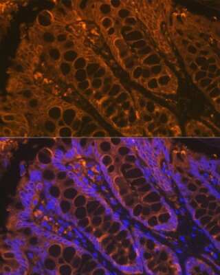
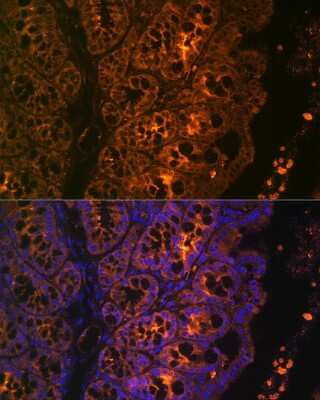
| Localization | Cytoplasmic |
| Marker | Epithelial marker |
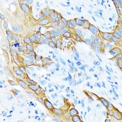
| Specificity | This reacts with cytokeratins of acidic as well as basic subfamilies, i.e. 1,2,3,4,5,6,7,8,10,12,13,14,15,16,17,18 and 19. It reacts with keratinized and corneal epidermis, stratified epithelia, hyperproliferative keratinocytes and simple epithelia. This antibody is a broad spectrum anti-pan cytokeratin antibody, which differentiates epithelial from non-epithelial tumors |
| Isotype | IgG1 |
| Clonality | Monoclonal |
| Host | Mouse |
| Gene | KRT1 |
| Purity | Protein A or G purified |
| Innovator's Reward | Test in a species/application not listed above to receive a full credit towards a future purchase. |
| Dilutions |
|
|
| Application Notes | IHC-P: 1/50 - 1/75 in an ABC method, we suggest an incubation period of 30 minutes at room temperature. Perform enzymatic antigen retrieval before commencing with IHC staining protocol.Not tested in other applications.Optimal dilutions/concentrations should be determined by the end user. |
|
| Publications |
|
| Storage | Store at 4C short term. Aliquot and store at -20C long term. Avoid freeze-thaw cycles. |
| Buffer | PBS |
| Preservative | Sodium Azide |
| Purity | Protein A or G purified |


![Simple Western Albumin Antibody (188835) [Unconjugated] - Serum](https://images.novusbio.com/images2/Albumin_MAB1455_Simple_Western_21888.jpg)
![Immunocytochemistry Albumin Antibody (188835) [Unconjugated] - Serum](https://images.novusbio.com/images2/Albumin_MAB1455_Immunocytochemistry__Immunofluorescence_17777.jpg)
![Immunohistochemistry Albumin Antibody (188835) [Unconjugated] - Serum](https://images.novusbio.com/images2/Albumin_MAB1455_Immunohistochemistry_23317.jpg)


| Publication using NB120-17155 | Applications | Species |
|---|---|---|
| Ordonez NG. Broad-spectrum immunohistochemical epithelial markers: a review. Hum Pathol. 2013-07-01 [PMID: 23427873] (IHC-P, Human) Details: IHC (P): Table 1 |
IHC-P | Human |
Secondary Antibodies |
Isotype Controls |
Research Areas for Cytokeratin, pan Antibody (NB120-17155)Find related products by research area.
|
The concentration calculator allows you to quickly calculate the volume, mass or concentration of your vial. Simply enter your mass, volume, or concentration values for your reagent and the calculator will determine the rest.


![N/A Endostatin [HRP]](https://images.novusbio.com/images2/DATA_Endostatin_DNST0_ELISA_778.jpg)
![N/A Endostatin [HRP]](https://images.novusbio.com/images2/Endostatin_DNST0_ELISA_152.jpg)


