![Immunohistochemistry-Paraffin: Cytokeratin, pan Antibody (C-11, PCK-26, CY-90, KS-1A3, M20, and A5) [NB120-11213] - Staining of FFPE human skin sections with 1:100 Monoclonal Anti-Cytokeratin, pan (Mixture) using Goat Anti-Mouse IgG, FITC-conjugate.](http://images.novusbio.com/fullsize/pan-Cytokeratin-Antibody-C-11-PCK-26-CY-90-KS-1A3-M20-and-A5-Immunohistochemistry-Paraffin-NB120-11213-img0002.jpg "Immunohistochemistry-Paraffin: Cytokeratin, pan Antibody (C-11, PCK-26, CY-90, KS-1A3, M20, and A5) [NB120-11213] - Staining of FFPE human skin sections with 1:100 Monoclonal Anti-Cytokeratin, pan (Mixture) using Goat Anti-Mouse IgG, FITC-conjugate.")
| Immunogen | This Cytokeratin, pan Antibody (C-11, PCK-26, CY-90, KS-1A3, M20, and A5) was developed against a mixture of human pan Cytokeratin. |
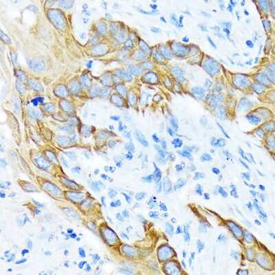
| Marker | Epithelial marker |
| Specificity | This is a mixture of monoclonal pan Cytokeratin antibodies. It recognizes human cytokeratins 1,4,5,6,8,10,13,18, and 19. It is a broad spectrum reagent, which reacts specifically with a wide variety of normal, reactive, and neoplastic epithelial tissues. The antibody mixture reacts with simple, cornifying and non-cornifying squamous epithelia and pseudostratified epithelia. It does not react with non-epithelial normal human tissues. Increased staining intensity is seen following proteolytic treatment (protease unmasking). |
| Isotype | IgG1/IgG2a |
| Clonality | Monoclonal |
| Host | Mouse |
| Gene | KRT |
| Purity | Unpurified |
| Innovator's Reward | Test in a species/application not listed above to receive a full credit towards a future purchase. |
| Dilutions |
|
|
| Publications |
|
| Storage | Store at 4C short term. Aliquot and store at -20C long term. Avoid freeze-thaw cycles. |
| Buffer | Ascites with 7% Equine serum |
| Preservative | 0.09% Sodium Azide |
| Purity | Unpurified |


![Simple Western Albumin Antibody (188835) [Unconjugated] - Serum](https://images.novusbio.com/images2/Albumin_MAB1455_Simple_Western_21888.jpg)
![Immunocytochemistry Albumin Antibody (188835) [Unconjugated] - Serum](https://images.novusbio.com/images2/Albumin_MAB1455_Immunocytochemistry__Immunofluorescence_17777.jpg)
![Immunohistochemistry Albumin Antibody (188835) [Unconjugated] - Serum](https://images.novusbio.com/images2/Albumin_MAB1455_Immunohistochemistry_23317.jpg)


Secondary Antibodies |
Isotype Controls |
Research Areas for Cytokeratin, pan Antibody (NB120-11213)Find related products by research area.
|
The concentration calculator allows you to quickly calculate the volume, mass or concentration of your vial. Simply enter your mass, volume, or concentration values for your reagent and the calculator will determine the rest.


![Immunoblotting: Cytokeratin, pan Antibody (C-11, PCK-26, CY-90, KS-1A3, M20, and A5) [NB120-11213] - Cell line lysates were separated on SDS-PAGE and probed with 1:500 Monoclonal Anti-Cytokeratin, pan (Mixture). The antibody was developed using Goat Anti-Mouse IgG-Peroxidase and a chemiluminescent substrate. Lane 1: HeLa cells . Lane 2: HepG2 cells. Lane 3: COS-7 cells.](http://images.novusbio.com/fullsize/pan-Cytokeratin-Antibody-C-11-PCK-26-CY-90-KS-1A3-M20-and-A5-Immunoblotting-NB120-11213-img0003.jpg "Immunoblotting: Cytokeratin, pan Antibody (C-11, PCK-26, CY-90, KS-1A3, M20, and A5) [NB120-11213] - Cell line lysates were separated on SDS-PAGE and probed with 1:500 Monoclonal Anti-Cytokeratin, pan (Mixture). The antibody was developed using Goat Anti-Mouse IgG-Peroxidase and a chemiluminescent substrate. Lane 1: HeLa cells . Lane 2: HepG2 cells. Lane 3: COS-7 cells.")
![Immunocytochemistry/Immunofluorescence: Cytokeratin, pan Antibody (C-11, PCK-26, CY-90, KS-1A3, M20, and A5) [NB120-11213] - Fetal rat lung epithelial cells were labeled with Monoclonal Anti-pan-Cytokeratin (mixture) (green) and DNA was counterstained with DAPI (blue).](http://images.novusbio.com/fullsize/pan-Cytokeratin-Antibody-C-11-PCK-26-CY-90-KS-1A3-M20-and-A5-Immunocytochemistry-Immunofluorescence-NB120-11213-img0001.jpg "Immunocytochemistry/Immunofluorescence: Cytokeratin, pan Antibody (C-11, PCK-26, CY-90, KS-1A3, M20, and A5) [NB120-11213] - Fetal rat lung epithelial cells were labeled with Monoclonal Anti-pan-Cytokeratin (mixture) (green) and DNA was counterstained with DAPI (blue).")


![N/A Endostatin [HRP]](https://images.novusbio.com/images2/DATA_Endostatin_DNST0_ELISA_778.jpg)
![N/A Endostatin [HRP]](https://images.novusbio.com/images2/Endostatin_DNST0_ELISA_152.jpg)


