 HAF109). A specific band was detected for CREB at approximately 43 kDa (as indicated). This experiment was conducted under reducing conditions and using Immunoblot Buffer Group 1." title="Western blot shows lysates of HT-29 human colon adenocarcinoma cell line, HepG2 human hepatocellular carcinoma cell line, MBA-MB-468 human breast cancer cell line, and MCF-7 human breast cancer cell line. PVDF membrane was probed with 0.5 µg/mL of Goat Anti-Human CREB Antigen Affinity-purified Polyclonal Antibody (Catalog # AF2989) followed by HRP-conjugated Anti-Goat IgG Secondary Antibody (Catalog # HAF109). A specific band was detected for CREB at approximately 43 kDa (as indicated). This experiment was conducted under reducing conditions and using Immunoblot Buffer Group 1." />
| Reactivity | HuSpecies Glossary |
| Applications | WB, IHC, ChIP, ICC/IF |
| Clonality | Polyclonal |
| Host | Goat |
| Conjugate | Unconjugated |
| Concentration | LYOPH |
| Immunogen | E. coli-derived recombinant human CREB Lys136-Asp341 Accession # P16220 |
| Specificity | Detects human CREB in Western blots. |
| Source | N/A |
| Isotype | IgG |
| Clonality | Polyclonal |
| Host | Goat |
| Gene | CREB1 |
| Purity Statement | Antigen Affinity-purified |
| Innovator's Reward | Test in a species/application not listed above to receive a full credit towards a future purchase. |
| Dilutions |
|
|
| Publications |
|
| Storage | Use a manual defrost freezer and avoid repeated freeze-thaw cycles.
|
| Buffer | Lyophilized from a 0.2 μm filtered solution in PBS with Trehalose. *Small pack size (SP) is supplied either lyophilized or as a 0.2 µm filtered solution in PBS. |
| Preservative | No Preservative |
| Concentration | LYOPH |
| Reconstitution Instructions | Reconstitute at 0.2 mg/mL in sterile PBS. |
![Western Blot EphB2 Antibody [Unconjugated]](https://images.novusbio.com/images/af467_human-mouse-ephb2-affinity-purified-polyclonal-ab-41202410485412.jpg)
![Immunohistochemistry EphB2 Antibody [Unconjugated]](https://images.novusbio.com/images/antibody/EphB2_AF467_Immunohistochemistry_20369.jpg)
![Western Blot EphB2 Antibody [Unconjugated]](https://images.novusbio.com/images/antibody/EphB2_AF467_Western_Blot_21511.jpg)

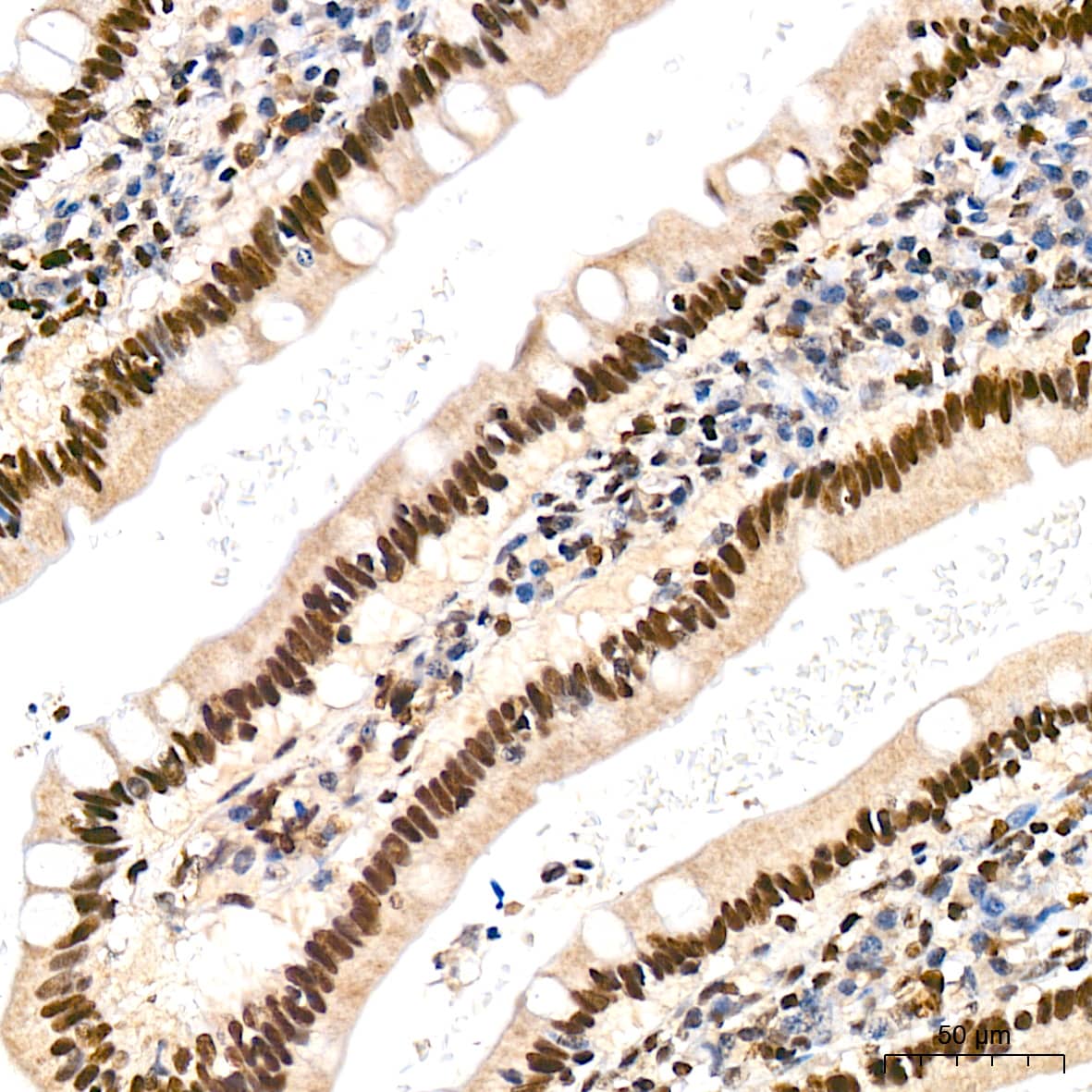

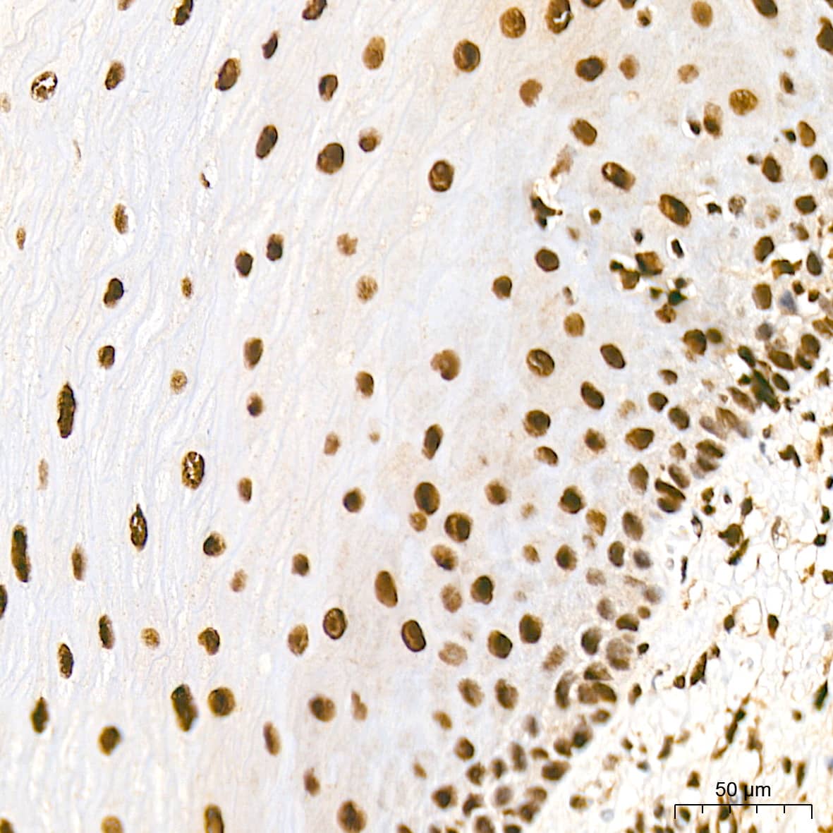

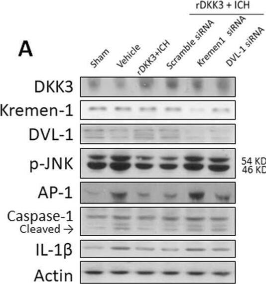

![Intracellular Staining by Flow Cytometry AKT [p Ser473] Antibody [Unconjugated] - Pan Specific](https://images.novusbio.com/images/antibody/Akt3_AF887_Flow_Cytometry_8283.jpg)
![Western Blot AKT [p Ser473] Antibody [Unconjugated] - Pan Specific](https://images.novusbio.com/images/af887_phospho-akt-s473-pan-specific-affinity-purified-pab-41202410485440.jpg)
![Western Blot AKT [p Ser473] Antibody [Unconjugated] - Pan Specific](https://images.novusbio.com/images/af887_phospho-akt-s473-pan-specific-affinity-purified-pab-8120255552843.jpg)


Secondary Antibodies |
Isotype Controls |
|
Detecting HIF alpha and beyond: Best controls for hypoxia Western blot analysis By Rosa Moreno, PhD. Detecting HIF alpha and beyond: Best controls for hypoxia Western blot analysisPhysiological low levels of oxygen induce normal hypoxic events across biological systems. This hypoxic state activ... Read full blog post. |
|
Exploring the Many Roles of PGC-1 alpha The peroxisome proliferator-activated receptor gamma, co-activator 1 (PGC-1 alpha or PPARGC1A) gene encodes a 91 kDa nuclear protein that acts as a transcriptional co-activator involved in energy metabolism. Interaction with PPAR gamma allows it to in... Read full blog post. |
The concentration calculator allows you to quickly calculate the volume, mass or concentration of your vial. Simply enter your mass, volume, or concentration values for your reagent and the calculator will determine the rest.


 AB-108-C) for 15 minutes in an ultrasonic bath, followed by Biotinylated Anti-Goat IgG Secondary Antibody (Catalog #
  CTS008) and counterstained with hematoxylin (blue). Specific labeling was localized to the nucleus in stromal cells. View our protocol for
  CTS013). Tissue was stained using the Anti-Goat HRP-DAB Cell & Tissue Staining Kit (brown; Catalog #
  NL001) and counterstained with DAPI (blue). Specific staining was localized to nuclei. View our protocol for
  NL001). Specific staining was localized to nuclei and cytoplasm. View our protocol for
 ![Immunocytochemistry/ Immunofluorescence c-jun [p Ser63] Antibody (SY0297)](https://images.novusbio.com/images/c-jun-p-Ser63-Antibody-SY0297-Immunocytochemistry-Immunofluorescence-NBP2-67471-img0006.jpg)
![Western Blot c-jun [p Ser63] Antibody (SY0297)](https://images.novusbio.com/images/c-jun-p-Ser63-Antibody-SY0297-Western-Blot-NBP2-67471-img0008.jpg)
![Western Blot c-jun [p Ser63] Antibody (SY0297)](https://images.novusbio.com/images/c-jun-p-Ser63-Antibody-SY0297-Western-Blot-NBP2-67471-img0009.jpg)


![Western Blot ERK2 Antibody [Unconjugated]](https://images.novusbio.com/images/antibody/ERK2_AF1230_Western_Blot_5097.jpg)
![Knockout Validated ERK2 Antibody [Unconjugated]](https://images.novusbio.com/images/antibody/ERK2_AF1230_Knockout_Validated_22864.jpg)
![Immunohistochemistry ERK2 Antibody [Unconjugated]](https://images.novusbio.com/images/antibody/ERK2_AF1230_Immunohistochemistry_20696.jpg)


![N/A BDNF [HRP]](https://images.novusbio.com/images/elisa/BDNF_DBD00_ELISA_36.jpg)
![N/A BDNF [HRP]](https://images.novusbio.com/images/elisa/DATA_BDNF_DBD00_ELISA_587.jpg)
![N/A BDNF [HRP]](https://images.novusbio.com/images/elisa/DATA_BDNF_DBD00_ELISA_588.jpg)
![Bioactivity GDNF [Unconjugated]](https://images.novusbio.com/images/protein/GDNF_212GD_2846.jpg)
![SDS-Page GDNF [Unconjugated]](https://images.novusbio.com/images/protein/GDNF_212-GD_62.jpg)
![SEC-MALS GDNF [Unconjugated]](https://images.novusbio.com/images/212-gd_recombinant-human-gdnf-protein-sec-mals-25202393740..jpg)
![Enzyme Activity u-Plasminogen Activator/Urokinase [Unconjugated]](https://images.novusbio.com/images/protein/uPlasminogen_Activator_1310SE_1837.jpg)
![Immunocytochemistry eIF4E Antibody (299910) [Unconjugated]](https://images.novusbio.com/images/antibody/eIF4E_MAB3228_Immunocytochemistry_6605.jpg)
![Western Blot eIF4E Antibody (299910) [Unconjugated]](https://images.novusbio.com/images/mab3228_human-mouse-rat-eif4e-mab-clone-299910-western-blot-12122025824121.jpg)
![Western Blot eIF4E Antibody (299910) [Unconjugated]](https://images.novusbio.com/images/mab3228_human-mouse-rat-eif4e-mab-clone-299910-western-blot-12122025745315.jpg)

![SDS-Page: CREB Recombinant Protein [H00001385-P01]](https://images.novusbio.com/images/CREB-Recombinant-Protein-SDS-Page-H00001385-P01-img0001.jpg "SDS-Page: CREB Recombinant Protein [H00001385-P01]")


