| Reactivity | BvSpecies Glossary |
| Applications | Bioactivity |
| Format | Carrier-Free |
| Details of Functionality | Measured by the ability of the immobilized protein to support the adhesion of B16‑F1 mouse melanoma cells. When 5 x 10⁴ cells/well are added to Vitronectin coated plates (5 µg/mL with 100 µL/well), approximately >55% will adhere after 30 minutes at 37 °C. Optimal concentration depends on cell type as well as the application or research objectives. |
| Source | Bovine plasma-derived Vitronectin protein |
| N-terminal Sequence | DQESCKGRCT |
| Protein/Peptide Type | Natural Proteins |
| Gene | VTN |
| Purity | >90%, by SDS-PAGE under reducing conditions and visualized by silver stain |
| Endotoxin Note | <0.10 EU per 1 μg of the protein by the LAL method. |
| Dilutions |
|
|
| SDS-PAGE | 58 kDa, 68 kDa and 80 kDa, reducing conditions |
|
| Publications |
|
| Storage | Use a manual defrost freezer and avoid repeated freeze-thaw cycles.
|
| Buffer | Lyophilized from a 0.2 μm filtered solution in PBS and Urea. |
| Purity | >90%, by SDS-PAGE under reducing conditions and visualized by silver stain |
| Reconstitution Instructions | Reconstitute at 100 μg/mL in sterile PBS. |
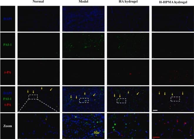
Vitronectin is a larger glycoprotein found in blood and in the extracellular matrix (ECM). The amino terminal segment of vitronectin harbors a binding site (aa 1 ‑ 44) for plasminogen activator inhibitor-1 (PAI‑1) and urokinase receptor, an Agr-Gly-ASP (RGD) sequence (aa 45 - 47) that provides a binding site for alpha v beta 3, alpha v beta 5, alpha v beta 1, alpha IIb beta 3, alpha v beta 6, and alpha v beta 8 integrins, a stretch of acidic amino acids including two sulfated tyrosine residues (aa 56 and 59) that provide a binding site for thrombin-anti-thrombin III complexes, and a collagen binding site. The major part of the vitronecitn molecule (aa 132 - 459) accommodate six hemopexin repeats. The carboxyl-terminal end of vitronectin containing a stretch of basic amino acids (aa 348 - 379) that binds the acidic stretch of acidic amino acids in the amino-terminal section and stabilized vitronectin’s three dimensional structure. The carboxyl-terminal end of vitronectin also contains a plaminogen binding site (aa 332 ‑ 348), a heparin binding site that can be bound by complement factor C7, C8 or C9 (aa 348 ‑ 376), a glycosaminoglycan binding site (aa 348 ‑ 361), and a second PAI-1 binding site (aa 348 ‑ 370). Vitronectin also contains an endogenous cleavage site, two elastase cleavage sites, two thrombin cleavage sites, and a plasmin cleavage site. Vitronectin also has been shown to bind insulin growth factor II (IGF‑II) and TGF-beta . The apparent molecular weight of bovine vitronectin is 70 kDa, with ~15% of its molecular mass being contributed to by glycosylation. In blood and plasma, vitronectin is found predominantly as a single chain monomer. It can also be found as a dimer after endogenous cleavage. The dimer is comprised of a 65 kDa and 10 kDa component held together by a disulfide bond. Binding of thrombin-anti-thrombin II complex or complement lead to an unfolding of vitronectin. Unfolding of vitronectin leads to the formation of disulfide-linked multimers that are found in platelet releasate and in the extracellular matrix. Vitronectin is produced at high levels by the liver and many tumors. Vitronectin is involved in a number of biological functions including cell adhesion, cell spreading and migration, cell proliferation, extracellular anchoring, fibrinolysis, hemostasis, and complement immune defense.


![Western Blot FAK [p Tyr397] Antibody](https://images.novusbio.com/images/nbp3-12897_rabbit-fak-p-tyr-397-pab-western-blot-2611202518381522.jpg)
![Western Blot FAK [p Tyr397] Antibody](https://images.novusbio.com/images/FAK-[p-Tyr397]-Antibody-Western-Blot-NBP3-12897-img0006.jpg)
![Western Blot FAK [p Tyr397] Antibody](https://images.novusbio.com/images/nbp3-12897_rabbit-fak-p-tyr-397-pab-western-blot-261120251843458.jpg)
![Western Blot Osteopontin/OPN Antibody [Unconjugated]](https://images.novusbio.com/images/af808_mouse-osteopontin-opn-affinity-purified-polyclonal-ab-western-blot-121220259133019.jpg)
![Western Blot Osteopontin/OPN Antibody [Unconjugated]](https://images.novusbio.com/images/af808_mouse-osteopontin-opn-affinity-purified-polyclonal-ab-western-blot-12122025932150.jpg)
![Western Blot Osteopontin/OPN Antibody [Unconjugated]](https://images.novusbio.com/images/af808_mouse-osteopontin-opn-affinity-purified-polyclonal-ab-western-blot-121220259224819.jpg)


![Immunohistochemistry Albumin Antibody (188835) [Unconjugated] - Serum](https://images.novusbio.com/images/antibody/Albumin_MAB1455_Immunohistochemistry_23317.jpg)
![Immunocytochemistry Albumin Antibody (188835) [Unconjugated] - Serum](https://images.novusbio.com/images/antibody/Albumin_MAB1455_Immunocytochemistry__Immunofluorescence_17777.jpg)
![Intracellular Staining by Flow Cytometry Albumin Antibody (188835) [Unconjugated] - Serum](https://images.novusbio.com/images/antibody/Albumin_MAB1455_Flow_Cytometry_15110.jpg)


The concentration calculator allows you to quickly calculate the volume, mass or concentration of your vial. Simply enter your mass, volume, or concentration values for your reagent and the calculator will determine the rest.
| Gene Symbol | VTN |


![Enzyme Activity u-Plasminogen Activator/Urokinase [Unconjugated]](https://images.novusbio.com/images/protein/uPlasminogen_Activator_1310SE_1837.jpg)
![N/A uPAR [HRP]](https://images.novusbio.com/images/elisa/uPAR_DUP00_ELISA_203.jpg)
![N/A uPAR [HRP]](https://images.novusbio.com/images/elisa/DATA_uPAR_DUP00_ELISA_866.jpg)


![Immunocytochemistry/ Immunofluorescence Integrin alpha V beta 5 Antibody (P5H9) [Unconjugated]](https://images.novusbio.com/images/mab2528_human-integrin-alpha-v-beta-5-mab-clone-p5h9-41202410481113.jpg)
![Immunocytochemistry Integrin alpha V beta 5 Antibody (P5H9) [Unconjugated]](https://images.novusbio.com/images/antibody/Integrin_beta_5_MAB2528_Immunocytochemistry_9856.jpg)
![Flow Cytometry Integrin alpha V beta 5 Antibody (P5H9) [Unconjugated]](https://images.novusbio.com/images/antibody/mab2528_human-integrin-alpha-v-beta-5-mab-clone-p5h9-flow-cytometry-1722026101014.jpg)

![Bioactivity CTLA-4 [Unconjugated]](https://images.novusbio.com/images/protein/CTLA4_7268CT_2293.jpg)
![SDS-Page TNF-alpha [Unconjugated]](https://images.novusbio.com/images/protein/TNF-alpha_210-TA_256.jpg)
![Bioactivity TNF-alpha [Unconjugated]](https://images.novusbio.com/images/protein/TNFalpha_210TA_1658.jpg)
![SEC-MALS TNF-alpha [Unconjugated]](https://images.novusbio.com/images/210-ta_recombinant-human-tnf-alpha-protein-sec-mals-35202312244..jpg)

![Western Blot Clusterin/APOJ Antibody [Unconjugated]](https://images.novusbio.com/images/af2747_mouse-clusterin-affinity-purified-polyclonal-ab-41202412403576.jpg)
![Western Blot Clusterin/APOJ Antibody [Unconjugated]](https://images.novusbio.com/images/af2747_mouse-clusterin-affinity-purified-polyclonal-ab-4120241240247.jpg)
![Western Blot Clusterin/APOJ Antibody [Unconjugated]](https://images.novusbio.com/images/af2747_mouse-clusterin-affinity-purified-polyclonal-ab-41202412402523.jpg)
![Flow Cytometry Integrin beta 1/CD29 Antibody (P5D2) [Unconjugated]](https://images.novusbio.com/images/antibody/Integrin_beta_1_MAB17781_Flow_Cytometry_17077.jpg)
![Flow Cytometry Integrin beta 1/CD29 Antibody (P5D2) [Unconjugated]](https://images.novusbio.com/images/antibody/Integrin_beta_1_MAB17781_Flow_Cytometry_23861.jpg)
![Western Blot ERK2 Antibody [Unconjugated]](https://images.novusbio.com/images/antibody/ERK2_AF1230_Western_Blot_5097.jpg)
![Knockout Validated ERK2 Antibody [Unconjugated]](https://images.novusbio.com/images/antibody/ERK2_AF1230_Knockout_Validated_22864.jpg)
![Immunohistochemistry ERK2 Antibody [Unconjugated]](https://images.novusbio.com/images/antibody/ERK2_AF1230_Immunohistochemistry_20696.jpg)