MAB8214) at 20 µg/mL at 37 ° Celsius for 4 minutes. Before incubation with the primary antibody, tissue underwent an all-in-one dewaxing and antigen retrieval preprocessing using PreTreatment Module (PT Module) and Dewax and HIER Buffer H (pH 9). Tissue was stained using the Alexa Fluor™ Plus 647 Goat anti-Rabbit IgG Secondary Antibody at 1:200 at 37 ° Celsius for 2 minutes. (Yellow; Lunaphore Catalog #
DR647RB) and counterstained with DAPI (blue; Lunaphore Catalog #
DR100). Specific staining was localized to the nucleus. Protocol available in
COMET™ Panel Builder." class="big_lightbox">

MAB8214) at 20 µg/mL at 37 ° Celsius for 4 minutes. Before incubation with the primary antibody, tissue underwent an all-in-one dewaxing and antigen retrieval preprocessing using PreTreatment Module (PT Module) and Dewax and HIER Buffer H (pH 9). Tissue was stained using the Alexa Fluor™ Plus 647 Goat anti-Rabbit IgG Secondary Antibody at 1:200 at 37 ° Celsius for 2 minutes. (Yellow; Lunaphore Catalog #
DR647RB) and counterstained with DAPI (blue; Lunaphore Catalog #
DR100). Specific staining was localized to the nucleus. Protocol available in
COMET™ Panel Builder." alt="FOXP3 was detected in immersion fixed paraffin-embedded sections of Hodgkin's Lymphoma using Rabbit Anti-Human FOXP3 Monoclonal Antibody (Catalog #
MAB8214) at 20 µg/mL at 37 ° Celsius for 4 minutes. Before incubation with the primary antibody, tissue underwent an all-in-one dewaxing and antigen retrieval preprocessing using PreTreatment Module (PT Module) and Dewax and HIER Buffer H (pH 9). Tissue was stained using the Alexa Fluor™ Plus 647 Goat anti-Rabbit IgG Secondary Antibody at 1:200 at 37 ° Celsius for 2 minutes. (Yellow; Lunaphore Catalog #
DR647RB) and counterstained with DAPI (blue; Lunaphore Catalog #
DR100). Specific staining was localized to the nucleus. Protocol available in
COMET™ Panel Builder." class="big_thumb" />
MAB8214) at 20ug/mL at 37 ° Celsius for 4 minutes. Before incubation with the primary antibody, tissue underwent an all-in-one dewaxing and antigen retrieval preprocessing using PreTreatment Module (PT Module) and Dewax and HIER Buffer H (pH 9; Epredia Catalog # TA-999-DHBH). Tissue was stained using the Alexa Fluor™ Plus 647 Goat anti-Rabbit IgG Secondary Antibody at 1:200 at 37 ° Celsius for 2 minutes. (Yellow; Lunaphore Catalog #
DR647RB) and counterstained with DAPI (blue; Lunaphore Catalog #
DR100). Specific staining was localized to the nucleus. Protocol available in
COMET™ Panel Builder." class="big_lightbox">
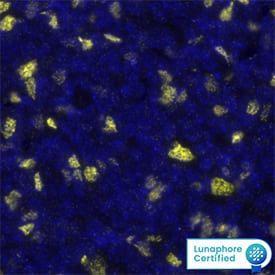
MAB8214) at 20ug/mL at 37 ° Celsius for 4 minutes. Before incubation with the primary antibody, tissue underwent an all-in-one dewaxing and antigen retrieval preprocessing using PreTreatment Module (PT Module) and Dewax and HIER Buffer H (pH 9; Epredia Catalog # TA-999-DHBH). Tissue was stained using the Alexa Fluor™ Plus 647 Goat anti-Rabbit IgG Secondary Antibody at 1:200 at 37 ° Celsius for 2 minutes. (Yellow; Lunaphore Catalog #
DR647RB) and counterstained with DAPI (blue; Lunaphore Catalog #
DR100). Specific staining was localized to the nucleus. Protocol available in
COMET™ Panel Builder." class="big_thumb" />
240-B) and Recombinant Human IL-2 (Catalog #
202-IL) for 2 days were stained with Rabbit Anti-Human/Mouse FoxP3 Monoclonal Antibody (Catalog # MAB8214) followed by Phycoerythrin-conjugated Anti-Rabbit IgG Secondary Antibody (Catalog #
F0110) and Mouse Anti-Human CD4 APC-conjugated Monoclonal Antibody (Catalog #
FAB3791A). Quadrant markers were set based on control antibody staining (Catalog #
AB-105-C). To facilitate intracellular staining, cells were fixed and permeabilized with FlowX FoxP3 Fixation & Permeabilization Buffer Kit (Catalog #
FC012)." class="big_lightbox">
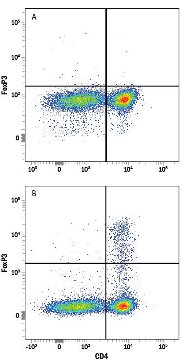
240-B) and Recombinant Human IL-2 (Catalog #
202-IL) for 2 days were stained with Rabbit Anti-Human/Mouse FoxP3 Monoclonal Antibody (Catalog # MAB8214) followed by Phycoerythrin-conjugated Anti-Rabbit IgG Secondary Antibody (Catalog #
F0110) and Mouse Anti-Human CD4 APC-conjugated Monoclonal Antibody (Catalog #
FAB3791A). Quadrant markers were set based on control antibody staining (Catalog #
AB-105-C). To facilitate intracellular staining, cells were fixed and permeabilized with FlowX FoxP3 Fixation & Permeabilization Buffer Kit (Catalog #
FC012)." class="big_thumb" />
FAB554A) and either (A) Rabbit Anti-Human/Mouse FoxP3 Monoclonal Antibody (Catalog # MAB8214) or (B) Normal Rabbit IgG Control (Catalog #
AB-105-C) followed by Phycoerythrin-conjugated Anti-Rabbit IgG Secondary Antibody (Catalog #
F0110). To facilitate intracellular staining, cells were fixed and permeabilized with FlowX FoxP3 Fixation & Permeabilization Buffer Kit (Catalog #
FC012)." class="big_lightbox">
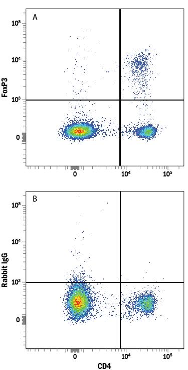
FAB554A) and either (A) Rabbit Anti-Human/Mouse FoxP3 Monoclonal Antibody (Catalog # MAB8214) or (B) Normal Rabbit IgG Control (Catalog #
AB-105-C) followed by Phycoerythrin-conjugated Anti-Rabbit IgG Secondary Antibody (Catalog #
F0110). To facilitate intracellular staining, cells were fixed and permeabilized with FlowX FoxP3 Fixation & Permeabilization Buffer Kit (Catalog #
FC012)." class="big_thumb" />
VC003). Tissue was stained using ACD RNAscope
®2.5 HD Duplex Detection Reagents (Catalog # 322500)." class="big_lightbox">
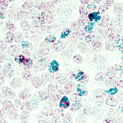
VC003). Tissue was stained using ACD RNAscope
®2.5 HD Duplex Detection Reagents (Catalog # 322500)." class="big_thumb" />


VC003). Before incubation with the primary antibody, tissue was subjected to heat-induced epitope retrieval using VisUCyte Antigen Retrieval Reagent-Basic (Catalog #
VCTS021). Tissue was stained using DAB (brown) and counterstained with hematoxylin (blue). Specific staining was localized to the nucleus. View our protocol for
IHC Staining with VisUCyte HRP Polymer Detection Reagents." class="big_lightbox">
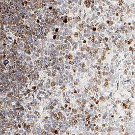
VC003). Before incubation with the primary antibody, tissue was subjected to heat-induced epitope retrieval using VisUCyte Antigen Retrieval Reagent-Basic (Catalog #
VCTS021). Tissue was stained using DAB (brown) and counterstained with hematoxylin (blue). Specific staining was localized to the nucleus. View our protocol for
IHC Staining with VisUCyte HRP Polymer Detection Reagents." class="big_thumb" />
 MAB8214) at 20 µg/mL at 37 ° Celsius for 4 minutes. Before incubation with the primary antibody, tissue underwent an all-in-one dewaxing and antigen retrieval preprocessing using PreTreatment Module (PT Module) and Dewax and HIER Buffer H (pH 9). Tissue was stained using the Alexa Fluor™ Plus 647 Goat anti-Rabbit IgG Secondary Antibody at 1:200 at 37 ° Celsius for 2 minutes. (Yellow; Lunaphore Catalog # DR647RB) and counterstained with DAPI (blue; Lunaphore Catalog # DR100). Specific staining was localized to the nucleus. Protocol available in COMET™ Panel Builder." alt="FOXP3 was detected in immersion fixed paraffin-embedded sections of Hodgkin's Lymphoma using Rabbit Anti-Human FOXP3 Monoclonal Antibody (Catalog # MAB8214) at 20 µg/mL at 37 ° Celsius for 4 minutes. Before incubation with the primary antibody, tissue underwent an all-in-one dewaxing and antigen retrieval preprocessing using PreTreatment Module (PT Module) and Dewax and HIER Buffer H (pH 9). Tissue was stained using the Alexa Fluor™ Plus 647 Goat anti-Rabbit IgG Secondary Antibody at 1:200 at 37 ° Celsius for 2 minutes. (Yellow; Lunaphore Catalog # DR647RB) and counterstained with DAPI (blue; Lunaphore Catalog # DR100). Specific staining was localized to the nucleus. Protocol available in COMET™ Panel Builder." class="big_thumb" />
 HAF019). A specific band was detected for FoxP3 at approximately 47 kDa (as indicated). This experiment was conducted under reducing conditions and using Immunoblot Buffer Group 8." alt="Western blot shows lysates of human CD4+CD25+T cells and human CD4+CD25-T cells. PVDF membrane was probed with 1 µg/mL of Goat Anti-Human FoxP3 Antigen Affinity-purified Polyclonal Antibody (Catalog # AF3240) followed by HRP-conjugated Anti-Goat IgG Secondary Antibody (Catalog # HAF019). A specific band was detected for FoxP3 at approximately 47 kDa (as indicated). This experiment was conducted under reducing conditions and using Immunoblot Buffer Group 8." class="big_thumb" />
 F0102B) and Mouse Anti-Human CD4 APC-conjugated Monoclonal Antibody (Catalog # FAB3791A). Quadrant markers were set based on control antibody staining (Catalog # MAB002). To facilitate intracellular staining, cells were fixed and permeabilized with FlowX FoxP3 Fixation & Permeabilization Buffer Kit (Catalog # FC012)." alt="Human peripheral blood mononuclear cells (PBMCs) were stained with Mouse Anti-Human/Mouse/Rat FoxP3 Monoclonal Antibody (Catalog # MAB8970) followed by Phycoerythrin-conjugated Anti-Mouse IgG Secondary Antibody (Catalog # F0102B) and Mouse Anti-Human CD4 APC-conjugated Monoclonal Antibody (Catalog # FAB3791A). Quadrant markers were set based on control antibody staining (Catalog # MAB002). To facilitate intracellular staining, cells were fixed and permeabilized with FlowX FoxP3 Fixation & Permeabilization Buffer Kit (Catalog # FC012)." class="big_thumb" />


![Immunohistochemistry-Paraffin: FoxP3 Antibody [NB100-39002] - Mouse spleen stained with FoxP3 antibody. IHC-P image submitted by a verified customer review.](http://images.novusbio.com/fullsize/FoxP3-Antibody-Immunohistochemistry-Paraffin-NB100-39002-img0011.jpg "Immunohistochemistry-Paraffin: FoxP3 Antibody [NB100-39002] - Mouse spleen stained with FoxP3 antibody. IHC-P image submitted by a verified customer review.")


![Flow Cytometry: FoxP3 Antibody [NB100-39002] - An intracellular stain was performed on SK-MEL-28 cells with NB100-39002 (blue) and a matched isotype control (orange). Cells were fixed with 4% PFA and then permeabilized with 0.1% saponin. Cells were incubated in an antibody dilution of 2.5 ug/mL for 30 minutes at room temperature, followed by Rabbit IgG (H+L) Cross-Adsorbed Secondary Antibody.](http://images.novusbio.com/fullsize/FoxP3-Antibody-Flow-Cytometry-NB100-39002-img0022.jpg)
![Immunohistochemistry-Paraffin: FoxP3 Antibody [NB100-39002] - Tissue section of human tonsil using 1:200 dilution of rabbit anti-FoxP3 antibody. The staining was developed with HRP-labeled anti-rabbit IgG secondary antibody and DAB reagent, and nuclei of cells were counter-stained with hematoxylin. This FoxP3 antibody generated an expected and a specific nuclear staining of FOXP3 in a subset of cells which are potentially the Treg cells.](http://images.novusbio.com/fullsize/FoxP3-Antibody-Immunohistochemistry-Paraffin-NB100-39002-img0017.jpg)
![Immunohistochemistry: FoxP3 Antibody [NB100-39002] - Antibody was tested in mouse spleen using DAB with hematoxylin counterstain.](http://images.novusbio.com/fullsize/FoxP3-Antibody-Immunohistochemistry-NB100-39002-img0014.jpg)
![Immunohistochemistry-Paraffin: FoxP3 Antibody [NB100-39002] - FFPE bovine spleen tissue. Deparaffinization and antigen retrieval was carried out with PTlink from Dako, citrate buffer pH 6. Anti-Foxp3 antibody was used at a 1:100 dilution. Secondary reagent: Envision, from Dako. Staining: DAB and hematoxylin. IHC-P image submitted by a verified customer review.](http://images.novusbio.com/fullsize/FoxP3-Antibody-Immunohistochemistry-Paraffin-NB100-39002-img0024.jpg)
![Western Blot: FoxP3 Antibody [NB100-39002] - Jurkat Tag cells were transfected with the expression vector (vector) or with the Foxp3 expression construct (Foxp3). 24 hours after transfection, cells were lysed and analyzed by Western blot with anti-Foxp3 antiserum (1:2000 dilution).](http://images.novusbio.com/fullsize/FoxP3-Antibody-Western-Blot-NB100-39002-img0018.jpg)
![Immunocytochemistry/Immunofluorescence: FoxP3 Antibody [NB100-39002] - Transfectants were stained with anti-Foxp3 antibody and HRPO-conjugated (upper panel) or Cy3-conjugated (lower panels) secondary antibody. Representative images (x100) are shown.](http://images.novusbio.com/fullsize/FoxP3-Antibody-Immunocytochemistry-Immunofluorescence-NB100-39002-img0012.jpg)
![Immunohistochemistry-Paraffin: FoxP3 Antibody [NB100-39002] - Tissue section of human tonsil using 1:200 dilution of rabbit anti-FoxP3 antibody. The staining was developed with HRP-labeled anti-rabbit IgG secondary antibody and DAB reagent, and nuclei of cells were counterstained with hematoxylin. This FoxP3 antibody generated an expected and a specific nuclear staining of FOXP3 in a subset of cells which are potentially the Treg cells.](http://images.novusbio.com/fullsize/FoxP3-Antibody-Immunohistochemistry-Paraffin-NB100-39002-img0015.jpg)
![Flow Cytometry: FoxP3 Antibody [NB100-39002] - Transfectants were permeabilized and stained with anti-Foxp3 antibody and FITC-conjugated secondary antibody. Cells were analyzed by flow cytometry. Percentage of positively stained cells are shown.](http://images.novusbio.com/fullsize/FoxP3-Antibody-Flow-Cytometry-NB100-39002-img0013.jpg)
![Flow Cytometry: FoxP3 Antibody [NB100-39002] - An intracellular stain was performed on MCF7 cells with FoxP3 Antibody NB100-39002PE (blue) and a matched isotype control (orange). Cells were fixed with 4% PFA and then permeablized with 0.1% saponin. Cells were incubated in an antibody dilution of 5 ug/mL for 30 minutes at room temperature. Both antibodies were conjugated to Phycoerythrin.](http://images.novusbio.com/fullsize/FoxP3-Antibody-Flow-Cytometry-NB100-39002-img0021.jpg)
![Dual RNAscope ISH-IHC: FoxP3 Antibody [NB100-39002] - CD8A mRNA (red) and FOXP3 protein (green) were detected in formalin-fixed paraffin-embedded tissue sections of human breast cancer. ACD's Integrated Co-Detection Workflow was performed using ACD RNAScope Probe Hs-CD8A and FoxP3 antibody at 1:400 dilution. Tissue was stained on Leica Bond RX using RNAscope (TM) 2.5 LS Reagent Kit-RED, BOND Polymer Refine Detection (DAB) and Hematoxylin, BOND Polymer Refine Red Detection and Hematoxylin and RNAscope (TM) 2.5 LS Green Accessory Pack. Tissue was counterstained with 50% hematoxylin (blue).](http://images.novusbio.com/fullsize/FoxP3-Antibody-Dual-RNAscope-ISH-IHC-NB100-39002-img0028.jpg)
![Dual RNAscope ISH-IHC: FoxP3 Antibody [NB100-39002] - CD4 mRNA (red) and FOXP3 protein (green) were detected in formalin-fixed paraffin-embedded tissue sections of human breast cancer. ACD's Integrated Co-Detection Workflow was performed using ACD RNAScope Probe Hs-CD4 and FOXP3 antibody at 1:200 dilution. Tissue was stained on Leica Bond RX using RNAscope (TM) 2.5 LS Reagent Kit-RED, BOND Polymer Refine Detection (DAB) and Hematoxylin, BOND Polymer Refine Red Detection and Hematoxylin and RNAscope (TM) 2.5 LS Green Accessory Pack. Tissue was counterstained with 50% hematoxylin (blue).](http://images.novusbio.com/fullsize/FoxP3-Antibody-Dual-RNAscope-ISH-IHC-NB100-39002-img0029.jpg)


![Immunocytochemistry/ Immunofluorescence: FoxP3 Antibody - BSA Free [NB100-39002] - CCR10-expressing CD4+CD25+FOXP3+ Treg cells are enriched in the spinal cord after SCI. (A, B) Representative flow cytometry dot plots (A) & percentage (B) of CD4+CD25+FOXP3+ cells among the CD4+ T cells in the spinal cord after sham or SCI surgery. Cells were gated on 7-AAD negative population & the numbers inside plots refer to % Treg cells (n=5). (C, D) Representative histogram of CCR10 (C) & CCR3 (D) expression in CD4+CD25+FOXP3+ cells as gated in (A). (E) CD4+CD25+FOXP3+ cells in the spinal cord from sham & SCI mice were sorted out & analyzed by Western blotting to detect the expression of CCR10 & CCR3. GAPDH was used as a loading control. (F) Double immunostaining of FOXP3 (red) & CCR10 (green) in the spinal cord sections after sham or SCI surgery. The merged images are also shown. Arrows indicate cells with positive staining & colocalization. Scale bar, 100 μm. Data are mean ± SD. The statistical analysis was performed using Student’s t-test. **, P<0.01. Image collected & cropped by CiteAb from the following publication (//pubmed.ncbi.nlm.nih.gov/31557129), licensed under a CC-BY license. Not internally tested by Novus Biologicals.](http://images.novusbio.com/fullsize/nb100-39002_rabbit-polyclonal-foxp3-antibody-310202415541911.jpg)
![Immunocytochemistry/ Immunofluorescence: FoxP3 Antibody - BSA Free [NB100-39002] - Effects of anaphylatoxins on transcription factor phosphorylation in ARPE-19 cells. (A–C) Protein phosphorylation of (A) CREB (Ser133); (B) FoxP3 (Ser418); & (C) FOXO1 (Ser256) was examined in ARPE-19 cells treated with C3a, C5a, or the combination of C3a/C5a at 1, 5, 10, & 15 min. Phosphorylation levels were normalized to beta -actin (relative p-protein levels), & slopes of the time course were determined. Anaphylatoxins were applied at concentrations of 300 nM (C3a) & 50 nM (C5a). Data are mean + SEM, n = 3, *p < 0.05, ***p < 0.001 (Student’s t-test). (D) Immunofluorescence staining of FoxP3. Upper panel: Lewis rat (albino) eye with experimental uveitis; lower panel: normal rat eye. Asterisks mark immune cells infiltrating the retina from the choroid through the retinal pigment epithelium (RPE) in the eye with uveitis (clinical score of 2, histological score of 1). Image collected & cropped by CiteAb from the following publication (//journal.frontiersin.org/article/10.3389/fimmu.2017.00703/full), licensed under a CC-BY license. Not internally tested by Novus Biologicals.](http://images.novusbio.com/fullsize/nb100-39002_rabbit-polyclonal-foxp3-antibody-31020241554547.jpg)
 MAB8214) at 20ug/mL at 37 ° Celsius for 4 minutes. Before incubation with the primary antibody, tissue underwent an all-in-one dewaxing and antigen retrieval preprocessing using PreTreatment Module (PT Module) and Dewax and HIER Buffer H (pH 9; Epredia Catalog # TA-999-DHBH). Tissue was stained using the Alexa Fluor™ Plus 647 Goat anti-Rabbit IgG Secondary Antibody at 1:200 at 37 ° Celsius for 2 minutes. (Yellow; Lunaphore Catalog #
  240-B) and Recombinant Human IL-2 (Catalog #
  FAB554A) and either (A) Rabbit Anti-Human/Mouse FoxP3 Monoclonal Antibody (Catalog # MAB8214) or (B) Normal Rabbit IgG Control (Catalog #
  NL004) and counterstained with DAPI (blue, lower panel). Specific staining was localized to nuclei. View our protocol for
  CTS005) and counterstained with hematoxylin (blue). Specific staining was localized to nuclei. View our protocol for
  VC003). Tissue was stained using DAB (brown) and counterstained with hematoxylin (blue). Specific staining was localized to nuclei. View our protocol for
  VC003). Tissue was stained using ACD RNAscope®2.5 HD Duplex Detection Reagents (Catalog # 322500)." class="big_thumb" />


 VC003). Before incubation with the primary antibody, tissue was subjected to heat-induced epitope retrieval using VisUCyte Antigen Retrieval Reagent-Basic (Catalog #
 ![Immunohistochemistry-Frozen: FoxP3 Antibody - BSA Free [NB600-245] - FoxP3 Antibody [NB600-245] - Healthy thymus from a C57BL/6 mouse stained with FoxP3 and CD4.](http://images.novusbio.com/fullsize/FoxP3-Antibody-Immunocytochemistry-Immunofluorescence-NB600-245-img0013.jpg "Immunohistochemistry-Frozen: FoxP3 Antibody - BSA Free [NB600-245] - FoxP3 Antibody [NB600-245] - Healthy thymus from a C57BL/6 mouse stained with FoxP3 and CD4.")
![Immunohistochemistry-Frozen: FoxP3 Antibody - BSA Free [NB600-245] - FoxP3 Antibody [NB600-245] - Healthy thymus from a C57BL/6 mouse incubated only with CD4 and no Foxp3.](http://images.novusbio.com/fullsize/FoxP3-Antibody-Immunocytochemistry-Immunofluorescence-NB600-245-img0012.jpg)
![Immunohistochemistry-Paraffin: FoxP3 Antibody [NB600-245] - Fox P3 was detected in immersion fixed paraffin-embedded sections of human tonsil using rabbit anti-human antibody (Catalog # NB600-245) at 1:100 dilution overnight at 4C. Tissue was stained using the VisuCyte anti-rabbit HRP polymer detection reagent (Catalog # VC003) with DAB chromogen (brown) and counterstained with hematoxylin (blue).<br/>Images may not be copied, printed or otherwise disseminated without express written permission of Novus Biologicals a bio-techne brand.](http://images.novusbio.com/fullsize/FoxP3-Antibody-Immunohistochemistry-Paraffin-NB600-245-img0011.jpg)
![Western Blot: FOXP3 Antibody [NB600-245] - Detection of human FOXP3 using NB 600-245. Lane 1: Human CD4+CD25+ PBL, lane 2: HEK293T transfected with human Foxp3 cDNA, lane 3: 293/mouse foxp3, lane 4: 293/empty vector](http://images.novusbio.com/fullsize/FOXP3-Antibody-Western-Blot-NB600-245-img0009.jpg)
![Dual RNAscope ISH-IHC: FoxP3 Antibody [NB600-245] - CD8A mRNA (red) and FOXP3 protein (green) were detected in formalin-fixed paraffin-embedded tissue sections of human breast cancer. ACD's Integrated Co-Detection Workflow was performed using ACD RNAScope Probe Hs-CD8A and FOXP3 antibody at 1:100 dilution. Tissue was stained on Leica Bond RX using RNAscope (TM) 2.5 LS Reagent Kit-RED, BOND Polymer Refine Detection (DAB) and Hematoxylin, BOND Polymer Refine Red Detection and Hematoxylin and RNAscope (TM) 2.5 LS Green Accessory Pack. Tissue was counterstained with 50% hematoxylin (blue).](http://images.novusbio.com/fullsize/FoxP3-Antibody-Dual-RNAscope-ISH-IHC-NB600-245-img0014.jpg)
![Dual RNAscope ISH-IHC: FoxP3 Antibody [NB600-245] - CD4 mRNA (red) and FOXP3 protein (green) were detected in formalin-fixed paraffin-embedded tissue sections of human breast cancer. ACD's Integrated Co-Detection Workflow was performed using ACD RNAScope Probe Hs-CD4 and FOXP3 antibody at 1:200 dilution. Tissue was stained on Leica Bond RX using RNAscope (TM) 2.5 LS Reagent Kit-RED, BOND Polymer Refine Detection (DAB) and Hematoxylin, BOND Polymer Refine Red Detection and Hematoxylin and RNAscope (TM) 2.5 LS Green Accessory Pack. Tissue was counterstained with 50% hematoxylin (blue).](http://images.novusbio.com/fullsize/FoxP3-Antibody-Dual-RNAscope-ISH-IHC-NB600-245-img0015.jpg)

![Western Blot: FoxP3 Antibody [NB600-242] - (1 ug/mL) staining of Human Muscle (A), (2 ug/mL) MOLT4 (B) and (1 ug/mL) negative control Pancreas (C) lysate (35 ug protein in RIPA buffer). Detected by chemiluminescence.](http://images.novusbio.com/fullsize/FoxP3-Antibody-Western-Blot-NB600-242-img0010.jpg "Western Blot: FoxP3 Antibody [NB600-242] - (1 ug/mL) staining of Human Muscle (A), (2 ug/mL) MOLT4 (B) and (1 ug/mL) negative control Pancreas (C) lysate (35 ug protein in RIPA buffer). Detected by chemiluminescence.")
![Immunocytochemistry: FoxP3 Antibody [NB600-242] - (Treg) human blood cells gathered by cytospin and detected by FITC (A) and in phase contrast (B).](http://images.novusbio.com/fullsize/FoxP3-Antibody-Immunocytochemistry-Immunofluorescence-NB600-242-img0008.jpg)
![Immunohistochemistry: FoxP3 Antibody [NB600-242] - (8ug/ml) staining of paraffin embedded Human Spleen. Heat induced antigen retrieval with citrate buffer Ph 6, HRP-staining.](http://images.novusbio.com/fullsize/FoxP3-Antibody-Immunohistochemistry-NB600-242-img0012.jpg)
![Flow Cytometry: FoxP3 Antibody [NB600-242] - Analysis of paraformaldehyde fixed Jurkat cells (blue line), permeabilized with 0.5% Triton. Primary incubation 1hr (10 ug/mL) followed by Alexa Fluor 488 secondary antibody (4 ug/mL). IgG control: Unimmunized goat IgG (black line) followed by Alexa Fluor 488 secondary antibody.](http://images.novusbio.com/fullsize/FoxP3-Antibody-Flow-Cytometry-NB600-242-img0009.jpg)
![Immunofluorescence: FoxP3 Antibody [NB600-242] - Paraformaldehyde fixed U2OS cells, permeabilized with 0.15% Triton. Primary incubation 1hr (10 ug/mL) followed by Alexa Fluor 488 secondary antibody (4 ug/mL), showing strong localization to nucleoplasm. The nuclear stain is DAPI (blue). Negative control: Unimmunized goat IgG (10 ug/mL) followed by Alexa Fluor 488 secondary antibody (4 ug/mL).](http://images.novusbio.com/fullsize/FoxP3-Antibody-Immunocytochemistry-Immunofluorescence-NB600-242-img0011.jpg)
![Control: FoxP3 Antibody [NB600-242] - Negative Control showing staining of paraffin embedded Human Spleen, with no primary antibody.](http://images.novusbio.com/fullsize/FoxP3-Antibody-Control-NB600-242-img0013.jpg)
![Western Blot: FoxP3 Antibody (3G3) [NB100-56582] - Analysis using the azide free version of NB100-56582. Detection of 1) full-length transfected and 2) mock transfected 293 cell lysate at 2 ug/mL.](http://images.novusbio.com/fullsize/FoxP3-Antibody-3G3-Western-Blot-NB100-56582-img0018.jpg "Western Blot: FoxP3 Antibody (3G3) [NB100-56582] - Analysis using the azide free version of NB100-56582. Detection of 1) full-length transfected and 2) mock transfected 293 cell lysate at 2 ug/mL.")
![<b>Biological Strategies Validation. </b>Flow Cytometry: FoxP3 Antibody (3G3) [NB100-56582] - Analysis using the Allophycocyanin conjugate of NB100-56582. Staining of FOXP3 in human PBMCs stimulated with anti-hCD3 and rhIL-2 for 48 hours using isotype control (left) and this antibody. 1 ug/10^6 cells. Products used: anti-CD4 FITC conjugate.](http://images.novusbio.com/fullsize/FoxP3-Antibody-3G3-Flow-Cytometry-NB100-56582-img0010.jpg)
![<b>Biological Strategies Validation. </b>Flow Cytometry: FoxP3 Antibody (3G3) [NB100-56582] - Analysis using the FITC conjugate of NB100-56582. Staining of FOXP3 in human PBMCs stimulated with anti-hCD3 and rhIL-2 for 48 hours using isotype control (left) and this antibody, 5 ug/10^6 cells. Products used: anti-CD4 PE conjugate, anti-CD25 APC conjugate, anti-FOXP3 FITC conjugate.](http://images.novusbio.com/fullsize/FoxP3-Antibody-3G3-Flow-Cytometry-NB100-56582-img0013.jpg)
![Flow Cytometry: FoxP3 Antibody (3G3) [NB100-56582] - Analysis using the azide free version of NB100-56582. Staining of 2 x 10^6 mouse lymph node cells using 0.1 ug of PE conjugated antibody.](http://images.novusbio.com/fullsize/FoxP3-Antibody-3G3-Flow-Cytometry-NB100-56582-img0012.jpg)
![Flow Cytometry: FoxP3 Antibody (3G3) [NB100-56582] - Analysis using the azide free version of NB100-56582. Staining of human PBMCs stimulated with anti-hCD3 and rhIL-2 for 48 hours using isotype control (left) and NBP2-33297PE (right) at 0.1 ug/10^6 cells. Products used: anti-CD4 FITC conjugate, anti-CD25 APC conjugate.](http://images.novusbio.com/fullsize/FoxP3-Antibody-3G3-Flow-Cytometry-NB100-56582-img0017.jpg)
![<b>Biological Strategies Validation. </b>Flow Cytometry: FoxP3 Antibody (3G3) [NB100-56582] - Analysis using the Allophycocyanin conjugate of NB100-56582. Staining of FOXP3 in mouse splenocytes stimulated with anti-mCD3 and rmIL-2 for 48 hours using isotype control (left) and (right) at 0.1 ug/10^6 cells. Anti-CD25 FITC conjugate this antibody, anti-FOXP3 FITC.](http://images.novusbio.com/fullsize/FoxP3-Antibody-3G3-Flow-Cytometry-NB100-56582-img0011.jpg)
![<b>Biological Strategies Validation. </b>Flow Cytometry: FoxP3 Antibody (3G3) [NB100-56582] - Analysis using the FITC conjugate of NB100-56582. Staining of FOXP3 in mouse splenocytes stimulated with anti-mCD3 and rmIL-2 for 48 hours using isotype control (left) and (right) at 0.5 ug/10^6 cells. Anti-CD25 APC conjugate this antibody, anti-FOXP3 FITC.](http://images.novusbio.com/fullsize/FoxP3-Antibody-3G3-Flow-Cytometry-NB100-56582-img0014.jpg)
![Immunohistochemistry-Paraffin: FoxP3 Antibody [NB600-243] - (5ug/ml) staining of paraffin embedded Mouse Spleen. Heat induced antigen retrieval with citrate buffer pH 6, HRP-staining.](http://images.novusbio.com/fullsize/NB600-243_Immunohistochemistry-Paraffin-FoxP3-Antibody-NB600-243-5ug-ml-staining-of-paraffin-embedded-Mouse-Spleen-Heat-induced-antigen-retrieval-with-citrate-buffer-pH-6-HRP-staining--29320239254016.jpg "Immunohistochemistry-Paraffin: FoxP3 Antibody [NB600-243] - (5ug/ml) staining of paraffin embedded Mouse Spleen. Heat induced antigen retrieval with citrate buffer pH 6, HRP-staining.")
![Flow Cytometry: FoxP3 Antibody [NB600-243] - Analysis of paraformaldehyde fixed NIH3T3 cells (blue line), permeabilized with 0.5% Triton. Primary incubation 1hr (10 ug/mL) followed by Alexa Fluor 488 secondary antibody (1 ug/mL). IgG control: Unimmunized goat IgG (black line) followed by Alexa Fluor 488 secondary antibody.](http://images.novusbio.com/fullsize/FoxP3-Antibody-Flow-Cytometry-NB600-243-img0003.jpg)
![Immunohistochemistry-Paraffin: FoxP3 Antibody [NB600-243] - Negative Control showing staining of paraffin embedded Mouse Spleen, with no primary antibody.](http://images.novusbio.com/fullsize/NB600-243_Immunohistochemistry-Paraffin-FoxP3-Antibody-NB600-243-Negative-Control-showing-staining-of-paraffin-embedded-Mouse-Spleen-with-no-primary-antibody--29320239254029.jpg)
 CTS008) and counterstained with hematoxylin (blue). View our protocol for
  AB-108-C) for 15 minutes in an ultrasonic bath, followed by Biotinylated Anti-Goat IgG Secondary Antibody (Catalog #
 ![Immunohistochemistry-Paraffin: FoxP3 Antibody [NBP2-24397] - IHC analysis of formalin fixed paraffin-embedded (FFPE) human tonsil using FOXP3 antibody at 1:50 on a Bond Rx autostainer (Leica Biosystems). The assay involved 20 minutes of heat induced antigen retrieval (HIER) using 10mM sodium citrate buffer (pH 6.0) and endogenous peroxidase quenching with peroxide block. The sections were incubated with primary antibody for 30 minutes and Bond Polymer Refine Detection (Leica Biosystems) with DAB was used for signal development followed by counterstaining with hematoxylin. Whole slide scanning and capturing of representative images was performed using Aperio AT2 (Leica Biosystems). Nuclear staining was observed. Staining was performed by Histowiz.](http://images.novusbio.com/fullsize/FoxP3-Antibody-Immunohistochemistry-Paraffin-NBP2-24397-img0002.jpg "Immunohistochemistry-Paraffin: FoxP3 Antibody [NBP2-24397] - IHC analysis of formalin fixed paraffin-embedded (FFPE) human tonsil using FOXP3 antibody at 1:50 on a Bond Rx autostainer (Leica Biosystems). The assay involved 20 minutes of heat induced antigen retrieval (HIER) using 10mM sodium citrate buffer (pH 6.0) and endogenous peroxidase quenching with peroxide block. The sections were incubated with primary antibody for 30 minutes and Bond Polymer Refine Detection (Leica Biosystems) with DAB was used for signal development followed by counterstaining with hematoxylin. Whole slide scanning and capturing of representative images was performed using Aperio AT2 (Leica Biosystems). Nuclear staining was observed. Staining was performed by Histowiz.")
![Western Blot: FoxP3 Antibody [NBP2-24397] - Analysis of FOXP3 in (A) FOXP3 transfected and (B) mock transfected cell lysate using NBP2-24397 at 5 ug/ml.](http://images.novusbio.com/fullsize/FoxP3-Antibody-Western-Blot-NBP2-24397-img0003.jpg)
![Western Blot: FoxP3 Antibody (BLR034F) [NBP3-14670] - Whole cell lysate (50 ug) from Jurkat, HEK293T, MJ, MOLT-4, and RPMI-8226 cells prepared using NETN lysis buffer. Rabbit anti-FOXP3 recombinant monoclonal antibody (NBP3-14670 lot 2) used at 1:1000. Secondary: HRP-conjugated goat anti-rabbit IgG (NB7156). Chemiluminescence with an exposure time of 30 seconds. Lower Panel: Rabbit anti-COPB2 antibody.](http://images.novusbio.com/fullsize/FoxP3-Antibody-Western-Blot-NBP3-14670-img0001.jpg "Western Blot: FoxP3 Antibody (BLR034F) [NBP3-14670] - Whole cell lysate (50 ug) from Jurkat, HEK293T, MJ, MOLT-4, and RPMI-8226 cells prepared using NETN lysis buffer. Rabbit anti-FOXP3 recombinant monoclonal antibody (NBP3-14670 lot 2) used at 1:1000. Secondary: HRP-conjugated goat anti-rabbit IgG (NB7156). Chemiluminescence with an exposure time of 30 seconds. Lower Panel: Rabbit anti-COPB2 antibody.")
![Immunohistochemistry: FoxP3 Antibody (BLR034F) [NBP3-14670] - FFPE section of human breast carcinoma. Antibody: Rabbit anti-FOXP3recombinant monoclonal antibody used at 1:250. Secondary: HRP-conjugated goat antirabbit IgG. Substrate: Opal™. Counterstain: DAPI (blue).](http://images.novusbio.com/fullsize/FoxP3-Antibody-Immunohistochemistry-NBP3-14670-img0006.jpg)
![Immunohistochemistry-Paraffin: FoxP3 Antibody (BLR034F) [NBP3-14670] - Antibody: Rabbit anti-FOXP3 recombinant monoclonal antibody. Secondary: HRP-conjugated goat anti-rabbit IgG. Substrate: DAB.](http://images.novusbio.com/fullsize/FoxP3-Antibody-Immunohistochemistry-NBP3-14670-img0003.jpg)
![Immunohistochemistry-Paraffin: FoxP3 Antibody (BLR034F) [NBP3-14670] - Antibody: Rabbit anti-FOXP3 recombinant monoclonal antibody. Secondary: HRP-conjugated goat anti-rabbit IgG. Substrate: DAB.](http://images.novusbio.com/fullsize/FoxP3-Antibody-Immunohistochemistry-NBP3-14670-img0004.jpg)
![Immunohistochemistry: FoxP3 Antibody (BLR034F) [NBP3-14670] - Antibody: Rabbit anti-FOXP3 recombinant monoclonal antibody. Secondary: HRP-conjugated goat anti-rabbit IgG. Substrate: DAB.](http://images.novusbio.com/fullsize/FoxP3-Antibody-Immunohistochemistry-NBP3-14670-img0005.jpg)
![Immunoprecipitation: FoxP3 Antibody (BLR034F) [NBP3-14670] - Samples: Whole cell lysate (1.0 mgper IP reaction; 20% of IP loaded) from MJ cells prepared using NETN lysis buffer. Antibodies: Rabbit anti-FOXP3 recombinant monoclonal antibody used for IP at 20 ul/mg lysate. FOXP3 was also immunoprecipitated by a previous lot of this antibody. For blotting immunoprecipitated FOXP3, was used at 1:1000. Chemiluminescence with an exposure time of 30 seconds.](http://images.novusbio.com/fullsize/FoxP3-Antibody-Immunoprecipitation-NBP3-14670-img0002.jpg)
![Immunohistochemistry-Paraffin: FoxP3 Antibody (FXP3/197) [NBP2-34282] - Formalin-fixed, paraffin-embedded human Testicular Carcinoma stained with FOXP3 Monoclonal Antibody (FXP3/197).](http://images.novusbio.com/fullsize/FoxP3-Antibody-FXP3-197-Immunohistochemistry-Paraffin-NBP2-34282-img0002.jpg "Immunohistochemistry-Paraffin: FoxP3 Antibody (FXP3/197) [NBP2-34282] - Formalin-fixed, paraffin-embedded human Testicular Carcinoma stained with FOXP3 Monoclonal Antibody (FXP3/197).")
![Immunohistochemistry-Paraffin: FoxP3 Antibody (FXP3/197) [NBP2-34282] - Formalin-fixed, paraffin-embedded human Tonsil stained with FOXP3 Monoclonal Antibody (FXP3/197).](http://images.novusbio.com/fullsize/FoxP3-Antibody-FXP3-197-Immunohistochemistry-Paraffin-NBP2-34282-img0001.jpg)
![Immunohistochemistry-Paraffin: FoxP3 Antibody (SPM579) [NBP2-34385] - Formalin-fixed, paraffin-embedded human Testicular Carcinoma stained with FoxP3 Antibody (SPM579).](http://images.novusbio.com/fullsize/FoxP3-Antibody-SPM579-Immunohistochemistry-Paraffin-NBP2-34385-img0002.jpg "Immunohistochemistry-Paraffin: FoxP3 Antibody (SPM579) [NBP2-34385] - Formalin-fixed, paraffin-embedded human Testicular Carcinoma stained with FoxP3 Antibody (SPM579).")


