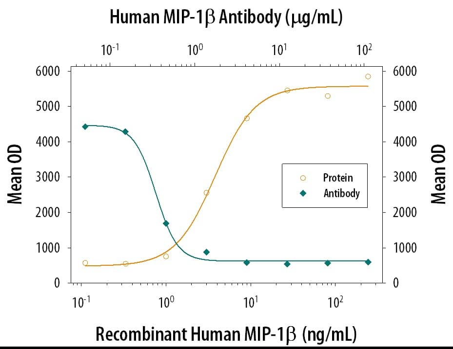
271-BME) chemoattracts the BaF3 mouse pro-B cell line transfected with human CCR5 in a dose-dependent manner (orange line). The amount of cells that migrated through to the lower chemotaxis chamber was measured by Resazurin (Catalog #
AR002). Chemotaxis elicited by Recombinant Human CCL4/MIP-1 beta (0.04 µg/mL) is neutralized (green line) by increasing concentrations of Mouse Anti-Human CCL4/MIP-1 beta Monoclonal Antibody (Catalog # MAB271). The ND
50 is typically 0.3-1.2 µg/mL." class="big_lightbox">
271-BME) chemoattracts the BaF3 mouse pro-B cell line transfected with human CCR5 in a dose-dependent manner (orange line). The amount of cells that migrated through to the lower chemotaxis chamber was measured by Resazurin (Catalog #
AR002). Chemotaxis elicited by Recombinant Human CCL4/MIP-1 beta (0.04 µg/mL) is neutralized (green line) by increasing concentrations of Mouse Anti-Human CCL4/MIP-1 beta Monoclonal Antibody (Catalog # MAB271). The ND
50 is typically 0.3-1.2 µg/mL." alt="Recombinant Human CCL4/MIP-1 beta (Catalog #
271-BME) chemoattracts the BaF3 mouse pro-B cell line transfected with human CCR5 in a dose-dependent manner (orange line). The amount of cells that migrated through to the lower chemotaxis chamber was measured by Resazurin (Catalog #
AR002). Chemotaxis elicited by Recombinant Human CCL4/MIP-1 beta (0.04 µg/mL) is neutralized (green line) by increasing concentrations of Mouse Anti-Human CCL4/MIP-1 beta Monoclonal Antibody (Catalog # MAB271). The ND
50 is typically 0.3-1.2 µg/mL." class="big_thumb" />
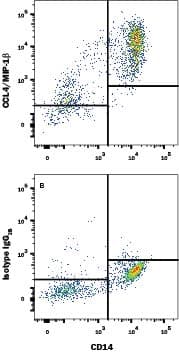
FAB3832A) and either (A) Mouse Anti-Human CCL4/MIP-1 beta Monoclonal Antibody (Catalog # MAB271) or (B) Mouse IgG
2BFlow Cytometry Isotype Control (Catalog #
MAB0041) followed by Allophycocyanin-conjugated Anti-Mouse IgG Secondary Antibody (Catalog #
F0101B). To facilitate intracellular staining, cells were fixed with Flow Cytometry Fixation Buffer (Catalog #
FC004) and permeabilized with Flow Cytometry Permeabilization/Wash Buffer I (Catalog #
FC005). View our protocol for
Staining Intracellular Molecules." class="big_lightbox">
FAB3832A) and either (A) Mouse Anti-Human CCL4/MIP-1 beta Monoclonal Antibody (Catalog # MAB271) or (B) Mouse IgG
2BFlow Cytometry Isotype Control (Catalog #
MAB0041) followed by Allophycocyanin-conjugated Anti-Mouse IgG Secondary Antibody (Catalog #
F0101B). To facilitate intracellular staining, cells were fixed with Flow Cytometry Fixation Buffer (Catalog #
FC004) and permeabilized with Flow Cytometry Permeabilization/Wash Buffer I (Catalog #
FC005). View our protocol for
Staining Intracellular Molecules." class="big_thumb" />
271-BME) was serially diluted and captured by Mouse Anti-Human CCL4/MIP‑1 beta Monoclonal Antibody (Catalog # MAB271) coated on a Clear Polystyrene Microplate (Catalog #
DY990). Goat Anti-Human CCL4/MIP‑1 beta Antigen Affinity-purified Polyclonal Antibody (Catalog #
AF-271-NA) was biotinylated and incubated with the protein captured on the plate. Detection of the standard curve was achieved by incubating Streptavidin-HRP (Catalog #
DY998)" class="big_lightbox">

271-BME) was serially diluted and captured by Mouse Anti-Human CCL4/MIP‑1 beta Monoclonal Antibody (Catalog # MAB271) coated on a Clear Polystyrene Microplate (Catalog #
DY990). Goat Anti-Human CCL4/MIP‑1 beta Antigen Affinity-purified Polyclonal Antibody (Catalog #
AF-271-NA) was biotinylated and incubated with the protein captured on the plate. Detection of the standard curve was achieved by incubating Streptavidin-HRP (Catalog #
DY998)" class="big_thumb" />
 271-BME) chemoattracts the BaF3 mouse pro-B cell line transfected with human CCR5 in a dose-dependent manner (orange line). The amount of cells that migrated through to the lower chemotaxis chamber was measured by Resazurin (Catalog # AR002). Chemotaxis elicited by Recombinant Human CCL4/MIP-1 beta (0.04 µg/mL) is neutralized (green line) by increasing concentrations of Mouse Anti-Human CCL4/MIP-1 beta Monoclonal Antibody (Catalog # MAB271). The ND50 is typically 0.3-1.2 µg/mL." alt="Recombinant Human CCL4/MIP-1 beta (Catalog # 271-BME) chemoattracts the BaF3 mouse pro-B cell line transfected with human CCR5 in a dose-dependent manner (orange line). The amount of cells that migrated through to the lower chemotaxis chamber was measured by Resazurin (Catalog # AR002). Chemotaxis elicited by Recombinant Human CCL4/MIP-1 beta (0.04 µg/mL) is neutralized (green line) by increasing concentrations of Mouse Anti-Human CCL4/MIP-1 beta Monoclonal Antibody (Catalog # MAB271). The ND50 is typically 0.3-1.2 µg/mL." class="big_thumb" />
 271-BME) chemoattracts the BaF3 mouse pro-B cell line transfected with human CCR5 in a dose-dependent manner (orange line). The amount of cells that migrated through to the lower chemotaxis chamber was measured by Resazurin (AR002). Chemotaxis elicited by Recombinant Human CCL4/MIP-1 beta (40 ng/mL) is neutralized (green line) by increasing concentrations of Goat Anti-Human CCL4/MIP-1 beta Antigen Affinity-purified Polyclonal Antibody (Catalog # AF-271-NA). The ND50 is typically 0.3-1.5 µg/mL." alt="Recombinant Human CCL4/MIP-1 beta (271-BME) chemoattracts the BaF3 mouse pro-B cell line transfected with human CCR5 in a dose-dependent manner (orange line). The amount of cells that migrated through to the lower chemotaxis chamber was measured by Resazurin (AR002). Chemotaxis elicited by Recombinant Human CCL4/MIP-1 beta (40 ng/mL) is neutralized (green line) by increasing concentrations of Goat Anti-Human CCL4/MIP-1 beta Antigen Affinity-purified Polyclonal Antibody (Catalog # AF-271-NA). The ND50 is typically 0.3-1.5 µg/mL." class="big_thumb" />
 HAF017). A specific band was detected for CCL4/MIP-1 beta at approximately 12 kDa (as indicated). This experiment was conducted under reducing conditions and using Immunoblot Buffer Group 1." alt="Western blot shows lysates of RAW 264.7 mouse monocyte/macrophage cell line untreated (-) or treated (+) with 10 µg/mL LPS for 4 hours. PVDF membrane was probed with 1 µg/mL of Goat Anti-Mouse CCL4/MIP-1 beta Antigen Affinity-purified Polyclonal Antibody (Catalog # AF-451-NA) followed by HRP-conjugated Anti-Goat IgG Secondary Antibody (Catalog # HAF017). A specific band was detected for CCL4/MIP-1 beta at approximately 12 kDa (as indicated). This experiment was conducted under reducing conditions and using Immunoblot Buffer Group 1." class="big_thumb" />
 451-MB) chemoattracts the BaF3 mouse pro-B cell line transfected with human CCR5 in a dose-dependent manner (orange line). The amount of cells that migrated through to the lower chemotaxis chamber was measured by Resazurin (Catalog # AR002). Chemotaxis elicited by Recombinant Mouse CCL4/MIP-1 beta (25 ng/mL) is neutralized (green line) by increasing concentrations of Rat Anti-Mouse CCL4/MIP-1 beta Monoclonal Antibody (Catalog # MAB451). The ND50 is typically 1-3 µg/mL." alt="Recombinant Mouse CCL4/MIP-1 beta (Catalog # 451-MB) chemoattracts the BaF3 mouse pro-B cell line transfected with human CCR5 in a dose-dependent manner (orange line). The amount of cells that migrated through to the lower chemotaxis chamber was measured by Resazurin (Catalog # AR002). Chemotaxis elicited by Recombinant Mouse CCL4/MIP-1 beta (25 ng/mL) is neutralized (green line) by increasing concentrations of Rat Anti-Mouse CCL4/MIP-1 beta Monoclonal Antibody (Catalog # MAB451). The ND50 is typically 1-3 µg/mL." class="big_thumb" />
 HAF016). A specific band was detected for CCL4/MIP-1 beta at approximately 12 kDa (as indicated). This experiment was conducted under reducing conditions and using Immunoblot Buffer Group 1." alt="Western blot shows lysates of NR8383 rat alveolar macrophage cell line untreated (-) or treated (+) with 10 µg/mL LPS for 4 hours. PVDF membrane was probed with 1 µg/mL of Sheep Anti-Rat CCL4/MIP-1 beta Antigen Affinity-purified Polyclonal Antibody (Catalog # AF6935) followed by HRP-conjugated Anti-Sheep IgG Secondary Antibody (Catalog # HAF016). A specific band was detected for CCL4/MIP-1 beta at approximately 12 kDa (as indicated). This experiment was conducted under reducing conditions and using Immunoblot Buffer Group 1." class="big_thumb" />
 1024-MB) chemo-attracts the BaF3 mouse pro-B cell line transfected with mouse CCR5 in a dose-dependent manner (orange line). The amount of cells that migrated through to the lower chemotaxis chamber was measured by Resazurin (Catalog # AR002). Chemotaxis elicited by Recombinant Cotton Rat CCL4/MIP-1 beta (10 ng/mL) is neutralized (green line) by increasing concentrations of Goat Anti-Cotton Rat CCL4/ MIP-1 beta Antigen Affinity-purified Polyclonal Antibody (Catalog # AF1024). The ND50 is typically 0.15-0.6 µg/mL." alt="Recombinant Cotton Rat CCL4/MIP-1 beta (Catalog # 1024-MB) chemo-attracts the BaF3 mouse pro-B cell line transfected with mouse CCR5 in a dose-dependent manner (orange line). The amount of cells that migrated through to the lower chemotaxis chamber was measured by Resazurin (Catalog # AR002). Chemotaxis elicited by Recombinant Cotton Rat CCL4/MIP-1 beta (10 ng/mL) is neutralized (green line) by increasing concentrations of Goat Anti-Cotton Rat CCL4/ MIP-1 beta Antigen Affinity-purified Polyclonal Antibody (Catalog # AF1024). The ND50 is typically 0.15-0.6 µg/mL." class="big_thumb" />


![N/A CCL4/MIP-1 beta [HRP]](http://images.novusbio.com/fullsize/elisa/DATA_CCL4_DMB00_ELISA_757.jpg "N/A CCL4/MIP-1 beta [HRP]")
![N/A CCL4/MIP-1 beta [HRP]](http://images.novusbio.com/fullsize/elisa/DATA_CCL4_DMB00_ELISA_758.jpg)
![N/A CCL4/MIP-1 beta [HRP]](http://images.novusbio.com/fullsize/elisa/CCL4_DMB00_ELISA_140.jpg)
![N/A CCL4/MIP-1 beta [HRP]](http://images.novusbio.com/fullsize/elisa/DATA_CCL4_MMB00_ELISA_1000.jpg "N/A CCL4/MIP-1 beta [HRP]")
![N/A CCL4/MIP-1 beta [HRP]](http://images.novusbio.com/fullsize/elisa/CCL4_MMB00_ELISA_292.jpg)
 FAB3832A) and either (A) Mouse Anti-Human CCL4/MIP-1 beta Monoclonal Antibody (Catalog # MAB271) or (B) Mouse IgG2BFlow Cytometry Isotype Control (Catalog #
  271-BME) was serially diluted and captured by Mouse Anti-Human CCL4/MIP‑1 beta Monoclonal Antibody (Catalog # MAB271) coated on a Clear Polystyrene Microplate (Catalog #
 ![N/A CCL4/MIP-1 beta [Biotin]](http://images.novusbio.com/fullsize/elisa/DATA_CCL4_DY271_ELISA_1877.jpg "N/A CCL4/MIP-1 beta [Biotin]")
 CTS009) and counterstained with hematoxylin (blue). View our protocol for
  NL001) and counterstained with DAPI (blue). View our protocol for
  271-BME) was serially diluted and captured by Mouse Anti-Human CCL4/MIP‑1 beta Monoclonal Antibody (Catalog #
 ![N/A CCL4/MIP-1 beta [Biotin]](http://images.novusbio.com/fullsize/elisa/DATA_CCL4_DY451_ELISA_2037.jpg "N/A CCL4/MIP-1 beta [Biotin]")
 NL001) and counterstained with DAPI (blue). Specific staining was localized to cytoplasm. View our protocol for
  CTS008) and counterstained with hematoxylin (blue). Specific staining was localized to Peyer's patches. View our protocol for
  451-MB) chemoattracts the BaF3 mouse pro-B cell line transfected with human CCR5 in a dose-dependent manner (orange line). The amount of cells that migrated through to the lower chemotaxis chamber was measured by Resazurin (Catalog #
  6935-MB) induces BaF3 mouse proB cell line transfected with human CCR5 in a dose-dependent manner (orange line). The amount of cells that migrated through to the lower chemotaxis chamber was measured by Resazurin (Catalog #


