 HAF016). A specific band was detected for Semaphorin 4C at approximately 110 kDa (as indicated). This experiment was conducted under reducing conditions and using Immunoblot Buffer Group 8." title="Western blot shows lysates of SW13 human adrenal cortex adenocarcinoma cell line. PVDF membrane was probed with 0.5 µg/mL of Sheep Anti-Human Semaphorin 4C Antigen Affinity-purified Polyclonal Antibody (Catalog # AF6125) followed by HRP-conjugated Anti-Sheep IgG Secondary Antibody (Catalog # HAF016). A specific band was detected for Semaphorin 4C at approximately 110 kDa (as indicated). This experiment was conducted under reducing conditions and using Immunoblot Buffer Group 8." />
| Reactivity | HuSpecies Glossary |
| Applications | WB, Simple Western, IHC |
| Clonality | Polyclonal |
| Host | Sheep |
| Conjugate | Unconjugated |
| Concentration | LYOPH |
| Immunogen | Mouse myeloma cell line NS0-derived recombinant human Semaphorin 4C Ala21-Gly663 Accession # Q9C0C4 |
| Specificity | Detects human Semaphorin 4C in direct ELISAs and Western blots. In direct ELISAs, less than 15% cross-reactivity with recombinant mouse (rm) Sema4C is observed and less than 5% cross-reactivity with recombinant human (rh) Sema4G, rhSema4D, rhSema4B and rhSema4A is observed. |
| Source | N/A |
| Isotype | IgG |
| Clonality | Polyclonal |
| Host | Sheep |
| Gene | SEMA4C |
| Purity Statement | Antigen Affinity-purified |
| Innovator's Reward | Test in a species/application not listed above to receive a full credit towards a future purchase. |
| Dilutions |
|
|
| Publications |
|
| Storage | Use a manual defrost freezer and avoid repeated freeze-thaw cycles.
|
| Buffer | Lyophilized from a 0.2 μm filtered solution in PBS with Trehalose. *Small pack size (SP) is supplied either lyophilized or as a 0.2 µm filtered solution in PBS. |
| Preservative | No Preservative |
| Concentration | LYOPH |
| Reconstitution Instructions | Reconstitute at 0.2 mg/mL in sterile PBS. |
Sema4C (Semaphorin 4C; also Sema I and M-SemaF) is a 100-105 kDa, class IV member of the semaphorin family of proteins. It is expressed by precursors to neurons and myocytes, and may regulate their differentiation into mature forms. Mature human Sema4C is a type I transmembrane glycoprotein that is 813 amino acids (aa) in length. It contains a 643 aa extracellular region (aa 21-663) that is characterized by the presence of one Sema domain (aa 30-497), a PSI region (aa 499‑551), and an Ig-like C2-type domain (aa 556-644). The cytoplasmic region interacts with PZD-domain containing proteins. There are three potential splice variants. One demonstrates an alternative start site at Met324, a second shows a deletion of aa 173-211, and a third contains an 89 aa substitution for aa 1-36. Over aa 21‑663, human Sema4C shares 85% aa identity with mouse Sema4C.








![Semaphorin 4C Antibody [Unconjugated]](/sites/all/modules/enterprise-tech/et_datasheets/images/novus_guarantee.png "Semaphorin 4C Antibody [Unconjugated]")
Secondary Antibodies |
Isotype Controls |
The concentration calculator allows you to quickly calculate the volume, mass or concentration of your vial. Simply enter your mass, volume, or concentration values for your reagent and the calculator will determine the rest.


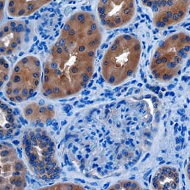
 CTS013). Tissue was stained using the Anti-Sheep HRP-DAB Cell & Tissue Staining Kit (brown; Catalog #
  HAF016). This experiment was conducted under reducing conditions and using the 12-230 kDa separation system. Non-specific interaction with the 230 kDa Simple Western standard may be seen with this antibody." title="Simple Western lane view shows lysates of SW13 human adrenal cortex adenocarcinoma cell line, loaded at 0.2 mg/mL. A specific band was detected for Semaphorin 4C at approximately 121 kDa (as indicated) using 25 µg/mL of Sheep Anti-Human Semaphorin 4C Antigen Affinity-purified Polyclonal Antibody (Catalog # AF6125) followed by 1:50 dilution of HRP-conjugated Anti-Sheep IgG Secondary Antibody (Catalog #
![Western Blot Plexin B2 Antibody [Unconjugated]](https://images.novusbio.com/images/antibody/Plexin_B2_AF5329_Western_Blot_5686.jpg)
![Immunohistochemistry Plexin B2 Antibody [Unconjugated]](https://images.novusbio.com/images/antibody/Plexin_B2_AF5329_Immunohistochemistry_7295.jpg)



![Bioactivity Semaphorin 3A [Unconjugated]](https://images.novusbio.com/images/protein/Semaphorin_3A_1250S3_1473.jpg)


![Immunocytochemistry CD55/DAF Antibody [Unconjugated]](https://images.novusbio.com/images/antibody/CD55_AF2009_Immunocytochemistry__Immunofluorescence_23052.jpg)
![Immunohistochemistry CD55/DAF Antibody [Unconjugated]](https://images.novusbio.com/images/antibody/CD55_AF2009_Immunohistochemistry_6728.jpg)
![Bioactivity GDNF [Unconjugated]](https://images.novusbio.com/images/protein/GDNF_212GD_2846.jpg)
![SDS-Page GDNF [Unconjugated]](https://images.novusbio.com/images/protein/GDNF_212-GD_62.jpg)
![SEC-MALS GDNF [Unconjugated]](https://images.novusbio.com/images/212-gd_recombinant-human-gdnf-protein-sec-mals-25202393740..jpg)


