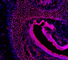
![Immunocytochemistry/Immunofluorescence: Melanopsin Antibody [NB100-74460] - Staining in rat retinal ganglion cells.](http://images.novusbio.com/fullsize/Melanopsin-Antibody-Immunocytochemistry-Immunofluorescence-NB100-74460-img0001.jpg "Immunocytochemistry/Immunofluorescence: Melanopsin Antibody [NB100-74460] - Staining in rat retinal ganglion cells.")
| Reactivity | Hu, Mu, RtSpecies Glossary |
| Applications | WB, ICC/IF, IHC, KO |
| Clonality | Polyclonal |
| Host | Rabbit |
| Conjugate | Unconjugated |
| Concentration | 1 mg/ml |
| Immunogen | Synthetic peptide corresponding to residues E(455) Q K S K T P K T K R H L P S L D R R M(474) of rat melanopsin. |
| Localization | Membrane; Multi-pass membrane protein. Note: Found in soma, dendrites and proximal part of axons of certain retinal ganglion cells. |
| Isotype | IgG |
| Clonality | Polyclonal |
| Host | Rabbit |
| Gene | OPN4 |
| Purity | Immunogen affinity purified |
| Innovator's Reward | Test in a species/application not listed above to receive a full credit towards a future purchase. |
| Dilutions |
|
|
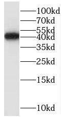
| Application Notes | Successful WB, IF, and ICC usages. WB: Detects approx. 53 and 85 kDa proteins representing overexpressed unglycosylated and glycosylated melanopsin from HEK 293 cells (does not work well on endogenously expressed protein). ICC/IF: NB100-74460 detects melanopsin from rat retinal ganglion cells (RGCs). Knockout data (PMID: 31924821). |
|
| Publications |
|
| Storage | Store at -20C. Avoid freeze-thaw cycles. |
| Buffer | PBS, 1 mg/mL BSA |
| Preservative | 0.05% Sodium Azide |
| Concentration | 1 mg/ml |
| Purity | Immunogen affinity purified |


Secondary Antibodies |
Isotype Controls |
Research Areas for Melanopsin Antibody (NB100-74460)Find related products by research area.
|
The concentration calculator allows you to quickly calculate the volume, mass or concentration of your vial. Simply enter your mass, volume, or concentration values for your reagent and the calculator will determine the rest.


![SDS-Page LIGHT/TNFSF14 [Unconjugated]](https://images.novusbio.com/images/protein/LIGHT_664-LI_719.jpg)
![Bioactivity LIGHT/TNFSF14 [Unconjugated]](https://images.novusbio.com/images/protein/LIGHT_664-LI_721.jpg)


![Immunohistochemistry mu Opioid R/OPRM1 Antibody (1126B) [Unconjugated]](https://images.novusbio.com/images/antibody/mu_Opioid_R_MAB8629_Immunohistochemistry_16797.jpg)
![Simple Western Pax6 Antibody [Unconjugated]](https://images.novusbio.com/images/antibody/Pax6_AF8150_Simple_Western_17126.jpg)
![Western Blot Pax6 Antibody [Unconjugated]](https://images.novusbio.com/images/antibody/Pax6_AF8150_Western_Blot_14743.jpg)
![Immunocytochemistry Pax6 Antibody [Unconjugated]](https://images.novusbio.com/images/antibody/Pax6_AF8150_Immunocytochemistry_14427.jpg)


![Immunocytochemistry S100A9 Antibody [Unconjugated]](https://images.novusbio.com/images/antibody/S100A9_AF2065_Immunocytochemistry__Immunofluorescence_22492.jpg)
![Immunohistochemistry S100A9 Antibody [Unconjugated]](https://images.novusbio.com/images/af2065_goat-s100a9-pab-alexa-fluor-594-immunohistochemistry-12122025852812.jpg)
![Western Blot: Goat anti-Rabbit IgG (H+L) Secondary Antibody [HRP] [NB7160] - Western blot showing vemurafenib treatment in BRAFV600E CRC cells inhibits fission mediator DRP1 with no significant effect on fusion proteins (Mfn1 & 2) using MFN-1 antibody (NBP1-51841) and corresponding secondary antibody, goat anti-rabbit IgG-HRP (NB7160). Image collected and cropped by CiteAb from the following publication (https://pubmed.ncbi.nlm.nih.gov/33738242).](https://images.novusbio.com/images/Goat-anti-Rabbit-IgG-H+L-Secondary-Antibody-HRP-Western-Blot-NB7160-img0001.jpg "Western Blot: Goat anti-Rabbit IgG (H+L) Secondary Antibody [HRP] [NB7160] - Western blot showing vemurafenib treatment in BRAFV600E CRC cells inhibits fission mediator DRP1 with no significant effect on fusion proteins (Mfn1 & 2) using MFN-1 antibody (NBP1-51841) and corresponding secondary antibody, goat anti-rabbit IgG-HRP (NB7160). Image collected and cropped by CiteAb from the following publication (https://pubmed.ncbi.nlm.nih.gov/33738242).")
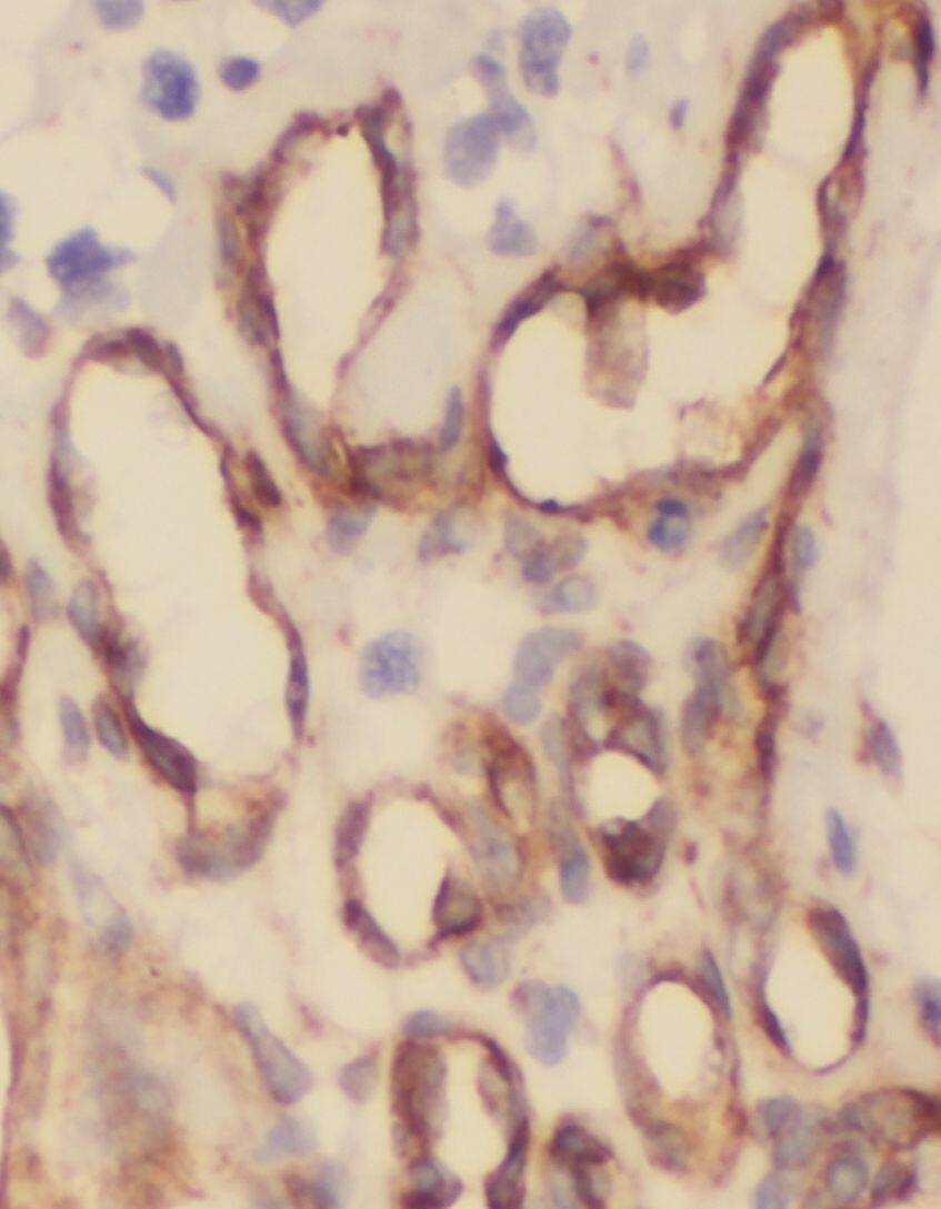
 followed by 30 min incubation with Goat anti Rabbit HRP conjugated secondary antibodies (Catalog # HAF008) at 1:20 dilution + DAB chromogen (brown). The tissue was counterstained with Hematoxylin (blue). Control was done by omitting primary antibody.")
![Flow Cytometry: Rabbit IgG Isotype Control [NBP2-24891] - An intracellular stain was performed on Raji cells with Adiponectin antibody NB100-65810 (blue) and a matched isotype control NBP2-24893 (orange). Cells were fixed with 4% PFA and then permeablized with 0.1% saponin. Cells were incubated in an antibody dilution of 1 ug/mL for 30 minutes at room temperature, followed by Dylight488-conjugated anti-rabbit secondary antibody. Image using the Azide Free form of this antibody.](https://images.novusbio.com/images/Rabbit--Mouse-IgG-Isotype-Control-Flow-Cytometry-NBP2-24891-img0006.jpg "Flow Cytometry: Rabbit IgG Isotype Control [NBP2-24891] - An intracellular stain was performed on Raji cells with Adiponectin antibody NB100-65810 (blue) and a matched isotype control NBP2-24893 (orange). Cells were fixed with 4% PFA and then permeablized with 0.1% saponin. Cells were incubated in an antibody dilution of 1 ug/mL for 30 minutes at room temperature, followed by Dylight488-conjugated anti-rabbit secondary antibody. Image using the Azide Free form of this antibody.")