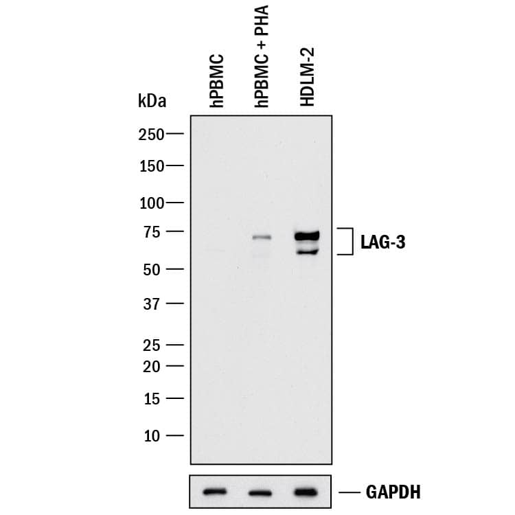
 HAF017). Specific bands were detected for LAG-3 at approximately 60-75 kDa (as indicated). GAPDH (Catalog # MAB5718) is shown as a loading control. This experiment was conducted under reducing conditions and using Immunoblot Buffer Group 1." title="Western blot shows lysates of human peripheral blood mononuclear cells (PBMC) untreated or treated (+) with 1 ug/mL PHA for 5 days and HDLM-2 human Hodgkin's lymphoma cell line. PVDF membrane was probed with 1 µg/mL of Goat Anti-Human LAG-3 Antigen Affinity-purified Polyclonal Antibody (Catalog # AF2319) followed by HRP-conjugated Anti-Goat IgG Secondary Antibody (Catalog # HAF017). Specific bands were detected for LAG-3 at approximately 60-75 kDa (as indicated). GAPDH (Catalog # MAB5718) is shown as a loading control. This experiment was conducted under reducing conditions and using Immunoblot Buffer Group 1." />
| Reactivity | HuSpecies Glossary |
| Applications | WB, Flow, IHC, CyTOF-ready |
| Clonality | Polyclonal |
| Host | Goat |
| Conjugate | Unconjugated |
| Concentration | LYOPH |
| Immunogen | Mouse myeloma cell line NS0-derived recombinant human LAG-3 Leu23-Leu450 Accession # P18627 |
| Specificity | Detects human LAG-3 in direct ELISAs and Western blots. In direct ELISAs, less than 10% cross-reactivity with recombinant mouse LAG-3 is observed. |
| Source | N/A |
| Isotype | IgG |
| Clonality | Polyclonal |
| Host | Goat |
| Gene | LAG3 |
| Purity Statement | Antigen Affinity-purified |
| Innovator's Reward | Test in a species/application not listed above to receive a full credit towards a future purchase. |
| Dilutions |
|
|
| Publications |
|
| Storage | Use a manual defrost freezer and avoid repeated freeze-thaw cycles.
|
| Buffer | Lyophilized from a 0.2 μm filtered solution in PBS with Trehalose. *Small pack size (SP) is supplied either lyophilized or as a 0.2 µm filtered solution in PBS. |
| Preservative | No Preservative |
| Concentration | LYOPH |
| Reconstitution Instructions | Reconstitute at 0.2 mg/mL in sterile PBS. |


Secondary Antibodies |
Isotype Controls |
|
Tired T cells: Hypoxia Drives T cell Exhaustion in the Tumor Microenvironment By Hunter MartinezThe paradigm shifting view of the immune system being leveraged to target cancer has led to numerous therapeutic breakthroughs. One major cell group responsible for this revelation is a T cell. ... Read full blog post. |
The concentration calculator allows you to quickly calculate the volume, mass or concentration of your vial. Simply enter your mass, volume, or concentration values for your reagent and the calculator will determine the rest.


 F0108) and Mouse Anti-Human CD3e PE-conjugated Monoclonal Antibody (Catalog #
  F0108). Quadrant markers were set based on control antibody staining(Catalog #
 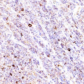 VC004). Before incubation with the primary antibody, tissue was subjected to heat-induced epitope retrieval using Antigen Retrieval Reagent-Basic (
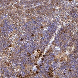 VC004). Before incubation with the primary antibody, tissue was subjected to heat-induced epitope retrieval using Antigen Retrieval Reagent-Basic (
![Bioactivity CTLA-4 [Unconjugated]](https://images.novusbio.com/images/protein/CTLA4_7268CT_2293.jpg)


![SDS-Page TNF-alpha [Unconjugated]](https://images.novusbio.com/images/protein/TNF-alpha_210-TA_256.jpg)
![Bioactivity TNF-alpha [Unconjugated]](https://images.novusbio.com/images/protein/TNFalpha_210TA_1658.jpg)
![SEC-MALS TNF-alpha [Unconjugated]](https://images.novusbio.com/images/210-ta_recombinant-human-tnf-alpha-protein-sec-mals-35202312244..jpg)
![Western Blot ICAM-1/CD54 Antibody [Unconjugated]](https://images.novusbio.com/images/af796_mouse-icam-1-cd54-affinity-purified-polyclonal-ab-41202410481192.jpg)
![Western Blot ICAM-1/CD54 Antibody [Unconjugated]](https://images.novusbio.com/images/af796_mouse-icam-1-cd54-affinity-purified-polyclonal-ab-4120241048112.jpg)
![Western Blot ICAM-1/CD54 Antibody [Unconjugated]](https://images.novusbio.com/images/af796_mouse-icam-1-cd54-affinity-purified-polyclonal-ab-4120241049436.jpg)
![Immunocytochemistry CD23/Fc epsilon RII Antibody [Unconjugated]](https://images.novusbio.com/images/antibody/CD23_AF123_Immunocytochemistry_6469.jpg)
![ELISA CD23/Fc epsilon RII Antibody [Unconjugated]](https://images.novusbio.com/images/af123_human-cd23-fc-epsilon-rii-affinity-purified-polyclonal-ab-elisa-639201850091061764.jpg)
![Bioactivity IL-4 [Unconjugated]](https://images.novusbio.com/images/protein/6507-ilcf_recombinant-human-il-4-cho-expressed-protein-cf-bioactivity-272020133214.jpg)

![Western Blot CD40/TNFRSF5 Antibody [Unconjugated]](https://images.novusbio.com/images/af632_human-cd40-tnfrsf5-affinity-purified-polyclonal-ab-822024950191.jpg)
![Western Blot CD40/TNFRSF5 Antibody [Unconjugated]](https://images.novusbio.com/images/af632_human-cd40-tnfrsf5-affinity-purified-polyclonal-ab-44202415425047.jpg)
![Simple Western CD40/TNFRSF5 Antibody [Unconjugated]](https://images.novusbio.com/images/antibody/af632_human-cd40-tnfrsf5-affinity-purified-polyclonal-ab-simple-western-58202111562.jpg)
![Bioactivity Lymphotoxin-alpha/TNF-beta [Unconjugated]](https://images.novusbio.com/images/protein/Lymphotoxin-alpha_211-TBB_CF_697.jpg)
![SDS-Page Lymphotoxin-alpha/TNF-beta [Unconjugated]](https://images.novusbio.com/images/protein/Lymphotoxin-alpha_211-TBB_CF_695.jpg)
![Immunohistochemistry CXCL13/BLC/BCA-1 Antibody [Unconjugated]](https://images.novusbio.com/images/af470_mouse-cxcl13-blc-bca-1-affinity-purified-polyclonal-ab-immunohistochemistry-121220259151521.jpg)
![Flow Cytometry CXCL13/BLC/BCA-1 Antibody [Unconjugated]](https://images.novusbio.com/images/af470_mouse-cxcl13-blc-bca-1-affinity-purified-polyclonal-ab-flow-cytometry-121220258541120.jpg)
![Immunohistochemistry CXCL13/BLC/BCA-1 Antibody [Unconjugated]](https://images.novusbio.com/images/af470_mouse-cxcl13-blc-bca-1-affinity-purified-polyclonal-ab-8120255531682.jpg)

![Immunohistochemistry CD45 Antibody [Unconjugated]](https://images.novusbio.com/images/antibody/CD45_AF114_Immunohistochemistry_23525.jpg)
![Immunocytochemistry CD45 Antibody [Unconjugated]](https://images.novusbio.com/images/antibody/af114_mouse-cd45-affinity-purified-polyclonal-ab-immunocytochemistry-6122021145449.jpg)