| Reactivity | HuSpecies Glossary |
| Applications | WB |
| Clone | 333210 |
| Clonality | Monoclonal |
| Host | Mouse |
| Conjugate | Alexa Fluor 405 |
| Immunogen | Mouse myeloma cell line NS0-derived recombinant human LAG-3 Leu23-Leu450 Accession # P18627 |
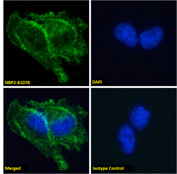
| Specificity | Detects human LAG‑3 in direct ELISAs and Western blots. In direct ELISAs and Western blots, no cross‑reactivity with recombinant mouse LAG-3 is observed. |
| Isotype | IgG2a |
| Clonality | Monoclonal |
| Host | Mouse |
| Purity Statement | Protein A or G purified |
| Innovator's Reward | Test in a species/application not listed above to receive a full credit towards a future purchase. |
| Storage | Protect from light. Do not freeze. 12 months from date of receipt, 2 to 8 °C as supplied |
| Buffer | Supplied 0.2 mg/mL in a saline solution containing BSA and Sodium Azide. |
LAG-3 (Lymphocyte activation gene-3), also known as CD223, is a member of the immunoglobulin superfamily (IgSF). The mature LAG-3 protein is a 496 amino acid (aa) membrane protein with a 421 aa extracellular region which contains four IgSF domains, a 21 aa transmembrane region and a 54 aa cytoplasmic region. The extracellular domain of human and mouse LAG-3 share 69% amino acid sequence identity. LAG-3 and CD4 molecules share 20% aa sequence homology but have a similar structure (1, 2). Both molecules bind to MHC class II. LAG-3 binds to MHC class II with higher affinity compared to CD4. LAG-3 is an activation-induced molecule, expressed on activated T cells and NK cells, but not on resting T cells. Studies using LAG-3 -/- mice have shown significant delay of T cell apoptosis following antigen stimulation and increased size of memory T cells pool following infection (3, 4). It also has been reported that anti-LAG-3 antibodies up-regulate T cell activation by blocking interaction of LAG-3 and MHC class II. The study has demonstrated that LAG-3 is selectively expressed on activated CD4+CD25+ TReg cells and plays a role in their suppressive activity (5). This evidence indicated, unlike the interaction of CD4 with MHC class II that plays a positive role in T cell activation, LAG-3 binds to MHC class II and negatively regulates T cell activation through LAG-3 signaling. On the other hand, studies have shown that binding of LAG-3 to MHC class II molecules on antigen presenting cells induce maturation of dendritic cells and cytokine secretion by monocytes through MHC class II signal transduction (6). Taken together, LAG-3 may have two major functions, it negatively regulates T cells activation through LAG-3 signaling and stimulates antigen presenting cells which express MHC class II.


Secondary Antibodies |
Isotype Controls |
|
Tired T cells: Hypoxia Drives T cell Exhaustion in the Tumor Microenvironment By Hunter MartinezThe paradigm shifting view of the immune system being leveraged to target cancer has led to numerous therapeutic breakthroughs. One major cell group responsible for this revelation is a T cell. ... Read full blog post. |
The concentration calculator allows you to quickly calculate the volume, mass or concentration of your vial. Simply enter your mass, volume, or concentration values for your reagent and the calculator will determine the rest.
| Uniprot |
|


![Bioactivity CTLA-4 [Unconjugated]](https://images.novusbio.com/images/protein/CTLA4_7268CT_2293.jpg)


![SDS-Page TNF-alpha [Unconjugated]](https://images.novusbio.com/images/protein/TNF-alpha_210-TA_256.jpg)
![Bioactivity TNF-alpha [Unconjugated]](https://images.novusbio.com/images/protein/TNFalpha_210TA_1658.jpg)
![SEC-MALS TNF-alpha [Unconjugated]](https://images.novusbio.com/images/210-ta_recombinant-human-tnf-alpha-protein-sec-mals-35202312244..jpg)
![Western Blot ICAM-1/CD54 Antibody [Unconjugated]](https://images.novusbio.com/images/af796_mouse-icam-1-cd54-affinity-purified-polyclonal-ab-41202410481192.jpg)
![Western Blot ICAM-1/CD54 Antibody [Unconjugated]](https://images.novusbio.com/images/af796_mouse-icam-1-cd54-affinity-purified-polyclonal-ab-4120241048112.jpg)
![Western Blot ICAM-1/CD54 Antibody [Unconjugated]](https://images.novusbio.com/images/af796_mouse-icam-1-cd54-affinity-purified-polyclonal-ab-4120241049436.jpg)
![Immunocytochemistry CD23/Fc epsilon RII Antibody [Unconjugated]](https://images.novusbio.com/images/antibody/CD23_AF123_Immunocytochemistry_6469.jpg)
![Bioactivity IL-4 [Unconjugated]](https://images.novusbio.com/images/protein/6507-ilcf_recombinant-human-il-4-cho-expressed-protein-cf-bioactivity-272020133214.jpg)

![Western Blot CD40/TNFRSF5 Antibody [Unconjugated]](https://images.novusbio.com/images/af632_human-cd40-tnfrsf5-affinity-purified-polyclonal-ab-822024950191.jpg)
![Western Blot CD40/TNFRSF5 Antibody [Unconjugated]](https://images.novusbio.com/images/af632_human-cd40-tnfrsf5-affinity-purified-polyclonal-ab-44202415425047.jpg)
![Simple Western CD40/TNFRSF5 Antibody [Unconjugated]](https://images.novusbio.com/images/antibody/af632_human-cd40-tnfrsf5-affinity-purified-polyclonal-ab-simple-western-58202111562.jpg)
![Bioactivity Lymphotoxin-alpha/TNF-beta [Unconjugated]](https://images.novusbio.com/images/protein/Lymphotoxin-alpha_211-TBB_CF_697.jpg)
![SDS-Page Lymphotoxin-alpha/TNF-beta [Unconjugated]](https://images.novusbio.com/images/protein/Lymphotoxin-alpha_211-TBB_CF_695.jpg)
![Immunohistochemistry CXCL13/BLC/BCA-1 Antibody [Unconjugated]](https://images.novusbio.com/images/af470_mouse-cxcl13-blc-bca-1-affinity-purified-polyclonal-ab-immunohistochemistry-121220259151521.jpg)
![Flow Cytometry CXCL13/BLC/BCA-1 Antibody [Unconjugated]](https://images.novusbio.com/images/af470_mouse-cxcl13-blc-bca-1-affinity-purified-polyclonal-ab-flow-cytometry-121220258541120.jpg)
![Immunohistochemistry CXCL13/BLC/BCA-1 Antibody [Unconjugated]](https://images.novusbio.com/images/af470_mouse-cxcl13-blc-bca-1-affinity-purified-polyclonal-ab-8120255531682.jpg)

![Immunohistochemistry CD45 Antibody [Unconjugated]](https://images.novusbio.com/images/antibody/CD45_AF114_Immunohistochemistry_23525.jpg)
![Immunocytochemistry CD45 Antibody [Unconjugated]](https://images.novusbio.com/images/antibody/af114_mouse-cd45-affinity-purified-polyclonal-ab-immunocytochemistry-6122021145449.jpg)