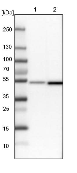
 followed by HRP-conjugated Anti-Sheep IgG Secondary Antibody (Catalog # HAF016). A specific band was detected for Drebrin 1 at approximately 120 kDa (as indicated). This experiment was conducted under reducing conditions and using Immunoblot Buffer Group 1.")
| Reactivity | HuSpecies Glossary |
| Applications | WB, ICC/IF |
| Clonality | Polyclonal |
| Host | Sheep |
| Conjugate | Unconjugated |
| Concentration | LYOPH |
| Immunogen | E. coli-derived recombinant human Drebrin 1 Asn482-Asp649 (Ser553Pro) Accession # Q16643 |
| Specificity | Detects human Drebrin 1 in direct ELISAs and Western blots. |
| Source | N/A |
| Isotype | IgG |
| Clonality | Polyclonal |
| Host | Sheep |
| Gene | DBN1 |
| Purity Statement | Antigen Affinity-purified |
| Innovator's Reward | Test in a species/application not listed above to receive a full credit towards a future purchase. |
| Storage | Use a manual defrost freezer and avoid repeated freeze-thaw cycles.
|
| Buffer | Lyophilized from a 0.2 μm filtered solution in PBS with Trehalose. *Small pack size (SP) is supplied either lyophilized or as a 0.2 µm filtered solution in PBS. |
| Preservative | No Preservative |
| Concentration | LYOPH |
| Reconstitution Instructions | Sterile PBS to a final concentration of 0.2 mg/mL. |
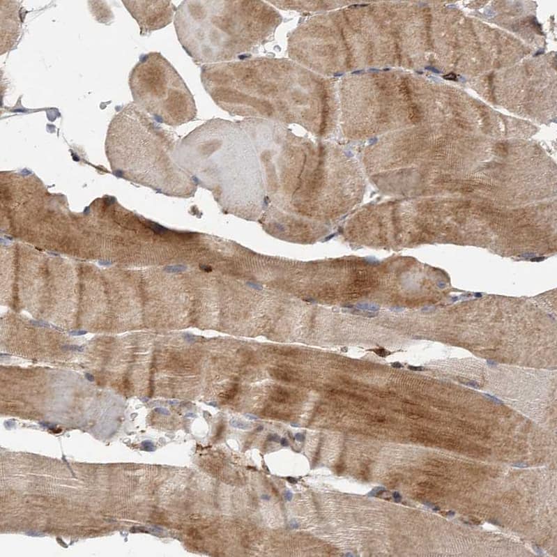
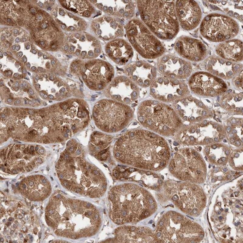
Drebrin 1 (DBN-1 [developmentally-regulated brain protein1]; also drebrin-E/E2 [Embryonic]) is an intracellular member of the ADF-H (actin-depolymerizing factor-H) family of actin binding proteins. Although its predicted MW is 72 kDa, it runs anomalously at 115-116 kDa in SDS-PAGE. It is expressed by neurons, gastric Parietal cells, astrocytes, distal convoluted tubule epithelium and proton-secreting intercalated cells of the renal collecting duct. Drebrin 1 interacts with multiple partners near the membrane. It links connexin-43 and F-actin, thereby stabilizing membrane gap junctions. It also binds to EB3 (end-binding protein 3) on microtubules, facilitating actin-microtubule interactions. Human Drebrin 1 is 649 amino acids (aa) in length. It contains one actin depolymerizing homology domain (aa 3-134), an actin-binding region (≈ aa 150-300), and two HOMER binding motifs (aa 539-543 and 617-621). There are at least 10 utilized Ser/Thr phosphorylation sites and one utilized Tyr phosphorylation site. Alternative splicing generates drebrin-A (Adult), a 124-126 kDa isoform that contains a 46 aa insert after Gly319. Drebrin-A is found in neurons and possibly podocytes, and is associated with dendritic spines where it inhibits the interaction of F-actin with alpha -actinin and tropomyosin. This favors the generation of excitatory impulses in neurons. Three other potential isoform variants are noted. One utilizes an alternative start site at Met64, a second shows a 60 aa substitution for aa 1-110, and a third contains a 28 aa substitution for aa 4-29. Over aa 482-649, human Drebrin 1 shares 84% aa sequence identity with mouse Drebrin 1.


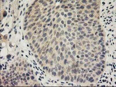


![Immunohistochemistry-Paraffin Tau [p Thr181] Antibody - BSA Free](https://images.novusbio.com/images/Tau-[p-Thr181]-Antibody-Immunohistochemistry-Paraffin-NB100-82245-img0005.jpg)
![Simple Western Tau [p Thr181] Antibody - BSA Free](https://images.novusbio.com/images/Tau-[p-Thr181]-Antibody-Simple-Western-NB100-82245-img0004.jpg)
![Western Blot Tau [p Thr181] Antibody - BSA Free](https://images.novusbio.com/images/Tau-[p-Thr181]-Antibody-Western-Blot-NB100-82245-img0001.jpg)


Secondary Antibodies |
Isotype Controls |
The concentration calculator allows you to quickly calculate the volume, mass or concentration of your vial. Simply enter your mass, volume, or concentration values for your reagent and the calculator will determine the rest.


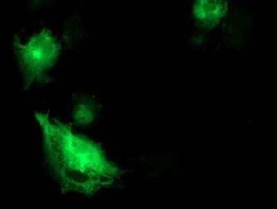
 at 15 µg/mL for 3 hours at room temperature. Cells were stained using the NorthernLights™ 557-conjugated Anti-Sheep IgG Secondary Antibody (red; Catalog # NL010) and counterstained with DAPI (blue). Specific staining was localized to plasma membranes. View our protocol for Fluorescent ICC Staining of Cells on Coverslips.")
![N/A BDNF [HRP]](https://images.novusbio.com/images/elisa/BDNF_DBD00_ELISA_36.jpg)
![N/A BDNF [HRP]](https://images.novusbio.com/images/elisa/DATA_BDNF_DBD00_ELISA_587.jpg)
![N/A BDNF [HRP]](https://images.novusbio.com/images/elisa/DATA_BDNF_DBD00_ELISA_588.jpg)


![Immunocytochemistry EGR1 Antibody [Unconjugated]](https://images.novusbio.com/images/antibody/af2818_human-egr1-affinity-purified-polyclonal-ab-immunocytochemistry-3102024831..jpg)
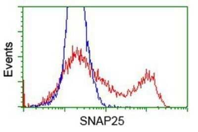
![Intracellular Staining by Flow Cytometry Nestin Antibody (196908) [Unconjugated]](https://images.novusbio.com/images/antibody/mab1259_human-nestin-mab-clone-196908-intracellular-staining-by-flow-cytometry-24120239959.jpg)
![Immunocytochemistry/ Immunofluorescence Nestin Antibody (196908) [Unconjugated]](https://images.novusbio.com/images/mab1259_human-nestin-mab-clone-196908-41202410481178.jpg)
) induced by Homo-BacPROTAC 8 (UdSBI-0545) compared to its enantiomer 8a (UdSBI-0966), matching monomer 5 and dCymC (3 independent experiments done in triplicates). b WES visualization of ClpC1-NTD degradation after titration of Homo-BacPROTACs, developed with anti-His antibody recognizing His6-tagged ClpC1-NTD and processed, His4-tagged ClpP1P2. Concentration-dependent degradation of ClpC1-NTD can be observed for 8 (UdSBI-0545) (lanes 10–13) but not for 8a (UdSBI-0966) (lanes 2–8). c Analogous to a, except that exit vector 7-based Homo-BacPROTAC 12 (UdSBI-4377), enantiomer 12a (UdSBI-0117) and monomer 10 were used. d WES-derived gel picture visualizing ClpC1-NTD degradation (lane 2–5) from representative experiment summarized in c. e SYPROTM Ruby-stained SDS-PAGE gel from exemplary cell-free degradation assay depicting efficient degradation (lane 3,4) of ClpC1-NTD by Homo-BacPROTAC 8 (UdSBI-0545), while full length ClpC1 is not significantly degraded. Green vertical lines indicate the DC50 (for 8,12). Error bars represent mean ± SD of n = 3 independent experiments in triplicates. The actual mean DC50 values for all cell-free degradation experiments conducted for this study are summarized in Supplementary Table 8. Source data are provided as a Source Data file. Image collected and cropped by CiteAb from the following open publication (https://www.nature.com/articles/s41467-024-46218-7), licensed under a CC-BY license. Not internally tested by R&D Systems.")