293-VE), Recombinant Human VEGF
111 (
5336-VE), Recombinant Human VEGF
121, aa 207-327 (
4644-VS), Recombinant Human VEGF
145 (aa 27-171) (
7626-VE), Recombinant Human VEGF
162 (
2347-VE), Recombinant Human VEGF
165b (
3045-VE), Recombinant Human VEGF
189 (aa 27-215) (
8147-VE), Recombinant Human VEGF
165 Extended Isoform (
9018-VE), Recombinant Human VEGF-B
167 (
751-VE), Recombinant Mouse VEGF
164 (
493-MV), and Recombinant Rat VEGF
164 (
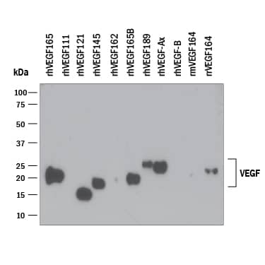
564-RV). PVDF Membrane was probed with 0.1 µg/mL of Mouse Anti-Human/Primate VEGF Monoclonal Antibody (Catalog # MAB293) followed by HRP-conjugated Anti-Mouse IgG Secondary Antibody (
HAF007). A specific band was detected for VEGF at approximately 15-25 kDa (as indicated). This experiment was conducted under reducing conditions and using
Immunoblot Buffer Group 3." class="big_lightbox">
293-VE), Recombinant Human VEGF
111 (
5336-VE), Recombinant Human VEGF
121, aa 207-327 (
4644-VS), Recombinant Human VEGF
145 (aa 27-171) (
7626-VE), Recombinant Human VEGF
162 (
2347-VE), Recombinant Human VEGF
165b (
3045-VE), Recombinant Human VEGF
189 (aa 27-215) (
8147-VE), Recombinant Human VEGF
165 Extended Isoform (
9018-VE), Recombinant Human VEGF-B
167 (
751-VE), Recombinant Mouse VEGF
164 (
493-MV), and Recombinant Rat VEGF
164 (
564-RV). PVDF Membrane was probed with 0.1 µg/mL of Mouse Anti-Human/Primate VEGF Monoclonal Antibody (Catalog # MAB293) followed by HRP-conjugated Anti-Mouse IgG Secondary Antibody (
HAF007). A specific band was detected for VEGF at approximately 15-25 kDa (as indicated). This experiment was conducted under reducing conditions and using
Immunoblot Buffer Group 3." alt="Western blot shows 25 ng of Recombinant Human VEGF
165 (
293-VE), Recombinant Human VEGF
111 (
5336-VE), Recombinant Human VEGF
121, aa 207-327 (
4644-VS), Recombinant Human VEGF
145 (aa 27-171) (
7626-VE), Recombinant Human VEGF
162 (
2347-VE), Recombinant Human VEGF
165b (
3045-VE), Recombinant Human VEGF
189 (aa 27-215) (
8147-VE), Recombinant Human VEGF
165 Extended Isoform (
9018-VE), Recombinant Human VEGF-B
167 (
751-VE), Recombinant Mouse VEGF
164 (
493-MV), and Recombinant Rat VEGF
164 (
564-RV). PVDF Membrane was probed with 0.1 µg/mL of Mouse Anti-Human/Primate VEGF Monoclonal Antibody (Catalog # MAB293) followed by HRP-conjugated Anti-Mouse IgG Secondary Antibody (
HAF007). A specific band was detected for VEGF at approximately 15-25 kDa (as indicated). This experiment was conducted under reducing conditions and using
Immunoblot Buffer Group 3." class="big_thumb" />
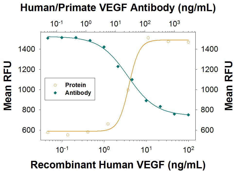
293-VE) stimulates proliferation in HUVEC human umbilical vein endothelial cells in a dose-dependent manner (orange line) as measured by Resazurin (
AR002). Proliferation elicited by Recombinant Human VEGF
165 (10 ng/mL) is neutralized (green line) by increasing concentrations of Mouse Anti-Human/Primate VEGF Monoclonal Antibody (Catalog # MAB293). The ND
50 is typically 10-60 ng/mL." class="big_lightbox">
293-VE) stimulates proliferation in HUVEC human umbilical vein endothelial cells in a dose-dependent manner (orange line) as measured by Resazurin (
AR002). Proliferation elicited by Recombinant Human VEGF
165 (10 ng/mL) is neutralized (green line) by increasing concentrations of Mouse Anti-Human/Primate VEGF Monoclonal Antibody (Catalog # MAB293). The ND
50 is typically 10-60 ng/mL." class="big_thumb" />
293-VE) was serially diluted and captured by Mouse Anti-Human/Primate VEGF Monoclonal Antibody (Catalog # MAB293) coated on a Clear Polystyrene Microplate (Catalog #
DY990). Goat Anti-Human VEGF 165 Antigen Affinity-purified Polyclonal Antibody (Catalog #
AF-293-NA) was biotinylated and incubated with the protein captured on the plate. Detection of the standard curve was achieved by incubating Streptavidin-HRP (Catalog #
DY998)" class="big_lightbox">

293-VE) was serially diluted and captured by Mouse Anti-Human/Primate VEGF Monoclonal Antibody (Catalog # MAB293) coated on a Clear Polystyrene Microplate (Catalog #
DY990). Goat Anti-Human VEGF 165 Antigen Affinity-purified Polyclonal Antibody (Catalog #
AF-293-NA) was biotinylated and incubated with the protein captured on the plate. Detection of the standard curve was achieved by incubating Streptavidin-HRP (Catalog #
DY998)" class="big_thumb" />
293-VE) was serially diluted and captured by Mouse Anti-Human/Primate VEGF Monoclonal Antibody (Catalog # MAB293) coated on a Clear Polystyrene Microplate (Catalog #
DY990). Goat Anti-Human VEGF 165 Antigen Affinity-purified Polyclonal Antibody (Catalog #
AF-293-NA) was biotinylated and incubated with the protein captured on the plate. Detection of the standard curve was achieved by incubating Streptavidin-HRP (Catalog #
DY998)" class="big_lightbox">
293-VE) was serially diluted and captured by Mouse Anti-Human/Primate VEGF Monoclonal Antibody (Catalog # MAB293) coated on a Clear Polystyrene Microplate (Catalog #
DY990). Goat Anti-Human VEGF 165 Antigen Affinity-purified Polyclonal Antibody (Catalog #
AF-293-NA) was biotinylated and incubated with the protein captured on the plate. Detection of the standard curve was achieved by incubating Streptavidin-HRP (Catalog #
DY998)" class="big_thumb" />
 293-VE), Recombinant Mouse VEGF164(Catalog # 493-MV), Recombinant Mouse VEGF120(Catalog # 494-VE), Recombinant Mouse VEGF-B167(Catalog # 2595-VE), Recombinant Mouse VEGF-B 186 (Catalog # 767-VE), Recombinant Mouse VEGF188(Catalog # 7916-MV), and Recombinant Rat VEGF164(Catalog # 564-RV). PVDF Membrane was probed with 0.1 µg/mL of Goat Anti-Mouse VEGF164Antigen Affinity-purified Polyclonal Antibody (Catalog # AF-493-NA) followed by HRP-conjugated Anti-Goat IgG Secondary Antibody (Catalog # HAF109). A specific band was detected for VEGF at approximately 18-27 kDa (as indicated). This experiment was conducted under reducing conditions and using Immunoblot Buffer Group 3." alt="Western blot shows 25 ng of Recombinant Human VEGF165(Catalog # 293-VE), Recombinant Mouse VEGF164(Catalog # 493-MV), Recombinant Mouse VEGF120(Catalog # 494-VE), Recombinant Mouse VEGF-B167(Catalog # 2595-VE), Recombinant Mouse VEGF-B 186 (Catalog # 767-VE), Recombinant Mouse VEGF188(Catalog # 7916-MV), and Recombinant Rat VEGF164(Catalog # 564-RV). PVDF Membrane was probed with 0.1 µg/mL of Goat Anti-Mouse VEGF164Antigen Affinity-purified Polyclonal Antibody (Catalog # AF-493-NA) followed by HRP-conjugated Anti-Goat IgG Secondary Antibody (Catalog # HAF109). A specific band was detected for VEGF at approximately 18-27 kDa (as indicated). This experiment was conducted under reducing conditions and using Immunoblot Buffer Group 3." class="big_thumb" />
 293-VE), Recombinant Human VEGF111 (5336-VE), Recombinant Human VEGF121, aa 207-327 (4644-VS), Recombinant Human VEGF145 (aa 27-171) (7626-VE), Recombinant Human VEGF162 (2347-VE), Recombinant Human VEGF165b (3045-VE), Recombinant Human VEGF189 (aa 27-215) (8147-VE), Recombinant Human VEGF165 Extended Isoform (9018-VE), Recombinant Human VEGF-B167 (751-VE), Recombinant Mouse VEGF164 (493-MV), and Recombinant Rat VEGF164 (564-RV). PVDF Membrane was probed with 0.1 µg/mL of Mouse Anti-Human/Primate VEGF Monoclonal Antibody (Catalog # MAB293) followed by HRP-conjugated Anti-Mouse IgG Secondary Antibody (HAF007). A specific band was detected for VEGF at approximately 15-25 kDa (as indicated). This experiment was conducted under reducing conditions and using Immunoblot Buffer Group 3." alt="Western blot shows 25 ng of Recombinant Human VEGF165 (293-VE), Recombinant Human VEGF111 (5336-VE), Recombinant Human VEGF121, aa 207-327 (4644-VS), Recombinant Human VEGF145 (aa 27-171) (7626-VE), Recombinant Human VEGF162 (2347-VE), Recombinant Human VEGF165b (3045-VE), Recombinant Human VEGF189 (aa 27-215) (8147-VE), Recombinant Human VEGF165 Extended Isoform (9018-VE), Recombinant Human VEGF-B167 (751-VE), Recombinant Mouse VEGF164 (493-MV), and Recombinant Rat VEGF164 (564-RV). PVDF Membrane was probed with 0.1 µg/mL of Mouse Anti-Human/Primate VEGF Monoclonal Antibody (Catalog # MAB293) followed by HRP-conjugated Anti-Mouse IgG Secondary Antibody (HAF007). A specific band was detected for VEGF at approximately 15-25 kDa (as indicated). This experiment was conducted under reducing conditions and using Immunoblot Buffer Group 3." class="big_thumb" />
 HAF017). A specific band was detected for VEGF at approximately 27 kDa (as indicated). This experiment was conducted under reducing conditions and using Immunoblot Buffer Group 1." alt="Western blot shows lysates of human lung tissue, human kidney tissue, and human liver tissue. PVDF membrane was probed with 5-10 µg/mL of Goat Anti-Human VEGF 165Antigen Affinity-purified Polyclonal Antibody (Catalog # AF-293-NA) followed by HRP-conjugated Anti-Goat IgG Secondary Antibody (HAF017). A specific band was detected for VEGF at approximately 27 kDa (as indicated). This experiment was conducted under reducing conditions and using Immunoblot Buffer Group 1." class="big_thumb" />
 CTS008) and counterstained with hematoxylin (blue). View our protocol for Chromogenic IHC Staining of Frozen Tissue Sections." alt="VEGF164was detected in perfusion fixed frozen sections of rat kidney using 15 µg/mL Goat Anti-Rat VEGF164Antigen Affinity-purified Polyclonal Antibody (Catalog # AF564) overnight at 4 °C. Tissue was stained with the Anti-Goat HRP-DAB Cell & Tissue Staining Kit (brown; Catalog # CTS008) and counterstained with hematoxylin (blue). View our protocol for Chromogenic IHC Staining of Frozen Tissue Sections." class="big_thumb" />


![N/A VEGF [HRP]](http://images.novusbio.com/fullsize/elisa/DATA_VEGF_DVE00_ELISA_871.jpg "N/A VEGF [HRP]")
![N/A VEGF [HRP]](http://images.novusbio.com/fullsize/elisa/DATA_VEGF_DVE00_ELISA_872.jpg)
![N/A VEGF [HRP]](http://images.novusbio.com/fullsize/elisa/VEGF_DVE00_ELISA_208.jpg)


![N/A VEGF [HRP]](http://images.novusbio.com/fullsize/elisa/DATA_VEGF_MMV00_ELISA_1008.jpg "N/A VEGF [HRP]")
![N/A VEGF [HRP]](http://images.novusbio.com/fullsize/elisa/VEGF_MMV00_ELISA_298.jpg)


![N/A VEGF [Biotin]](http://images.novusbio.com/fullsize/elisa/DATA_VEGF_DY293_ELISA_2316.jpg "N/A VEGF [Biotin]")

 Fluorescent IHC Staining of Frozen Tissue Sections." class="big_thumb" />
 493-MV) stimulates proliferation in HUVEC human umbilical vein endothelial cells in a dose-dependent manner (orange line), as measured by Resazurin (
 493-MV) was serially diluted and captured by Goat Anti-Mouse VEGF 164 Antigen Affinity-purified Polyclonal Antibody (Catalog # AF-493-NA) coated on a Clear Polystyrene Microplate (Catalog #
  493-MV) was serially diluted and captured by Goat Anti-Mouse VEGF 164 Antigen Affinity-purified Polyclonal Antibody (Catalog #
  293-VE) stimulates proliferation in HUVEC human umbilical vein endothelial cells in a dose-dependent manner (orange line) as measured by Resazurin (
 293-VE) was serially diluted and captured by Mouse Anti-Human/Primate VEGF Monoclonal Antibody (Catalog # MAB293) coated on a Clear Polystyrene Microplate (Catalog #
  293-VE) was serially diluted and captured by Mouse Anti-Human/Primate VEGF Monoclonal Antibody (Catalog # MAB293) coated on a Clear Polystyrene Microplate (Catalog #


![Western Blot: VEGF Antibody (VG1) - BSA Free [NB100-664] - Regorafenib reduced TNBC cell migration & targeted p-STAT3/VEGF-A signaling.(A) Chemical structure of regorafenib. (B,C), Transwell assay (B) & wound-healing assay (C) were performed in TNBC cell lines after regorafenib treatment for 24 h. *p < 0.05, **p < 0.01. (D) dose-dependent effects of regorafenib on p-STAT3 & VEGF-A proteins were analyzed by western blot. (E) Dose-dependent effects of regorafenib on VEGF-A mRNA were analyzed by qPCR. TNBC cells were exposed to the indicated doses for 24 hours. *p < 0.05, **p < 0.01. Image collected & cropped by CiteAb from the following publication (//pubmed.ncbi.nlm.nih.gov/27364975), licensed under a CC-BY license. Not internally tested by Novus Biologicals.](http://images.novusbio.com/fullsize/nb100-664_mouse-monoclonal-vegf-antibody-vg1-310202415382436.jpg)
![Western Blot: VEGF Antibody (VG1) - BSA Free [NB100-664] - SC-78, a derivative of regorafenib, exhibited more potent anti-migratory effects than regorafenib.(A) Chemical structure of SC-78. (B) The protein levels of p-VEGFR2, VEGFR2, p-PDGFR, & PDGFR in EAhy926 treated with various doses of regorafenib or SC-78 were determined by western blot. (C,D), The effect of SC-78 on migration of human TNBC cells was analyzed by Transwell migration assay (C) & wound-healing assay (D). *p < 0.05, **p < 0.01. (E) Human TNBC cells were treated with SC-78 dose dependently for 24 h, & the cell lysates were subjected to western blot assay. (F) MDA-MB-231 cells were transfected with vector control or VEGF-A overexpression plasmid (left), STAT3 overexpression plasmid (middle), scramble or SHP-1 siRNA (right), respectively. After 48 h transfection, cells were treated with SC-78 (2.5 μM) for 24 h & subjected to western blot assay or cells analyzed by Transwell migration assay. *p < 0.05, **p < 0.01. (G) SHP-1 activity assay was performed in MDA-MB-231 & MDA-MB-468 cells treated with SC-78 dose dependently. *p < 0.05, **p < 0.01. Image collected & cropped by CiteAb from the following publication (//pubmed.ncbi.nlm.nih.gov/27364975), licensed under a CC-BY license. Not internally tested by Novus Biologicals.](http://images.novusbio.com/fullsize/nb100-664_mouse-monoclonal-vegf-antibody-vg1-310202415535199.jpg)
![Western Blot: VEGF Antibody (VG1) - BSA Free [NB100-664] - Anti-tumor activity of regorafenib & SC-78 in a murine TNBC metastasis model.(A) Nude mice were subcutaneously injected with MDA-MB-468 cells (2 × 10⁶). Mice were treated with vehicle, bevacizumab (125 μg/100 μl, twice a week, IP), regorafenib, or SC-78 (10 mg/kg, everyday, oral) for 43 days (n = 6), & the tumor size (left, bottom) was measured. In vivo protein levels were analyzed (Middle, top). Middle, bottom, the in vivo SHP-1 activity. Right, Representative images of IHC staining (100 × ). *p < 0.05, **p < 0.01. (B) Luciferase-expressing MDA-MB-231 (1 × 10⁶) cells were injected orthotopically into the mammary fat pad of the mice. After two weeks, mice received regorafenib & SC-78, or vehicle orally at 10 mg/kg/every day (n = 5). Tumor growth was monitored by IVIS imaging system at the indicated times. Left, top, visualized by IVIS analysis. Left, bottom, quantification analysis from the IVIS total flux. Right, Representative images of IHC staining (100×). *p < 0.05, **p < 0.01. (C) In vivo bioluminescence images of nude mice injected i.v. with MDA-MB-231/Luc2 cells (1 × 10⁶). After bioluminescence was observed, mice received vehicle or SC-78 orally at 10 mg/kg/every day. left, visualized by IVIS analysis. Right, Kaplan–Meier plot showing animal survival after treatment with vehicle or SC-78 (n = 8). The survival endpoint was set at 64 days after drug administration. (D) Schematic displays the drug mechanism of regorafenib & SC-78 on VEGF-A autocrine & paracrine inhibition & cell migration. SC-78 suppressed cancer metastasis dominantly through SHP-1 dependent-STAT3 dephosphorylation. Image collected & cropped by CiteAb from the following publication (//pubmed.ncbi.nlm.nih.gov/27364975), licensed under a CC-BY license. Not internally tested by Novus Biologicals.](http://images.novusbio.com/fullsize/nb100-664_mouse-monoclonal-vegf-antibody-vg1-310202415523928.jpg)
![Western Blot: VEGF Antibody (VG1) - BSA Free [NB100-664] - Regorafenib transcriptionally inhibited VEGF-A expression through decreasing binding of STAT3 on promoter of VEGF-A.(A) Top, MDA-MB-231 cells co-transfected w/ a Renilla control vector & plasmids containing firefly luciferase gene driven by wild-type or STAT3 binding site-mutated VEGF-A promoter. After transfection for 48 h, cells treated w/ regorafenib for 24 h. Promoter activity analyzed by luciferase assay after regorafenib treatment. Bottom, MDA-MB-231 cells transfected w/ vector-control or STAT3-overexpression plasmid for 48 h. After , cells further co-transfected w/ Renilla & wild-type VEGF-A promoter & detect promoter activity as mentioned above. *p < 0.05, **p < 0.01. (B) After regorafenib treatment for 24 h, STAT3 binding site fragment detected by PCR in ChIP samples precipitated w/ STAT3 & rabbit IgG control antibodies in MDA-MB-231 cells. (C,D), MDA-MB-231 cells transfected, respectively, w/ control vector or VEGF-A overexpression plasmid (C) or STAT3 overexpression plasmid (D) for 48 h. After transfection, cells treated w/wo regorafenib for 24 h & subjected to WB assay or seeded to Transwell to analyze migration ability. *p < 0.05, **p < 0.01. (E) Cells treated w/ regorafenib at indicated dosages for 24 h & cell lysates analyzed by SHP-1 phosphatase activity assay. *p < 0.05, **p < 0.01. (F) MDA-MB-231 cells pretreated w/ pan-phosphatase inhibitor (left), or specific SHP-1 inhibitor (Middle) for 1 h before regorafenib treatment. Right, MDA-MB-231 cells transfected, respectively, w/ control siRNA or SHP-1 siRNA for 48 h. After transfection, cells treated w/wo regorafenib (10 μM) for 24 h. The protein levels analyzed by WB assay or cells seeded to Transwell to analyze migration ability. *p < 0.05, **p < 0.01. Image collected & cropped by CiteAb from following publication (//pubmed.ncbi.nlm.nih.gov/27364975), licensed under a CC-BY license. Not internally tested by Novus Biologicals.](http://images.novusbio.com/fullsize/nb100-664_mouse-monoclonal-vegf-antibody-vg1-31020241555228.jpg)
![Western Blot: VEGF Antibody (VG1) - BSA Free [NB100-664] - Notch-1 is identified as an intermediate molecule in the miR-1956-VEGF axis. (A) Notch-1 is a target gene for miR-1956. The sequence of miR-1956, the potential binding site at the 3' UTR of Notch-1 mRNA, & the nucleotides mutated in the Notch-1-3' UTR mutant are shown. HEK293 cells were transiently cotransfected with miR-1956 mimic or NC using luciferase reporter vectors. The luciferase activity was normalized to the activity of Renilla luciferase. Data are shown as mean ± SD from three independent experiments. *, significantly different from NC; **, p < 0.01. After transfection with 50 nmol/L of miR-1956 mimic for 3 days, the mRNA level of Notch-1 was determined using qRT-PCR (B), & the protein expression of Notch-1 was measured using western blotting (C). For B, data are shown as mean ± SD from three independent experiments. *, significantly different from control; **, p < 0.01. For C, representative images of two independent experiments are shown. Data are shown as mean ± SD. *, significantly different from control; **, p < 0.01. ADMSCs were transfected with 2.5 μg Notch-1 plasmid & 50 nmol/L miR-1956 mimic or 100 nmol/L miR-1956 inhibitor for 3 days, the intracellular expression of Notch-1 (D) & VEGF (E) was detected by western blotting. For D & E, representative images of four independent experiments are shown. Data are shown as mean ± SD. *, significantly different from the control group transfected with control plasmid & NC mimic or inhibitor; *, p < 0.01; **, p < 0.01. #, significantly different from the group transfected with Notch-1 plasmid & NC mimic or inhibitor; #, p < 0.05. One-way ANOVA followed by Tukey's post-test was performed (A-E). Image collected & cropped by CiteAb from the following publication (//pubmed.ncbi.nlm.nih.gov/31938051), licensed under a CC-BY license. Not internally tested by Novus Biologicals.](http://images.novusbio.com/fullsize/nb100-664_mouse-monoclonal-vegf-antibody-vg1-310202415535177.jpg)
![Western Blot: VEGF Antibody (VG1) - BSA Free [NB100-664] - SHP-1 is negatively associated with p-STAT3/VEGF-A signaling & metastasis in TNBC cells & clinical samples.(A) Protein expression pattern of SHP-1, p-STAT3, & VEGF-A in nine TNBC cell lines were analyzed by western blot. (B) The migration abilities of nine TNBC cell lines were analyzed by Transwell assay. DAPI stains the nuclei. BT-20 cells were used as a normalization control. (C) The correlation (linear regression model) of VEGF-A (top), p-STAT3 (middle), & SHP-1 (bottom) & migration ability in nine TNBC cell lines. (D) TNBC cells from representative two patients with SHP-1, p-STAT3, & VEGF-A staining. (200×) (E) Kaplan-Meier graph was prepared to compare DMFS (left) & DFS (right) in patients with high VEGF-A (H score > 160) or low VEGF-A (H score < = 160) levels for the indicated time of follow up. Chi-square test indicated a significant difference between VEGF-A high (N = 21) & low (N = 76) patients. Image collected & cropped by CiteAb from the following publication (//pubmed.ncbi.nlm.nih.gov/27364975), licensed under a CC-BY license. Not internally tested by Novus Biologicals.](http://images.novusbio.com/fullsize/nb100-664_mouse-monoclonal-vegf-antibody-vg1-3102024155374.jpg)
![Western Blot: VEGF Antibody (VG1) - BSA Free [NB100-664] - Regorafenib transcriptionally inhibited VEGF-A expression through decreasing binding of STAT3 on promoter of VEGF-A.(A) Top, MDA-MB-231 cells co-transfected w/ a Renilla control vector & plasmids containing firefly luciferase gene driven by wild-type or STAT3 binding site-mutated VEGF-A promoter. After transfection for 48 h, cells treated w/ regorafenib for 24 h. Promoter activity analyzed by luciferase assay after regorafenib treatment. Bottom, MDA-MB-231 cells transfected w/ vector-control or STAT3-overexpression plasmid for 48 h. After , cells further co-transfected w/ Renilla & wild-type VEGF-A promoter & detect promoter activity as mentioned above. *p < 0.05, **p < 0.01. (B) After regorafenib treatment for 24 h, STAT3 binding site fragment detected by PCR in ChIP samples precipitated w/ STAT3 & rabbit IgG control antibodies in MDA-MB-231 cells. (C,D), MDA-MB-231 cells transfected, respectively, w/ control vector or VEGF-A overexpression plasmid (C) or STAT3 overexpression plasmid (D) for 48 h. After transfection, cells treated w/wo regorafenib for 24 h & subjected to WB assay or seeded to Transwell to analyze migration ability. *p < 0.05, **p < 0.01. (E) Cells treated w/ regorafenib at indicated dosages for 24 h & cell lysates analyzed by SHP-1 phosphatase activity assay. *p < 0.05, **p < 0.01. (F) MDA-MB-231 cells pretreated w/ pan-phosphatase inhibitor (left), or specific SHP-1 inhibitor (Middle) for 1 h before regorafenib treatment. Right, MDA-MB-231 cells transfected, respectively, w/ control siRNA or SHP-1 siRNA for 48 h. After transfection, cells treated w/wo regorafenib (10 μM) for 24 h. The protein levels analyzed by WB assay or cells seeded to Transwell to analyze migration ability. *p < 0.05, **p < 0.01. Image collected & cropped by CiteAb from following publication (//pubmed.ncbi.nlm.nih.gov/27364975), licensed under a CC-BY license. Not internally tested by Novus Biologicals.](http://images.novusbio.com/fullsize/nb100-664_mouse-monoclonal-vegf-antibody-vg1-31020241682159.jpg)

![Analysis of a FFPE tissue section of human liver using 1:200 dilution of VEGF antibody [VG1]. The staining was developed using HRP labeled anti-rabbit secondary antibody and DAB reagent, and nuclei of cells were counter-stained with hematoxylin.](http://images.novusbio.com/fullsize/antibody/nb100-664_mouse-monoclonal-vegf-antibody-vg1-immunohistochemistry-paraffin-1211202510571.jpg)
![Analysis of a FFPE tissue section of mouse liver using 1:200 dilution of VEGF antibody [VG1]. The staining was developed using HRP labeled anti-rabbit secondary antibody and DAB reagent, and nuclei of cells were counter-stained with hematoxylin.](http://images.novusbio.com/fullsize/antibody/nb100-664_mouse-monoclonal-vegf-antibody-vg1-immunohistochemistry-paraffin-12112025105832.jpg)


 NL001) and counter-stained with DAPI (blue). View our protocol for
  VC004). Before incubation with the primary antibody, tissue was subjected to heat-induced epitope retrieval using Antigen Retrieval Reagent-Basic (
 293-VE) stimulates proliferation in HUVEC human umbilical vein endothelial cells in a dose-dependent manner (orange line). Proliferation elicited by Recombinant Human VEGF165 (10 ng/mL) is neutralized (green line) by increasing concentrations of Goat Anti-Human VEGF165 Antigen Affinity-purified Polyclonal Antibody (Catalog # AF-293-NA). The ND50 is typically 0.02‑0.12 µg/mL." class="big_thumb" />


 AF-293-NA) at 5ug/mL with overnight incubation at 4 degrees Celsius followed by incubation with anti-goat IgG VisUCyte HRP Polymer Antibody (Catalog #
  293-VE) was serially diluted and captured by Mouse Anti-Human/Primate VEGF Monoclonal Antibody (Catalog #
  293-VE) was serially diluted and captured by Mouse Anti-Human/Primate VEGF Monoclonal Antibody (Catalog #
 ![N/A VEGF [HRP]](http://images.novusbio.com/fullsize/elisa/DATA_VEGF_RRV00_ELISA_1097.jpg "N/A VEGF [HRP]")
![N/A VEGF [HRP]](http://images.novusbio.com/fullsize/elisa/DATA_VEGF_RRV00_ELISA_1098.jpg)
![N/A VEGF [HRP]](http://images.novusbio.com/fullsize/elisa/VEGF_RRV00_ELISA_364.jpg)
![N/A VEGF [Biotin]](http://images.novusbio.com/fullsize/elisa/DATA_VEGF_DY493_ELISA_2066.jpg "N/A VEGF [Biotin]")
 564-RV) stimulates proliferation in HUVEC human umbilical vein endothelial cells in a dose-dependent manner (orange line). Proliferation elicited by Recombinant Rat VEGF164(20 ng/mL) is neutralized (green line) by increasing concentrations of Goat Anti-Rat VEGF 164 Antigen Affinity-purified Polyclonal Antibody (Catalog # AF564). The ND50 is typically 0.2-0.6 µg/mL." class="big_thumb" />
 564-RV), Recombinant Human VEGF 165 (Catalog #


 564-RV) was serially diluted and captured by Mouse Anti-Rat VEGF 164 Monoclonal Antibody (Catalog #


