279-MC), Recombinant Mouse CCL2/JE/MCP-1 (Catalog #
479-JE), Recombinant Rat CCL2/JE/MCP-1 (Catalog #
3144-JE), Recombinant Human CCL7/MCP-3/MARC (Catalog #
282-P3), Recombinant Human CCL11/Eotaxin (Catalog #
320-EO), Recombinant Human CCL8/MCP-2 (Catalog #
281-CP), and Recombinant Human CCL13/MCP-4 (Catalog #
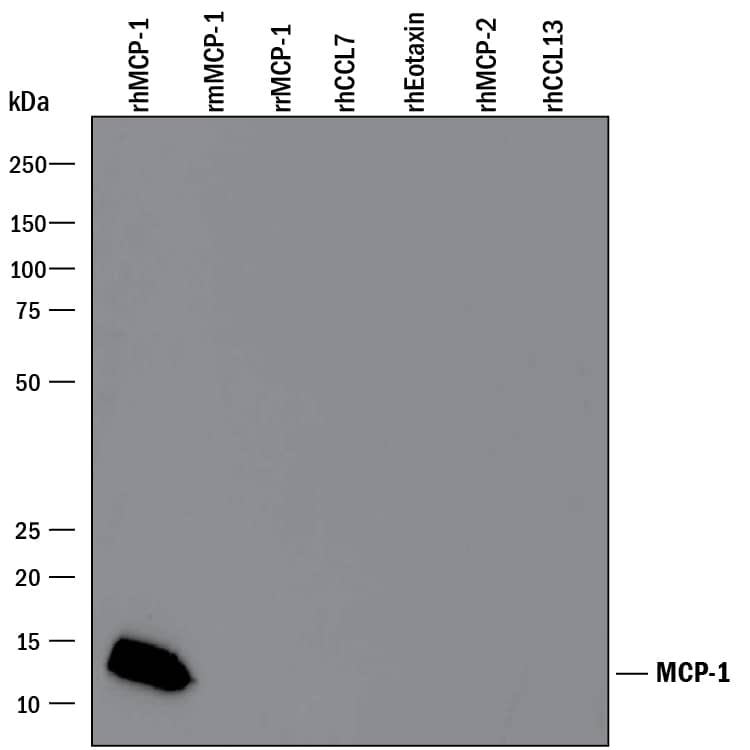
327-P4). PVDF Membrane was probed with 0.1 µg/mL of Goat Anti-Human CCL2/JE/MCP-1 Antigen Affinity-purified Polyclonal Antibody (Catalog # AF-279-NA) followed by HRP-conjugated Anti-Goat IgG Secondary Antibody (Catalog #
HAF109). A specific band was detected for CCL2/JE/MCP-1 at approximately 12 kDa (as indicated). This experiment was conducted under reducing conditions and using
Immunoblot Buffer Group 3." class="big_lightbox">
279-MC), Recombinant Mouse CCL2/JE/MCP-1 (Catalog #
479-JE), Recombinant Rat CCL2/JE/MCP-1 (Catalog #
3144-JE), Recombinant Human CCL7/MCP-3/MARC (Catalog #
282-P3), Recombinant Human CCL11/Eotaxin (Catalog #
320-EO), Recombinant Human CCL8/MCP-2 (Catalog #
281-CP), and Recombinant Human CCL13/MCP-4 (Catalog #
327-P4). PVDF Membrane was probed with 0.1 µg/mL of Goat Anti-Human CCL2/JE/MCP-1 Antigen Affinity-purified Polyclonal Antibody (Catalog # AF-279-NA) followed by HRP-conjugated Anti-Goat IgG Secondary Antibody (Catalog #
HAF109). A specific band was detected for CCL2/JE/MCP-1 at approximately 12 kDa (as indicated). This experiment was conducted under reducing conditions and using
Immunoblot Buffer Group 3." alt="Western blot shows 25 ng of Recombinant Human CCL2/JE/MCP-1 (Catalog #
279-MC), Recombinant Mouse CCL2/JE/MCP-1 (Catalog #
479-JE), Recombinant Rat CCL2/JE/MCP-1 (Catalog #
3144-JE), Recombinant Human CCL7/MCP-3/MARC (Catalog #
282-P3), Recombinant Human CCL11/Eotaxin (Catalog #
320-EO), Recombinant Human CCL8/MCP-2 (Catalog #
281-CP), and Recombinant Human CCL13/MCP-4 (Catalog #
327-P4). PVDF Membrane was probed with 0.1 µg/mL of Goat Anti-Human CCL2/JE/MCP-1 Antigen Affinity-purified Polyclonal Antibody (Catalog # AF-279-NA) followed by HRP-conjugated Anti-Goat IgG Secondary Antibody (Catalog #
HAF109). A specific band was detected for CCL2/JE/MCP-1 at approximately 12 kDa (as indicated). This experiment was conducted under reducing conditions and using
Immunoblot Buffer Group 3." class="big_thumb" />
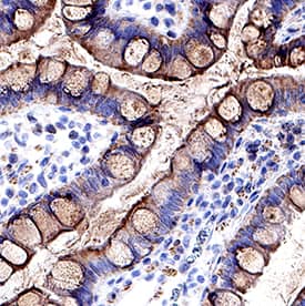
VC004). Before incubation with the primary antibody, tissue was subjected to heat-induced epitope retrieval using Antigen Retrieval Reagent-Basic (Catalog #
CTS013). Tissue was stained using DAB (brown) and counterstained with hematoxylin (blue). Specific staining was localized to epithelial cells. View our protocol for
IHC Staining with VisUCyte HRP Polymer Detection Reagents." class="big_lightbox">
VC004). Before incubation with the primary antibody, tissue was subjected to heat-induced epitope retrieval using Antigen Retrieval Reagent-Basic (Catalog #
CTS013). Tissue was stained using DAB (brown) and counterstained with hematoxylin (blue). Specific staining was localized to epithelial cells. View our protocol for
IHC Staining with VisUCyte HRP Polymer Detection Reagents." class="big_thumb" />
279-MC) chemoattracts the BaF3 mouse pro-B cell line transfected with human CCR2A in a dose-dependent manner (orange line). The amount of cells that migrated through to the lower chemotaxis chamber was measured by Resazurin (Catalog #
AR002). Chemotaxis elicited by Recombinant Human CCL2/MCP-1 (150 ng/mL) is neutralized (green line) by increasing concentrations of Goat Anti-Human CCL2/MCP-1 Antigen Affinity-purified Polyclonal Antibody (Catalog # AF-279-NA). The ND
50 is typically 1.5-4.5 µg/mL." class="big_lightbox">

279-MC) chemoattracts the BaF3 mouse pro-B cell line transfected with human CCR2A in a dose-dependent manner (orange line). The amount of cells that migrated through to the lower chemotaxis chamber was measured by Resazurin (Catalog #
AR002). Chemotaxis elicited by Recombinant Human CCL2/MCP-1 (150 ng/mL) is neutralized (green line) by increasing concentrations of Goat Anti-Human CCL2/MCP-1 Antigen Affinity-purified Polyclonal Antibody (Catalog # AF-279-NA). The ND
50 is typically 1.5-4.5 µg/mL." class="big_thumb" />
279-MC) was serially diluted and captured by Mouse Anti-Human CCL2/JE/MCP‑1 Monoclonal Antibody (Catalog #
MAB679) coated on a Clear Polystyrene Microplate (Catalog #
DY990). Goat Anti-Human CCL2/JE/MCP‑1 Antigen Affinity-purified Polyclonal Antibody (Catalog # AF-279-NA) was biotinylated and incubated with the protein captured on the plate. Detection of the standard curve was achieved by incubating Streptavidin-HRP (Catalog #
DY998)" class="big_lightbox">

279-MC) was serially diluted and captured by Mouse Anti-Human CCL2/JE/MCP‑1 Monoclonal Antibody (Catalog #
MAB679) coated on a Clear Polystyrene Microplate (Catalog #
DY990). Goat Anti-Human CCL2/JE/MCP‑1 Antigen Affinity-purified Polyclonal Antibody (Catalog # AF-279-NA) was biotinylated and incubated with the protein captured on the plate. Detection of the standard curve was achieved by incubating Streptavidin-HRP (Catalog #
DY998)" class="big_thumb" />
 479-JE), Recombinant Human CCL2/JE/MCP-1 (Catalog # 279-MC), Recombinant Rat CCL2/JE/MCP 3144-JE), Recombinant Mouse CCL11/Eotaxin (Catalog # 420-ME), Recombinant Mouse CCL12/MCP-5 (Catalog # 428-P5), and Recombinant Mouse CCL7/MCP-3/MARC (456-MC). PVDF Membrane was probed with 0.1 µg/mL of Goat Anti-Mouse CCL2/JE/MCP-1 Antigen Affinity-purified Polyclonal Antibody (Catalog # AF-479-NA) followed by HRP-conjugated Anti-Goat IgG Secondary Antibody (HAF109). A specific band was detected for CCL2/JE/ MCP-1 at approximately 10 kDa (as indicated). This experiment was conducted under reducing conditions and using Immunoblot Buffer Group 3." alt="Western blot shows 25 ng of Recombinant Mouse CCL2/JE/MCP-1 (479-JE), Recombinant Human CCL2/JE/MCP-1 (Catalog # 279-MC), Recombinant Rat CCL2/JE/MCP 3144-JE), Recombinant Mouse CCL11/Eotaxin (Catalog # 420-ME), Recombinant Mouse CCL12/MCP-5 (Catalog # 428-P5), and Recombinant Mouse CCL7/MCP-3/MARC (456-MC). PVDF Membrane was probed with 0.1 µg/mL of Goat Anti-Mouse CCL2/JE/MCP-1 Antigen Affinity-purified Polyclonal Antibody (Catalog # AF-479-NA) followed by HRP-conjugated Anti-Goat IgG Secondary Antibody (HAF109). A specific band was detected for CCL2/JE/ MCP-1 at approximately 10 kDa (as indicated). This experiment was conducted under reducing conditions and using Immunoblot Buffer Group 3." class="big_thumb" />
 VC001). Before incubation with the primary antibody, tissue was subjected to heat-induced epitope retrieval using Antigen Retrieval Reagent-Basic (CTS013). Tissue was stained using DAB (brown) and counterstained with hematoxylin (blue). Specific staining was localized to cell membranes and cytoplasm. View our protocol for IHC Staining with VisUCyte HRP Polymer Detection Reagents." alt="CCL2/JE/MCP-1 was detected in immersion fixed paraffin-embedded sections of human Crohn's disease intestine using Mouse Anti-Human CCL2/JE/MCP-1 Monoclonal Antibody (Catalog # MAB679) at 15 µg/mL for 1 hour at room temperature followed by incubation with the Anti-Mouse IgG VisUCyte™ HRP Polymer Antibody (VC001). Before incubation with the primary antibody, tissue was subjected to heat-induced epitope retrieval using Antigen Retrieval Reagent-Basic (CTS013). Tissue was stained using DAB (brown) and counterstained with hematoxylin (blue). Specific staining was localized to cell membranes and cytoplasm. View our protocol for IHC Staining with VisUCyte HRP Polymer Detection Reagents." class="big_thumb" />
 279-MC), Recombinant Human CCL8/ MCP-2 (Catalog # 281-CP), Recombinant Human CCL7/MCP-3/MARC (Catalog # 282-P3), Recombinant Human CCL13/ MCP-4 (Catalog # 327-P4), Recombinant Human CCL11/Eotaxin (Catalog # 320-EO), Recombinant Mouse CCL2/JE/MCP-1 (Catalog # 479-JE), and Recombinant Rat CCL2/JE/MCP-1 (Catalog # 3144-JE). PVDF Membrane was probed with 1 µg/mL of Mouse Anti-Human CCL2/JE/MCP-1 Monoclonal Antibody (Catalog # MAB279) followed by HRP-conjugated Anti-Mouse IgG Secondary Antibody (Catalog # HAF018). A specific band was detected for CCL2/JE/ MCP-1 at approximately 11 kDa (as indicated). This experiment was conducted under reducing conditions and using Immunoblot Buffer Group 3." alt="Western blot shows 25 ng of Recombinant Human CCL2/JE/MCP-1 (Catalog # 279-MC), Recombinant Human CCL8/ MCP-2 (Catalog # 281-CP), Recombinant Human CCL7/MCP-3/MARC (Catalog # 282-P3), Recombinant Human CCL13/ MCP-4 (Catalog # 327-P4), Recombinant Human CCL11/Eotaxin (Catalog # 320-EO), Recombinant Mouse CCL2/JE/MCP-1 (Catalog # 479-JE), and Recombinant Rat CCL2/JE/MCP-1 (Catalog # 3144-JE). PVDF Membrane was probed with 1 µg/mL of Mouse Anti-Human CCL2/JE/MCP-1 Monoclonal Antibody (Catalog # MAB279) followed by HRP-conjugated Anti-Mouse IgG Secondary Antibody (Catalog # HAF018). A specific band was detected for CCL2/JE/ MCP-1 at approximately 11 kDa (as indicated). This experiment was conducted under reducing conditions and using Immunoblot Buffer Group 3." class="big_thumb" />
 479-JE) chemoattracts the BaF3 mouse pro‑B cell line transfected with human CCR2A in a dose-dependent manner (orange line). The amount of cells that migrated through to the lower chemotaxis chamber was measured by Resazurin (Catalog # AR002). Chemotaxis elicited by Recombinant Mouse CCL2/JE (40 ng/mL) is neutralized (green line) by increasing concentrations of Mouse CCL2/JE Monoclonal Antibody (Catalog # MAB479). The ND50 is typically 0.75-3.0 µg/mL." alt="Recombinant Mouse CCL2/JE (Catalog # 479-JE) chemoattracts the BaF3 mouse pro‑B cell line transfected with human CCR2A in a dose-dependent manner (orange line). The amount of cells that migrated through to the lower chemotaxis chamber was measured by Resazurin (Catalog # AR002). Chemotaxis elicited by Recombinant Mouse CCL2/JE (40 ng/mL) is neutralized (green line) by increasing concentrations of Mouse CCL2/JE Monoclonal Antibody (Catalog # MAB479). The ND50 is typically 0.75-3.0 µg/mL." class="big_thumb" />
 279-MC), Recombinant Mouse CCL2/JE/MCP-1 (Catalog # 479-JE), Recombinant Rat CCL2/JE/MCP-1 (Catalog # 3144-JE), Recombinant Human CCL7/MCP-3/MARC (Catalog # 282-P3), Recombinant Human CCL11/Eotaxin (Catalog # 320-EO), Recombinant Human CCL8/MCP-2 (Catalog # 281-CP), and Recombinant Human CCL13/MCP-4 (Catalog # 327-P4). PVDF Membrane was probed with 0.1 µg/mL of Goat Anti-Human CCL2/JE/MCP-1 Antigen Affinity-purified Polyclonal Antibody (Catalog # AF-279-NA) followed by HRP-conjugated Anti-Goat IgG Secondary Antibody (Catalog # HAF109). A specific band was detected for CCL2/JE/MCP-1 at approximately 12 kDa (as indicated). This experiment was conducted under reducing conditions and using Immunoblot Buffer Group 3." alt="Western blot shows 25 ng of Recombinant Human CCL2/JE/MCP-1 (Catalog # 279-MC), Recombinant Mouse CCL2/JE/MCP-1 (Catalog # 479-JE), Recombinant Rat CCL2/JE/MCP-1 (Catalog # 3144-JE), Recombinant Human CCL7/MCP-3/MARC (Catalog # 282-P3), Recombinant Human CCL11/Eotaxin (Catalog # 320-EO), Recombinant Human CCL8/MCP-2 (Catalog # 281-CP), and Recombinant Human CCL13/MCP-4 (Catalog # 327-P4). PVDF Membrane was probed with 0.1 µg/mL of Goat Anti-Human CCL2/JE/MCP-1 Antigen Affinity-purified Polyclonal Antibody (Catalog # AF-279-NA) followed by HRP-conjugated Anti-Goat IgG Secondary Antibody (Catalog # HAF109). A specific band was detected for CCL2/JE/MCP-1 at approximately 12 kDa (as indicated). This experiment was conducted under reducing conditions and using Immunoblot Buffer Group 3." class="big_thumb" />


![N/A CCL2/JE/MCP-1 [HRP]](http://images.novusbio.com/fullsize/elisa/DATA_CCL2_DCP00_ELISA_633.jpg "N/A CCL2/JE/MCP-1 [HRP]")
![N/A CCL2/JE/MCP-1 [HRP]](http://images.novusbio.com/fullsize/elisa/DATA_CCL2_DCP00_ELISA_634.jpg)
![N/A CCL2/JE/MCP-1 [HRP]](http://images.novusbio.com/fullsize/elisa/CCL2_DCP00_ELISA_67.jpg)
![N/A CCL2/JE/MCP-1 [Biotin]](http://images.novusbio.com/fullsize/elisa/DATA_CCL2_DY479_ELISA_2324.jpg "N/A CCL2/JE/MCP-1 [Biotin]")
![N/A CCL2/JE/MCP-1 [Biotin]](http://images.novusbio.com/fullsize/elisa/DATA_CCL2_DY279_ELISA_1887.jpg "N/A CCL2/JE/MCP-1 [Biotin]")


 479-JE) chemoattracts the BaF3 mouse pro-B cell line transfected with human CCR2A in a dose-dependent manner (orange line). The amount of cells that migrated through to the lower chemotaxis chamber was measured by Resazurin (Catalog #
  479-JE) was serially diluted and captured by Goat Anti-Mouse CCL2/JE/MCP‑1 Antigen Affinity-purified Polyclonal Antibody (Catalog # AF-479-NA) coated on a Clear Polystyrene Microplate (Catalog #
  479-JE) was serially diluted and captured by Goat Anti-Mouse CCL2/JE/MCP‑1 Antigen Affinity-purified Polyclonal Antibody (Catalog #
  279-MC) chemoattracts the BaF3 mouse pro-B cell line transfected with human CCR2A in a dose-dependent manner (orange line). The amount of cells that migrated through to the lower chemotaxis chamber was measured by Resazurin (


 MAB679) at 20ug/mL with 1 hour incubation at room temperature followed by incubation with anti-mouse IgG VisUCyte HRP Polymer Antibody (
 279-MC) was serially diluted and captured by Mouse Anti-Human CCL2/JE/MCP‑1 Monoclonal Antibody (Catalog # MAB679) coated on a Clear Polystyrene Microplate (Catalog #
 ![Western Blot: CCL2/MCP1 Antibody (2D8) - BSA Free [NBP2-22115] - Analysis using MCP1 mouse mAb against A549 (1), HeLa (2), Raw264.7 (3), L1210 (4), C6 (5), and COS-7 (6) cell lysates.](http://images.novusbio.com/fullsize/CCL2-MCP1-Antibody-2D8-BSA-Free-Western-Blot-NBP2-22115-img0003.jpg "Western Blot: CCL2/MCP1 Antibody (2D8) - BSA Free [NBP2-22115] - Analysis using MCP1 mouse mAb against A549 (1), HeLa (2), Raw264.7 (3), L1210 (4), C6 (5), and COS-7 (6) cell lysates.")
![Immunocytochemistry/Immunofluorescence: CCL2/MCP1 Antibody (2D8) - BSA Free [NBP2-22115] - Analysis of HepG2 cells using A) MCP1 mAb (green), DRAQ5 fluorescent DNA dye (blue), and Actin filaments have been labeled with Alexa Fluor-555 phalloidin (red). B) Staining without MCP1 mAb](http://images.novusbio.com/fullsize/CCL2-MCP1-Antibody-2D8-BSA-Free-Immunocytochemistry-Immunofluorescence-NBP2-22115-img0011.jpg)

![Flow Cytometry: CCL2/MCP1 Antibody (2D8) - BSA Free [NBP2-22115] - Analysis of A549 cells using MCP1 mouse mAb (green) and negative control (red).](http://images.novusbio.com/fullsize/CCL2-MCP1-Antibody-2D8-BSA-Free-Flow-Cytometry-NBP2-22115-img0004.jpg)
![Western Blot: CCL2/MCP1 Antibody (2D8) - BSA Free [NBP2-22115] - Analysis using MCP1 mAb against human MCP1 (AA: 1-99) recombinant protein. (Expected MW is 36.5 kDa)](http://images.novusbio.com/fullsize/CCL2-MCP1-Antibody-2D8-BSA-Free-Western-Blot-NBP2-22115-img0002.jpg)


![Immunohistochemistry-Paraffin: CCL2/MCP1 Antibody (2D8) - BSA Free [NBP2-22115] - Analysis of paraffin-embedded liver cancer tissues using MCP1 mouse mAb with DAB staining.](http://images.novusbio.com/fullsize/CCL2-MCP1-Antibody-2D8-BSA-Free-Immunohistochemistry-Paraffin-NBP2-22115-img0007.jpg)
![ELISA: CCL2/MCP1 Antibody (2D8) - BSA Free [NBP2-22115] - Red: Control Antigen (100ng), Purple: Antigen (10ng), Green: Antigen (50ng), Blue: Antigen (100ng).](http://images.novusbio.com/fullsize/CCL2-MCP1-Antibody-2D8-BSA-Free-ELISA-NBP2-22115-img0001.jpg)
![Immunocytochemistry/ Immunofluorescence: CCL2/MCP1 Antibody (2D8) - BSA Free [NBP2-22115] - Expression patterns of CCL2 & CCR2 in unwounded & wounded corneas of WT & MMP12 KO mice. (A) Immunofluorescence of CCL2 chemokine & (B) its receptor CCR2 in unwounded & chemically wounded (2-days after injury) WT & Mmp12−/− mouse corneas. Control images represent mouse corneas stained with secondary antibody only & without primary antibody. Nuclei were visualized by staining with DAPI (blue). Scale bars: 50 µm. A magnified image of a wounded WT cornea shows perinuclear expression of CCL2 (orange box). CCL2 staining was visualized in epithelial, stromal, & endothelial layers of wounded WT & Mmp12−/− corneas. CCR2 staining was visualized in epithelial & stromal layers of unwounded & wounded WT & Mmp12−/− corneas. Image collected & cropped by CiteAb from the following publication (//pubmed.ncbi.nlm.nih.gov/31399604), licensed under a CC-BY license. Not internally tested by Novus Biologicals.](http://images.novusbio.com/fullsize/nbp2-22115_mouse-monoclonal-ccl2-mcp1-antibody-2d8-310202415384136.jpg)
![Immunocytochemistry/ Immunofluorescence: CCL2/MCP1 Antibody (2D8) - BSA Free [NBP2-22115] - LIF represses CXCL9 through epigenetic silencing. a, b qRT-PCR analysis of the indicated genes in BMDMs. BMDMs were pre-incubated with 20 ng/ml LIF for 72 h & then stimulated with 5 ng/ml IFN gamma or 10 μg/ml IL4 during 6 h (a) or with 0.1, 0.5, 1 & 5 ng/ml IFN gamma for 24 h (b). c CXCL9 ELISA from BMDMs pre-incubated with 20 ng/ml LIF & then stimulated with 0.1 ng/ml IFN gamma for 24 h. d CXCL9 ELISA from human CD11b+sorted cells (77% CD11b+ CD14+, see Supplementary Fig. 7b, c) from human GBM cultured with 20 ng/ml LIF for 72 h & then with 0.1 ng/ml IFN gamma for 24 h. e ChIP of Tri-methyl-histone H3 (H3K27me3), EZH2 & acetyl-histone4 (H4ac) was performed in BMDMs treated with 20 ng/ml LIF for 72 h. Scheme shows the analysed CXCL9 promoter region. Representative data are presented as mean ± SD. f Representative images of IF of the indicated markers in human GBM organotypic slices (patients 1, 2, 3) incubated with 10 μg/ml anti-LIF for 3 days. Scale bar, 20 μm. (see Supplementary Fig. 8). Bottom, percentage of double positive cells relative to Iba1+ cells & percentage of CXCL9+ cells relative to the total number of cells. Data are mean of all patients ± SEM. Statistical analyses by Student’s t-test or Mann–Whitney T-test. *P < 0.05, **P < 0.01; ***P < 0.001; ****P < 0.0001 Image collected & cropped by CiteAb from the following publication (//pubmed.ncbi.nlm.nih.gov/31186412), licensed under a CC-BY license. Not internally tested by Novus Biologicals.](http://images.novusbio.com/fullsize/nbp2-22115_mouse-monoclonal-ccl2-mcp1-antibody-2d8-3102024161360.jpg)
![Immunohistochemistry: CCL2/MCP1 Antibody (2D8) - BSA Free [NBP2-22115] - LIF regulates CXCL9, CCL2, CD206, & CD163 in TAMs. a Differential expression analysis of isolated CD11b+ cells from anti-LIF treated ID8 mice vs. control. Volcano plot representing the genes significantly (Q-value < 0.1) overexpressed (brown) & significantly underexpressed (turquoise). Heatmap representing the expression values of the indicated genes, each column represents a sample & each row a gene. The last column represents the log2 fold change (log2 FC) of gene expression. b mRNA expression for the indicated genes in isolated CD11b+ cells from anti-LIF treated or untreated ID8 & GL261N tumors. c Percentage & mean fluorescence intensity (MFI) of CCL2 & CXCL9 in TAMs (CD11b+ Ly6G− Ly6C−) from anti-LIF treated or untreated GL261N tumors. d Representative IF images of Iba1 & the indicated markers stainings of GL261N tumors (see Supplementary Fig. 4). Scale bar, 20 μm. Bottom, percentage of double positive cells relative to the TAM marker positive cells. CXCL9 quantification is relative to the total number of cells. e Tumor growth of GL261N in WT, CXCL9−/−, & CCL2−/− mice or mice treated with the indicated antibodies is shown as total flux (p/s). f Fold change (FC) of tumor infiltrating CD8+ T cells in the indicated treatments. Data are mean ± SEM. g, h Representative IHC of the indicated markers from 20 GBM tumors. The degree of staining was quantified using H-score method. Correlations between LIF & CCL2, CD206, CD163, & CXCL9 with the R-squared coefficients (R2) were calculated (h). Statistical analyses by Mann–Whitney T-test. *P < 0.05; **P < 0.01; ***P < 0.001; ****P < 0.0001 Image collected & cropped by CiteAb from the following publication (//pubmed.ncbi.nlm.nih.gov/31186412), licensed under a CC-BY license. Not internally tested by Novus Biologicals.](http://images.novusbio.com/fullsize/nbp2-22115_mouse-monoclonal-ccl2-mcp1-antibody-2d8-310202416114760.jpg)
![Immunocytochemistry/ Immunofluorescence: CCL2/MCP1 Antibody (2D8) - BSA Free [NBP2-22115] - LIF regulates CXCL9, CCL2, CD206, & CD163 in TAMs. a Differential expression analysis of isolated CD11b+ cells from anti-LIF treated ID8 mice vs. control. Volcano plot representing the genes significantly (Q-value < 0.1) overexpressed (brown) & significantly underexpressed (turquoise). Heatmap representing the expression values of the indicated genes, each column represents a sample & each row a gene. The last column represents the log2 fold change (log2 FC) of gene expression. b mRNA expression for the indicated genes in isolated CD11b+ cells from anti-LIF treated or untreated ID8 & GL261N tumors. c Percentage & mean fluorescence intensity (MFI) of CCL2 & CXCL9 in TAMs (CD11b+ Ly6G− Ly6C−) from anti-LIF treated or untreated GL261N tumors. d Representative IF images of Iba1 & the indicated markers stainings of GL261N tumors (see Supplementary Fig. 4). Scale bar, 20 μm. Bottom, percentage of double positive cells relative to the TAM marker positive cells. CXCL9 quantification is relative to the total number of cells. e Tumor growth of GL261N in WT, CXCL9−/−, & CCL2−/− mice or mice treated with the indicated antibodies is shown as total flux (p/s). f Fold change (FC) of tumor infiltrating CD8+ T cells in the indicated treatments. Data are mean ± SEM. g, h Representative IHC of the indicated markers from 20 GBM tumors. The degree of staining was quantified using H-score method. Correlations between LIF & CCL2, CD206, CD163, & CXCL9 with the R-squared coefficients (R2) were calculated (h). Statistical analyses by Mann–Whitney T-test. *P < 0.05; **P < 0.01; ***P < 0.001; ****P < 0.0001 Image collected & cropped by CiteAb from the following publication (//pubmed.ncbi.nlm.nih.gov/31186412), licensed under a CC-BY license. Not internally tested by Novus Biologicals.](http://images.novusbio.com/fullsize/nbp2-22115_mouse-monoclonal-ccl2-mcp1-antibody-2d8-31020241613617.jpg)


![<b>Biological Strategies Validation. </b>Immunohistochemistry-Paraffin: CCL2/MCP1 Antibody [NBP1-07035] - IHC analysis of a formalin-fixed paraffin-embedded (FFPE) human breast carcinoma tissue section using 1:1000 dilution of CCL2/MCP1 antibody (NBP1-07035) on a Bond Rx autostainer (Leica Biosystems). The assay involved 20 minutes of heat induced antigen retrieval (HIER) with 10mM sodium citrate buffer (pH 6.0) and endogenous peroxidase quenching using peroxide block. The sections were incubated with primary antibody for 30 minutes. Bond Polymer Refine Detection (Leica Biosystems) and DAB were used for signal detection which followed counterstaining with hematoxylin. Whole slide scanning and capturing of representative images (20X) were performed using Aperio AT2 (Leica Biosystems). This antibody generated a diffused cytoplasmic staining of CCL2 antigen in the cancer cells, stromal cells as well as the endothelial cells. The stroma itself showed a weak immunopositivity for CCL2. Staining was performed by Histowiz.](http://images.novusbio.com/fullsize/CCL2-MCP1-Antibody-Immunohistochemistry-Paraffin-NBP1-07035-img0007.jpg)

![Immunocytochemistry/Immunofluorescence: CCL2/MCP1 Antibody [NBP1-07035] - MCP1 antibody was tested in HeLa cells with Dylight 488 (green). Nuclei and alpha-tubulin were counterstained with DAPI (blue) and Dylight 550 (red).](http://images.novusbio.com/fullsize/CCL2-MCP1-Antibody-Immunocytochemistry-Immunofluorescence-NBP1-07035-img0006.jpg)
![Immunohistochemistry-Paraffin: CCL2/MCP1 Antibody [NBP1-07035] - IHC analysis of a formalin-fixed paraffin-embedded (FFPE) human breast carcinoma tissue section using 1:1000 dilution of CCL2/MCP1 antibody (NBP1-07035) on a Bond Rx autostainer (Leica Biosystems). The assay involved 20 minutes of heat induced antigen retrieval (HIER) with 10mM sodium citrate buffer (pH 6.0) and endogenous peroxidase quenching using peroxide block. The sections were incubated with primary antibody for 30 minutes. Bond Polymer Refine Detection (Leica Biosystems) and DAB were used for signal detection which followed counterstaining with hematoxylin. Whole slide scanning and capturing of representative images (20X) were performed using Aperio AT2 (Leica Biosystems). This antibody generated a diffused cytoplasmic staining of CCL2 antigen in the cancer cells, stromal cells as well as the endothelial cells. The stroma itself showed a weak immunopositivity for CCL2. Staining was performed by Histowiz.](http://images.novusbio.com/fullsize/CCL2-MCP1-Antibody-Immunohistochemistry-Paraffin-NBP1-07035-img0008.jpg)
![Western Blot: CCL2/MCP1 Antibody [NBP1-07035] - CCL2 expression level was evaluated in control group and aLAG-3 group.](http://images.novusbio.com/fullsize/CCL2-MCP1-Antibody-Western-Blot-NBP1-07035-img0009.jpg)
![Western Blot: CCL2/MCP1 Antibody - BSA Free [NBP1-07035] - Ox-LDL induced macrophage foam cell formation, SIRT1 inhibition, autophagy impairment, & MCP-1 production in THP-1 cellsHuman THP-1 macrophages were exposed to 0, 20, 40, 60, & 80 μg/mL of ox-LDL for 24 hrs. Treated cells were photographed using light microscopy (A). The THP-1 macrophage-derived foam cell formation was determined using ORO staining method (B) & (C). Western blot for SIRT1, LC3, Beclin1, p62, & MCP-1 proteins were analyzed from the ox-LDL-stimulated THP-1 cells. beta -actin was used as loading control (D-I). Scale bar: 20 μm. Bar graph indicates the mean ± SD (n = 3). *P < 0.05 & **P < 0.01 vs. Cont group (0 μg/mL of ox-LDL). Image collected & cropped by CiteAb from the following publication (//www.oncotarget.com/lookup/doi/10.18632/oncotarget.17691), licensed under a CC-BY license. Not internally tested by Novus Biologicals.](http://images.novusbio.com/fullsize/nbp1-07035_rabbit-polyclonal-ccl2-mcp1-antibody-310202415304238.jpg)
![Western Blot: CCL2/MCP1 Antibody - BSA Free [NBP1-07035] - Inhibition of autophagy with 3-MA led to increased foam cell formation & MCP-1 productionHuman THP-1 macrophages were pretreated with 3-MA (5 μM) for 2 hrs, & then exposed to 80 μg/mL of ox-LDL for an additional 24 hrs. Western blot for LC3, Beclin1, p62, & MCP-1 proteins were analyzed from the ox-LDL-stimulated THP-1 cells. beta -actin was used as loading control (A-E). THP-1 macrophage-derived foam cell formation was determined using ORO staining method (F-G). Scale bar: 40 μm. Bar graph indicates the mean ± SD (n = 3). *P < 0.05 & **P < 0.01 vs. Cont group; #P < 0.05 & ##P < 0.01 vs. 3-MA group; &P < 0.05 & &&P < 0.01 represent significant differences between ox-LDL group & 3-MA+ox-LDL group. Image collected & cropped by CiteAb from the following publication (//www.oncotarget.com/lookup/doi/10.18632/oncotarget.17691), licensed under a CC-BY license. Not internally tested by Novus Biologicals.](http://images.novusbio.com/fullsize/nbp1-07035_rabbit-polyclonal-ccl2-mcp1-antibody-310202415384154.jpg)
![Immunocytochemistry/ Immunofluorescence: CCL2/MCP1 Antibody - BSA Free [NBP1-07035] - CCL2-ir cellular distribution in ependymal layers attached to the CC. A, B CCL2-ir (CCL2 panels, arrows) & GFAP (GFAP panels, arrowheads) double immunostained structures (Merged panels, arrow-arrowheads) are seen inlaid in the ependymal layers attached to the CC in both saline & LPS injected mice. C A CCL2-ir & Iba-1 double labeled soma (Merged, arrow-arrowhead) is seen at a corner of lateral ventricle, which seems be just next to an invaginated choroid plexus between the CC & basal ganglion (framed areas & insets). D, E Hyper-ramified & amoeba like Iba-1 labeled cells (Iba-1 panel, opened arrowheads) are observed in the CC of LPS injected mice, indicating their activated states. The CCL2-ir & Iba-1 double labeled structures are also regarded in the ependymal layers (Merged, arrow-arrowheads). Scare bar = 100 µm in all A– E Image collected & cropped by CiteAb from the following publication (//pubmed.ncbi.nlm.nih.gov/35354428), licensed under a CC-BY license. Not internally tested by Novus Biologicals.](http://images.novusbio.com/fullsize/nbp1-07035_rabbit-polyclonal-ccl2-mcp1-antibody-310202416161849.jpg)
![Western Blot: CCL2/MCP1 Antibody - BSA Free [NBP1-07035] - Inhibition of autophagy using Atg5 siRNA aggravated foam cell formation & MCP-1 expressionHuman THP-1 macrophages were pretreated with Atg5 siRNA (20 μM) for 24 hrs, & then exposed to 80 μg/mL of ox-LDL for an additional 24 hrs. Western blot forAtg5, LC3, Beclin1, p62, & MCP-1 proteins were analyzed from the ox-LDL-stimulated THP-1 cells. beta -actin was used as loading control (A-F) & (I). THP-1 macrophage-derived foam cell formation was determined using ORO staining method (G) & (H). Scale bar: 40 μm. Bar graph indicates the mean ± SD (n = 3). *P < 0.05 & **P < 0.01 vs. NC siRNA group; #P < 0.05 & ##P < 0.01 vs. Atg5 siRNA group; &P < 0.05 & &&P < 0.01 represent significant differences between NC siRNA+ox-LDL group & Atg5 siRNA+ox-LDL group. Image collected & cropped by CiteAb from the following publication (//www.oncotarget.com/lookup/doi/10.18632/oncotarget.17691), licensed under a CC-BY license. Not internally tested by Novus Biologicals.](http://images.novusbio.com/fullsize/nbp1-07035_rabbit-polyclonal-ccl2-mcp1-antibody-31020241618266.jpg)
![Western Blot: CCL2/MCP1 Antibody - BSA Free [NBP1-07035] - Inhibition of SIRT1 using EX-527 or SIRT1 siRNA transfection enhanced MCP-1 expression & foam cell formationHuman THP-1 macrophages were pretreated with EX-527 (2 μM, for 2 hrs) or SIRT1 siRNA (20 μM, for 24 hrs), & then exposed to 80 μg/mL of ox-LDL for an additional 24 hrs. Western blot for SIRT1 & MCP-1 proteins were analyzed from the ox-LDL-stimulated THP-1 cells. beta -actin was used as loading control (A-C) & (F-H). THP-1 macrophage-derived foam cell formation was determined using ORO staining method (D-E) & (I-J). Scale bar: 40 μm. Bar graph indicates the mean ± SD (n = 3). *P < 0.05 & **P < 0.01 vs. Cont group (NC siRNA group); #P < 0.05 & ##P < 0.01 vs. EX-527 group (SIRT1 siRNA group); &P < 0.05 & &&P < 0.01 represent significant differences between ox-LDL group (NC siRNA+ox-LDL group) & EX-527+ox-LDL group (SIRT1 siRNA+ox-LDL group). Image collected & cropped by CiteAb from the following publication (//www.oncotarget.com/lookup/doi/10.18632/oncotarget.17691), licensed under a CC-BY license. Not internally tested by Novus Biologicals.](http://images.novusbio.com/fullsize/nbp1-07035_rabbit-polyclonal-ccl2-mcp1-antibody-310202416171476.jpg)
![Western Blot: CCL2/MCP1 Antibody - BSA Free [NBP1-07035] - Overexpression of SIRT1 using adenoviral transfection reversed ox-LDL-induced macrophage foam cell formation & autophagy impairment in THP-1 cellsHuman THP-1 macrophages were transfected by SIRT1 over-expressing adenovirus (HBAD-SIRT1) or NC adenovirus (HBAD-GFP) for 24 hrs & then exposed to 80 μg/mL of ox-LDL for an additional 24 hrs. The transfected THP-1 cells were observed using an inverted fluorescence microscope (A) & then were harvested for transfection efficiency analysis by Western blot method (B). THP-1 macrophage-derived foam cell formation was determined using ORO staining method (C) & (D). Western blot for LC3, Beclin1, p62, & Atg5 proteins & immunoprecipitation for acetyl-Lys Atg5 were analyzed from the ox-LDL-stimulated THP-1 cells. beta -actin was used as loading control (E-L). Scale bar: 40 μm. Bar graph indicates the mean ± SD (n = 3). *P < 0.05 & **P < 0.01 vs. HBAD-GFP group; #P < 0.05 & ##P < 0.01 vs. HBAD-SIRT1 group; &P < 0.05 & &&P < 0.01 represent significant differences between HBAD-GFP+ox-LDL group & HBAD-SIRT1+ox-LDL group. Image collected & cropped by CiteAb from the following publication (//www.oncotarget.com/lookup/doi/10.18632/oncotarget.17691), licensed under a CC-BY license. Not internally tested by Novus Biologicals.](http://images.novusbio.com/fullsize/nbp1-07035_rabbit-polyclonal-ccl2-mcp1-antibody-310202416163742.jpg)
 279-MC) chemoattracts the BaF3 mouse pro-B cell line transfected with human CCR2A in a dose-dependent manner (orange line). The amount of cells that migrated through to the lower chemotaxis chamber was measured by Resazurin (Catalog #
 VC004). Before incubation with the primary antibody, tissue was subjected to heat-induced epitope retrieval using Antigen Retrieval Reagent-Basic (Catalog #
  279-MC) chemoattracts the BaF3 mouse pro-B cell line transfected with human CCR2A in a dose-dependent manner (orange line). The amount of cells that migrated through to the lower chemotaxis chamber was measured by Resazurin (Catalog #
  279-MC) was serially diluted and captured by Mouse Anti-Human CCL2/JE/MCP‑1 Monoclonal Antibody (Catalog #


