MAB006, open histogram), followed by Phycoerythrin-conjugated Anti-Rat IgG Secondary Antibody (F0105B). View our protocol for Staining Membrane-associated Proteins." title="RAW 264.7 mouse monocyte/macrophage cell line was stained with Rat Anti-Mouse MDL-1/CLEC5A Monoclonal Antibody (MAB1639, filled histogram) or isotype control antibody (MAB006, open histogram), followed by Phycoerythrin-conjugated Anti-Rat IgG Secondary Antibody (F0105B). View our protocol for Staining Membrane-associated Proteins." />
| Reactivity | MuSpecies Glossary |
| Applications | Flow, CyTOF-ready |
| Clone | 226402 |
| Clonality | Monoclonal |
| Host | Rat |
| Conjugate | Unconjugated |
| Concentration | LYOPH |
| Immunogen | BaF3 mouse pro-B cell line transfected with mouse MDL-1/CLEC5A |
| Specificity | Detects mouse MDL-1/CLEC5A. In ELISAs, approximately 50% cross-reactivity with recombinant human MDL‑1 is observed. |
| Source | N/A |
| Isotype | IgG2a |
| Clonality | Monoclonal |
| Host | Rat |
| Gene | CLEC5A |
| Purity Statement | Protein A or G purified from hybridoma culture supernatant |
| Innovator's Reward | Test in a species/application not listed above to receive a full credit towards a future purchase. |
| Dilutions |
|
|
| Publications |
|
| Storage | Use a manual defrost freezer and avoid repeated freeze-thaw cycles.
|
| Buffer | Lyophilized from a 0.2 μm filtered solution in PBS with Trehalose. See Certificate of Analysis for details. *Small pack size (-SP) is supplied either lyophilized or as a 0.2 µm filtered solution in PBS. |
| Preservative | No Preservative |
| Concentration | LYOPH |
| Reconstitution Instructions | Reconstitute at 0.5 mg/mL in sterile PBS. For liquid material, refer to CoA for concentration. |
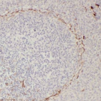
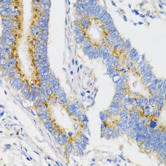
![Immunohistochemistry-Paraffin DAP10/HCST Antibody (982101) [Unconjugated] - BSA Free](https://images.novusbio.com/images/DAP10-HCST-Antibody-982101-Unconjugated-Immunohistochemistry-Paraffin-MAB97861-img0001.jpg)


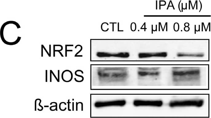
![Western Blot STAT1 [p Tyr701] Antibody [Unconjugated]](https://images.novusbio.com/images/antibody/STAT1_AF2894_Western_Blot_5226.jpg)
![Intracellular Staining by Flow Cytometry STAT1 [p Tyr701] Antibody [Unconjugated]](https://images.novusbio.com/images/antibody/STAT1_AF2894_Flow_Cytometry_8189.jpg)
![Western Blot STAT1 [p Tyr701] Antibody [Unconjugated]](https://images.novusbio.com/images/af2894_human-mouse-phospho-stat1-y701-affinity-purified-pab-western-blot-121220258554112.jpg)





Secondary Antibodies |
Isotype Controls |
The concentration calculator allows you to quickly calculate the volume, mass or concentration of your vial. Simply enter your mass, volume, or concentration values for your reagent and the calculator will determine the rest.
| Gene Symbol | CLEC5A |


![Western Blot TREM1 Antibody (193015) [Unconjugated]](https://images.novusbio.com/images/mab1278_human-trem-1-mab-clone-193015-western-blot-12122025914018.jpg)
![Flow Cytometry TREM1 Antibody (193015) [Unconjugated]](https://images.novusbio.com/images/antibody/TREM1_MAB1278_Flow_Cytometry_19636.jpg)
![Western Blot TREM1 Antibody (193015) [Unconjugated]](https://images.novusbio.com/images/mab1278_human-trem-1-mab-clone-193015-western-blot-1212202592082.jpg)
![SDS-Page TNF-alpha [Unconjugated]](https://images.novusbio.com/images/protein/TNF-alpha_210-TA_256.jpg)
![Bioactivity TNF-alpha [Unconjugated]](https://images.novusbio.com/images/protein/TNFalpha_210TA_1658.jpg)
![SEC-MALS TNF-alpha [Unconjugated]](https://images.novusbio.com/images/210-ta_recombinant-human-tnf-alpha-protein-sec-mals-35202312244..jpg)
![ELISA TREM2 Antibody [Unconjugated]](https://images.novusbio.com/images/antibody/TREM2_AF1729_ELISA_23734.jpg)
![Immunocytochemistry/ Immunofluorescence TREM2 Antibody [Unconjugated]](https://images.novusbio.com/images/af1729_mouse-trem-2-affinity-purified-polyclonal-ab-412024103388.jpg)
![Immunocytochemistry/ Immunofluorescence TREM2 Antibody [Unconjugated]](https://images.novusbio.com/images/af1729_mouse-trem-2-affinity-purified-polyclonal-ab-4120241033417.jpg)