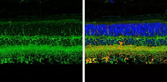
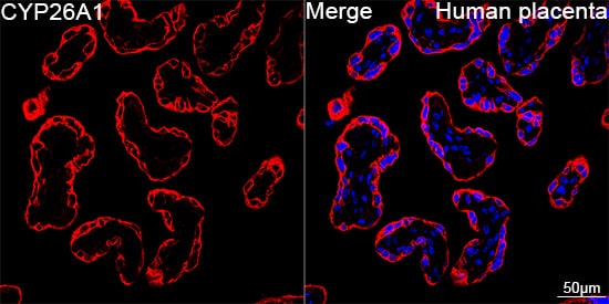
![Immunocytochemistry/Immunofluorescence: CRABP1 Antibody (C-1) [NB300-539] - Analysis of CRABPI using Anti-CRABPI Monoclonal Antibody (C-1) shows staining in NIH-3T3 Cells. CRABPI staining (green), F-Actin staining with Phalloidin (red) and nuclei with DAPI (blue) is shown. Cells were grown on chamber slides and fixed with formaldehyde prior to staining. Cells were probed without (control) or with or an antibody recognizing CRABPI at a dilution of 1:200 over night at 4C, washed with PBS and incubated with a DyLight-488 conjugated.](http://images.novusbio.com/fullsize/CRABP1-Antibody-C-1-Immunocytochemistry-Immunofluorescence-NB300-539-img0005.jpg "Immunocytochemistry/Immunofluorescence: CRABP1 Antibody (C-1) [NB300-539] - Analysis of CRABPI using Anti-CRABPI Monoclonal Antibody (C-1) shows staining in NIH-3T3 Cells. CRABPI staining (green), F-Actin staining with Phalloidin (red) and nuclei with DAPI (blue) is shown. Cells were grown on chamber slides and fixed with formaldehyde prior to staining. Cells were probed without (control) or with or an antibody recognizing CRABPI at a dilution of 1:200 over night at 4C, washed with PBS and incubated with a DyLight-488 conjugated.")
| Immunogen | Oxidized CRABPI from bovine retina. |
| Specificity | Detects cellular Retinoic Acid Binding Protein 1 (CRABP 1). This does not cross-react with Cellular Retinol Binding Protein (CRBP), performic acid oxidized CRBP, interphotoreceptor retinoid binding protein or retinol binding protein. |
| Isotype | IgG2b |
| Clonality | Monoclonal |
| Host | Mouse |
| Gene | CRABP1 |
| Purity | Unpurified |
| Innovator's Reward | Test in a species/application not listed above to receive a full credit towards a future purchase. |
| Dilutions |
|
|
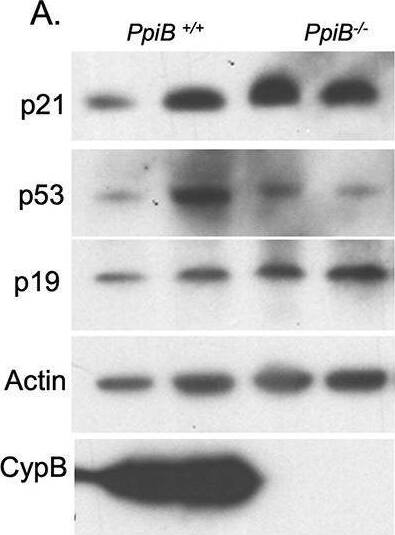
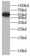
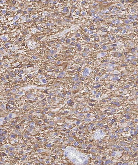
| Application Notes | WB: Detects an approx. 16 kDa protein representing CRABP 1 from rat retinal supernatant. IHC: Staining of CRABP 1 in rat retina results in its localization to amacrine somata and laminae in the inner plexiform layer. |
|
| Publications |
|
| Storage | Store at -20C. Avoid freeze-thaw cycles. |
| Buffer | Ascites diluted with PBS |
| Preservative | 0.05% Sodium Azide |
| Concentration | This product is unpurified. The exact concentration of antibody is not quantifiable. |
| Purity | Unpurified |


")
Secondary Antibodies |
Isotype Controls |
Research Areas for CRABP1 Antibody (NB300-539)Find related products by research area.
|
The concentration calculator allows you to quickly calculate the volume, mass or concentration of your vial. Simply enter your mass, volume, or concentration values for your reagent and the calculator will determine the rest.


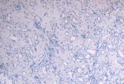
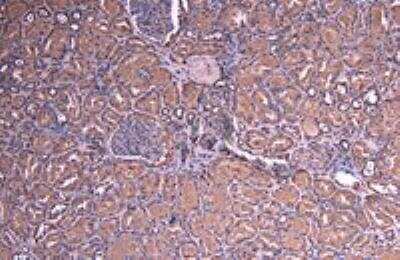
![Immunohistochemistry-Paraffin: CRABP1 Antibody (C-1) [NB300-539] - Both normal and cancer biopsies of deparaffinized Human thyroid tissues.](http://images.novusbio.com/fullsize/CRABP1-Antibody-C-1-Immunohistochemistry-Paraffin-NB300-539-img0009.jpg "Immunohistochemistry-Paraffin: CRABP1 Antibody (C-1) [NB300-539] - Both normal and cancer biopsies of deparaffinized Human thyroid tissues.")
![Flow Cytometry: CRABP1 Antibody (C-1) [NB300-539] - Analysis of CRABPI in Hela cells (green) compared to an isotype control (blue).](http://images.novusbio.com/fullsize/CRABP1-Antibody-C-1-Flow-Cytometry-NB300-539-img0008.jpg "Flow Cytometry: CRABP1 Antibody (C-1) [NB300-539] - Analysis of CRABPI in Hela cells (green) compared to an isotype control (blue).")
![Immunocytochemistry/Immunofluorescence: CRABP1 Antibody (C-1) [NB300-539] - Analysis of CRABPI using Anti-CRABPI Monoclonal Antibody (C-1) shows staining in Hela Cells. CRABPI staining (green), F-Actin staining with Phalloidin (red) and nuclei with DAPI (blue) is shown. Cells were grown on chamber slides and fixed with formaldehyde prior to staining. Cells were probed without (control) or with or an antibody recognizing CRABPI at a dilution of 1:200 over night at 4C, washed with PBS and incubated with a DyLight-488 conjugated.](http://images.novusbio.com/fullsize/CRABP1-Antibody-C-1-Immunocytochemistry-Immunofluorescence-NB300-539-img0003.jpg "Immunocytochemistry/Immunofluorescence: CRABP1 Antibody (C-1) [NB300-539] - Analysis of CRABPI using Anti-CRABPI Monoclonal Antibody (C-1) shows staining in Hela Cells. CRABPI staining (green), F-Actin staining with Phalloidin (red) and nuclei with DAPI (blue) is shown. Cells were grown on chamber slides and fixed with formaldehyde prior to staining. Cells were probed without (control) or with or an antibody recognizing CRABPI at a dilution of 1:200 over night at 4C, washed with PBS and incubated with a DyLight-488 conjugated.")
![Immunocytochemistry/Immunofluorescence: CRABP1 Antibody (C-1) [NB300-539] - Analysis of CRABPI using Anti-CRABPI Monoclonal Antibody (C-1) shows staining in MCF-7 Cells. CRABPI staining (green), F-Actin staining with Phalloidin (red) and nuclei with DAPI (blue) is shown. Cells were grown on chamber slides and fixed with formaldehyde prior to staining. Cells were probed without (control) or with or an antibody recognizing CRABPI at a dilution of 1:100 over night at 4C, washed with PBS and incubated with a DyLight-488 conjugated.](http://images.novusbio.com/fullsize/CRABP1-Antibody-C-1-Immunocytochemistry-Immunofluorescence-NB300-539-img0004.jpg "Immunocytochemistry/Immunofluorescence: CRABP1 Antibody (C-1) [NB300-539] - Analysis of CRABPI using Anti-CRABPI Monoclonal Antibody (C-1) shows staining in MCF-7 Cells. CRABPI staining (green), F-Actin staining with Phalloidin (red) and nuclei with DAPI (blue) is shown. Cells were grown on chamber slides and fixed with formaldehyde prior to staining. Cells were probed without (control) or with or an antibody recognizing CRABPI at a dilution of 1:100 over night at 4C, washed with PBS and incubated with a DyLight-488 conjugated.")
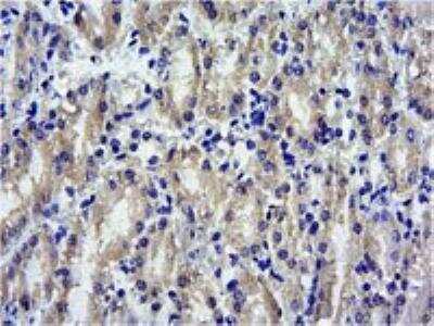
![Immunohistochemistry-Paraffin: CRABP1 Antibody (C-1) [NB300-539] - Both normal and cancer biopsies of deparaffinized Human breast carcinoma tissues.](http://images.novusbio.com/fullsize/CRABP1-Antibody-C-1-Immunohistochemistry-Paraffin-NB300-539-img0010.jpg "Immunohistochemistry-Paraffin: CRABP1 Antibody (C-1) [NB300-539] - Both normal and cancer biopsies of deparaffinized Human breast carcinoma tissues.")
![Flow Cytometry: CRABP1 Antibody (C-1) [NB300-539] - Analysis of CRABPI in NIH-3T3 cells (green) compared to an isotype control (blue).](http://images.novusbio.com/fullsize/CRABP1-Antibody-C-1-Flow-Cytometry-NB300-539-img0006.jpg "Flow Cytometry: CRABP1 Antibody (C-1) [NB300-539] - Analysis of CRABPI in NIH-3T3 cells (green) compared to an isotype control (blue).")
![Flow Cytometry: CRABP1 Antibody (C-1) [NB300-539] - Analysis of CRABPI in MCF-7 cells (green) compared to an isotype control (blue).](http://images.novusbio.com/fullsize/CRABP1-Antibody-C-1-Flow-Cytometry-NB300-539-img0007.jpg "Flow Cytometry: CRABP1 Antibody (C-1) [NB300-539] - Analysis of CRABPI in MCF-7 cells (green) compared to an isotype control (blue).")


![SDS-Page IGF-II/IGF2 [Unconjugated]](https://images.novusbio.com/images/protein/IGF-II_292-G2_668.jpg)
![Bioactivity IGF-II/IGF2 [Unconjugated]](https://images.novusbio.com/images/protein/IGF-II_292-G2_669.jpg)
![N/A RBP4/Retinol-Binding Protein 4 [HRP]](https://images.novusbio.com/images/elisa/DATA_RBP4_DRB400_ELISA_804.jpg)
![N/A RBP4/Retinol-Binding Protein 4 [HRP]](https://images.novusbio.com/images/elisa/RBP4_DRB400_ELISA_169.jpg)


![Intracellular Staining by Flow Cytometry RUNX3/CBFA3 Antibody (527327) [Unconjugated]](https://images.novusbio.com/images/antibody/RUNX3_MAB3765_Flow_Cytometry_8418.jpg)