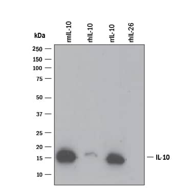
417-ML), Recombinant Human IL-10 (Catalog #
217-IL), Recombinant Rat IL-10 (Catalog #
522-RLB), and Recombinant Human IL-26/AK155 Monomer (Catalog #
1375-IL). PVDF Membrane was probed with 0.1 µg/mL of Goat Anti-Mouse/Rat IL-10 Antigen Affinity-purified Polyclonal Antibody (Catalog # AF519) followed by HRP-conjugated Anti-Goat IgG Secondary Antibody (Catalog #
HAF109). A specific band was detected for IL-10 at approximately 16 kDa (as indicated). This experiment was conducted under reducing conditions and using
Immunoblot Buffer Group 3." class="big_lightbox">
417-ML), Recombinant Human IL-10 (Catalog #
217-IL), Recombinant Rat IL-10 (Catalog #
522-RLB), and Recombinant Human IL-26/AK155 Monomer (Catalog #
1375-IL). PVDF Membrane was probed with 0.1 µg/mL of Goat Anti-Mouse/Rat IL-10 Antigen Affinity-purified Polyclonal Antibody (Catalog # AF519) followed by HRP-conjugated Anti-Goat IgG Secondary Antibody (Catalog #
HAF109). A specific band was detected for IL-10 at approximately 16 kDa (as indicated). This experiment was conducted under reducing conditions and using
Immunoblot Buffer Group 3." alt="Western blot shows 25 ng of Recombinant Mouse IL-10 (Catalog #
417-ML), Recombinant Human IL-10 (Catalog #
217-IL), Recombinant Rat IL-10 (Catalog #
522-RLB), and Recombinant Human IL-26/AK155 Monomer (Catalog #
1375-IL). PVDF Membrane was probed with 0.1 µg/mL of Goat Anti-Mouse/Rat IL-10 Antigen Affinity-purified Polyclonal Antibody (Catalog # AF519) followed by HRP-conjugated Anti-Goat IgG Secondary Antibody (Catalog #
HAF109). A specific band was detected for IL-10 at approximately 16 kDa (as indicated). This experiment was conducted under reducing conditions and using
Immunoblot Buffer Group 3." class="big_thumb" />
522-RL) stimulates proliferation in the MC/9-2 mouse mast cell line in a dose-dependent manner (orange line). Proliferation elicited by Recombinant Rat IL-10 (40 ng/mL) is neutralized (green line) by increasing concentrations of Goat Anti-Rat IL-10 Antigen Affinity-purified Polyclonal Antibody (Catalog # AF519). The ND
50 is typically 0.3-1.0 µg/mL." class="big_lightbox">

522-RL) stimulates proliferation in the MC/9-2 mouse mast cell line in a dose-dependent manner (orange line). Proliferation elicited by Recombinant Rat IL-10 (40 ng/mL) is neutralized (green line) by increasing concentrations of Goat Anti-Rat IL-10 Antigen Affinity-purified Polyclonal Antibody (Catalog # AF519). The ND
50 is typically 0.3-1.0 µg/mL." class="big_thumb" />
NL001) and counterstained with DAPI (blue). Specific staining was localized to cytoplasm. Staining was performed using our protocol for Fluorescent ICC Staining of Non-adherent Cells." class="big_lightbox">
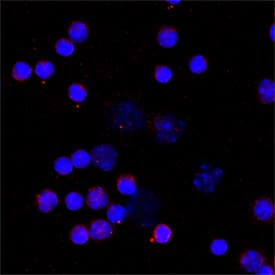
NL001) and counterstained with DAPI (blue). Specific staining was localized to cytoplasm. Staining was performed using our protocol for Fluorescent ICC Staining of Non-adherent Cells." class="big_thumb" />
MAB519) coated on a Clear Polystyrene Microplate (Catalog #
DY990). Goat Anti-Mouse/Rat IL‑10 Antigen Affinity-purified Polyclonal Antibody (Catalog # AF519) was biotinylated and incubated with the protein captured on the plate. Detection of the standard curve was achieved by incubating Streptavidin-HRP (Catalog #
DY998)" class="big_lightbox">

MAB519) coated on a Clear Polystyrene Microplate (Catalog #
DY990). Goat Anti-Mouse/Rat IL‑10 Antigen Affinity-purified Polyclonal Antibody (Catalog # AF519) was biotinylated and incubated with the protein captured on the plate. Detection of the standard curve was achieved by incubating Streptavidin-HRP (Catalog #
DY998)" class="big_thumb" />
 417-ML), Recombinant Human IL-10 (Catalog # 217-IL), Recombinant Rat IL-10 (Catalog # 522-RLB), and Recombinant Human IL-26/AK155 Monomer (Catalog # 1375-IL). PVDF Membrane was probed with 1 µg/mL of Rat Anti-Mouse IL-10 Monoclonal Antibody (Catalog # MAB417) followed by HRP-conjugated Anti-Rat IgG Secondary Antibody (Catalog # HAF005). A specific band was detected for IL-10 at approximately 15 kDa (as indicated). This experiment was conducted under reducing conditions and using Immunoblot Buffer Group 3." alt="Western blot shows 25 ng of Recombinant Mouse IL-10 (Catalog # 417-ML), Recombinant Human IL-10 (Catalog # 217-IL), Recombinant Rat IL-10 (Catalog # 522-RLB), and Recombinant Human IL-26/AK155 Monomer (Catalog # 1375-IL). PVDF Membrane was probed with 1 µg/mL of Rat Anti-Mouse IL-10 Monoclonal Antibody (Catalog # MAB417) followed by HRP-conjugated Anti-Rat IgG Secondary Antibody (Catalog # HAF005). A specific band was detected for IL-10 at approximately 15 kDa (as indicated). This experiment was conducted under reducing conditions and using Immunoblot Buffer Group 3." class="big_thumb" />
 417-ML), Recombinant Human IL-10 (Catalog # 217-IL), Recombinant Rat IL-10 (Catalog # 522-RLB), and Recombinant Human IL-26/AK155 Monomer (Catalog # 1375-IL). PVDF Membrane was probed with 0.1 µg/mL of Goat Anti-Mouse/Rat IL-10 Antigen Affinity-purified Polyclonal Antibody (Catalog # AF519) followed by HRP-conjugated Anti-Goat IgG Secondary Antibody (Catalog # HAF109). A specific band was detected for IL-10 at approximately 16 kDa (as indicated). This experiment was conducted under reducing conditions and using Immunoblot Buffer Group 3." alt="Western blot shows 25 ng of Recombinant Mouse IL-10 (Catalog # 417-ML), Recombinant Human IL-10 (Catalog # 217-IL), Recombinant Rat IL-10 (Catalog # 522-RLB), and Recombinant Human IL-26/AK155 Monomer (Catalog # 1375-IL). PVDF Membrane was probed with 0.1 µg/mL of Goat Anti-Mouse/Rat IL-10 Antigen Affinity-purified Polyclonal Antibody (Catalog # AF519) followed by HRP-conjugated Anti-Goat IgG Secondary Antibody (Catalog # HAF109). A specific band was detected for IL-10 at approximately 16 kDa (as indicated). This experiment was conducted under reducing conditions and using Immunoblot Buffer Group 3." class="big_thumb" />
 HAF017). A specific band was detected for IL-10 at approximately 16 kDa (as indicated). This experiment was conducted under reducing conditions and using Immunoblot Buffer Group 1." alt="Western blot shows conditioned media of HEK293 human embryonic kidney cell line either mock transfected or transfected with human IL-10. PVDF membrane was probed with 1 µg/mL of Goat Anti-Human IL-10 Antigen Affinity-purified Polyclonal Antibody (Catalog # AF-217-NA) followed by HRP-conjugated Anti-Goat IgG Secondary Antibody (Catalog # HAF017). A specific band was detected for IL-10 at approximately 16 kDa (as indicated). This experiment was conducted under reducing conditions and using Immunoblot Buffer Group 1." class="big_thumb" />


![N/A IL-10 [Biotin]](http://images.novusbio.com/fullsize/elisa/DATA_IL10_DY417_ELISA_2014.jpg "N/A IL-10 [Biotin]")
![N/A IL-10 [HRP]](http://images.novusbio.com/fullsize/elisa/IL-10_M1000_ELISA_223.jpg "N/A IL-10 [HRP]")
![N/A IL-10 [HRP]](http://images.novusbio.com/fullsize/elisa/DATA_IL10_M1000_ELISA_919.jpg)
![N/A IL-10 [HRP]](http://images.novusbio.com/fullsize/elisa/IL10_M1000_ELISA_439.jpg)
![N/A IL-10 [Biotin]](http://images.novusbio.com/fullsize/elisa/DATA_IL10_DY217_ELISA_1777.jpg "N/A IL-10 [Biotin]")
![N/A IL-10 [Biotin]](http://images.novusbio.com/fullsize/elisa/DATA_IL10_DY217_ELISA_2308.jpg)
![N/A IL-10 [HRP]](http://images.novusbio.com/fullsize/elisa/DATA_IL10_D1000_ELISA_527.jpg "N/A IL-10 [HRP]")
![N/A IL-10 [HRP]](http://images.novusbio.com/fullsize/elisa/DATA_IL10_D1000_ELISA_528.jpg)
![N/A IL-10 [HRP]](http://images.novusbio.com/fullsize/elisa/IL-10_D1000_ELISA_374.jpg)
![N/A IL-10 [HRP]](http://images.novusbio.com/fullsize/elisa/DATA_IL10_R1000_ELISA_1080.jpg "N/A IL-10 [HRP]")
![N/A IL-10 [HRP]](http://images.novusbio.com/fullsize/elisa/DATA_IL10_R1000_ELISA_1081.jpg)
![N/A IL-10 [HRP]](http://images.novusbio.com/fullsize/elisa/IL-10_PR1000_ELISA_347.jpg)


 417-ML) stimulates proliferation in the MC/9-2 mouse mast cell line in a dose-dependent manner (orange line). Proliferation elicited by Recombinant Mouse IL-10 (2.5 ng/mL) is neutralized (green line) by increasing concen-trations of Rat Anti-Mouse IL-10 Monoclonal Antibody (Catalog # MAB417). The ND50 is typically 0.005-0.015 µg/mL." class="big_thumb" />


 417-ML) was serially diluted and captured by Rat Anti-Mouse IL‑10 Monoclonal Antibody (Catalog # MAB417) coated on a Clear Polystyrene Microplate (Catalog #
 ![N/A IL-10 [HRP]](http://images.novusbio.com/fullsize/elisa/DATA_IL10_HS100_ELISA_883.jpg "N/A IL-10 [HRP]")
![N/A IL-10 [HRP]](http://images.novusbio.com/fullsize/elisa/IL-10_HS100_ELISA_398.jpg)
![N/A IL-10 [Biotin]](http://images.novusbio.com/fullsize/elisa/DATA_IL10_DY522_ELISA_2087.jpg "N/A IL-10 [Biotin]")
 522-RL) stimulates proliferation in the MC/9-2 mouse mast cell line in a dose-dependent manner (orange line). Proliferation elicited by Recombinant Rat IL-10 (40 ng/mL) is neutralized (green line) by increasing concentrations of Goat Anti-Rat IL-10 Antigen Affinity-purified Polyclonal Antibody (Catalog # AF519). The ND50 is typically 0.3-1.0 µg/mL." class="big_thumb" />
 NL001) and counterstained with DAPI (blue). Specific staining was localized to cytoplasm. Staining was performed using our protocol for Fluorescent ICC Staining of Non-adherent Cells." class="big_thumb" />
 MAB519) coated on a Clear Polystyrene Microplate (Catalog #
  CTS008) and counterstained with hematoxylin (blue). View our protocol for
  CTS008) and counterstained with hematoxylin (blue). Lower panel shows a lack of labeling if primary antibodies are omitted and tissue is stained only with secondary antibody followed by incubation with detection reagents. View our protocol for
  HAF109). This experiment was conducted under reducing conditions and using the 12-230 kDa separation system." class="big_thumb" />
 217-IL) stimulates proliferation in the MC/9-2 mouse mast cell line in a dose-dependent manner (orange line). Proliferation elicited by Recombinant Human IL-10 (5 ng/mL) is neutralized (green line) by increasing concentrations of Goat Anti-Human IL-10 Antigen Affinity-purified Polyclonal Antibody (Catalog # AF-217-NA). The ND50 is typically 0.2-1.0 µg/mL." class="big_thumb" />


