HAF018). A specific band was detected for HMGB1/HMG-1 at approximately 26 kDa (as indicated). This experiment was conducted under reducing conditions and using
Immunoblot Buffer Group 1." class="big_lightbox">
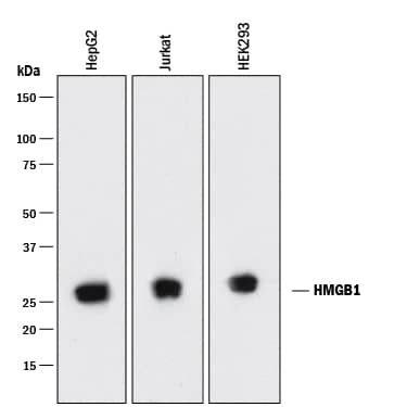
HAF018). A specific band was detected for HMGB1/HMG-1 at approximately 26 kDa (as indicated). This experiment was conducted under reducing conditions and using
Immunoblot Buffer Group 1." alt="Western blot shows lysates of HepG2 human hepatocellular carcinoma cell line, Jurkat human acute T cell leukemia cell line, and HEK293 human embryonic kidney cell line. PVDF membrane was probed with 0.05 µg/mL of Mouse Anti-Human HMGB1/HMG-1 Monoclonal Antibody (Catalog # MAB1690) followed by HRP-conjugated Anti-Mouse IgG Secondary Antibody (Catalog #
HAF018). A specific band was detected for HMGB1/HMG-1 at approximately 26 kDa (as indicated). This experiment was conducted under reducing conditions and using
Immunoblot Buffer Group 1." class="big_thumb" />
MAB004) for 15 minutes in an ultrasonic bath, followed by Biotinylated Anti-Mouse IgG Secondary Antibody (Catalog #
BAF007). Immunocomplexes were captured using 50 µL of MagCellect Streptavidin Ferrofluid (Catalog #
MAG999) and DNA was purified using chelating resin solution. The
AFPpromoter was detected by standard PCR." class="big_lightbox">
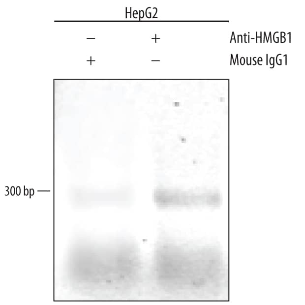
MAB004) for 15 minutes in an ultrasonic bath, followed by Biotinylated Anti-Mouse IgG Secondary Antibody (Catalog #
BAF007). Immunocomplexes were captured using 50 µL of MagCellect Streptavidin Ferrofluid (Catalog #
MAG999) and DNA was purified using chelating resin solution. The
AFPpromoter was detected by standard PCR." class="big_thumb" />
MAB0041, open histogram), followed by Phycoerythrin-conjugated Anti-Mouse IgG F(ab')
2Secondary Antibody (Catalog #
F0102B). To facilitate intracellular staining, cells were fixed with Flow Cytometry Fixation Buffer (Catalog #
FC004) and permeabilized with Flow Cytometry Permeabilization/Wash Buffer I (Catalog #
FC005). View our protocol for
Staining Intracellular Molecules." class="big_lightbox">

MAB0041, open histogram), followed by Phycoerythrin-conjugated Anti-Mouse IgG F(ab')
2Secondary Antibody (Catalog #
F0102B). To facilitate intracellular staining, cells were fixed with Flow Cytometry Fixation Buffer (Catalog #
FC004) and permeabilized with Flow Cytometry Permeabilization/Wash Buffer I (Catalog #
FC005). View our protocol for
Staining Intracellular Molecules." class="big_thumb" />
 HAF018). A specific band was detected for HMGB1/HMG-1 at approximately 26 kDa (as indicated). This experiment was conducted under reducing conditions and using Immunoblot Buffer Group 1." alt="Western blot shows lysates of HepG2 human hepatocellular carcinoma cell line, Jurkat human acute T cell leukemia cell line, and HEK293 human embryonic kidney cell line. PVDF membrane was probed with 0.05 µg/mL of Mouse Anti-Human HMGB1/HMG-1 Monoclonal Antibody (Catalog # MAB1690) followed by HRP-conjugated Anti-Mouse IgG Secondary Antibody (Catalog # HAF018). A specific band was detected for HMGB1/HMG-1 at approximately 26 kDa (as indicated). This experiment was conducted under reducing conditions and using Immunoblot Buffer Group 1." class="big_thumb" />
 HAF005). A specific band was detected for HMGB1/HMG-1 at approximately 28 kDa (as indicated). This experiment was conducted under reducing conditions and using Immunoblot Buffer Group 1." alt="Western blot shows lysates of Jurkat human acute T cell leukemia cell line, Hepa 1-6 mouse hepatoma cell line, H4-II-E-C3 rat hepatoma cell line, HEK293 human embryonic kidney cell line, and HeLa human cervical epithelial carcinoma cell line. PVDF membrane was probed with 0.1 µg/mL of Rat Anti-Human HMGB1/HMG-1 Monoclonal Antibody (Catalog # MAB16901) followed by HRP-conjugated Anti-Rat IgG Secondary Antibody (HAF005). A specific band was detected for HMGB1/HMG-1 at approximately 28 kDa (as indicated). This experiment was conducted under reducing conditions and using Immunoblot Buffer Group 1." class="big_thumb" />


![Western Blot: HMGB1/HMG-1 Antibody [NB100-2322] - Western blot shows lysates of HEK293T human embryonic kidney parental cell line and HMGB1 knockout (KO) HEK293T cell line. PVDF membrane was probed with 1.0 ug/ml of Rabbit Anti-Human HMGB1 Polyclonal Antibody (Catalog # NB100-2322) followed by HRP-conjugated Anti-Rabbit IgG Secondary Antibody (Catalog #HAF008). Specific band was detected for HMGB1 at approximately 30 kDa (as indicated) in the parental HEK293T cell line, but is not detectable in the knockout HEK293T cell line. This experiment was conducted under reducing conditions.](http://images.novusbio.com/fullsize/HMGB1-HMG-1-Antibody-Knockdown-Validated-NB100-2322-img0025.jpg "<b>Genetic Strategies Validation. </b>Western Blot: HMGB1/HMG-1 Antibody [NB100-2322] - Western blot shows lysates of HEK293T human embryonic kidney parental cell line and HMGB1 knockout (KO) HEK293T cell line. PVDF membrane was probed with 1.0 ug/ml of Rabbit Anti-Human HMGB1 Polyclonal Antibody (Catalog # NB100-2322) followed by HRP-conjugated Anti-Rabbit IgG Secondary Antibody (Catalog #HAF008). Specific band was detected for HMGB1 at approximately 30 kDa (as indicated) in the parental HEK293T cell line, but is not detectable in the knockout HEK293T cell line. This experiment was conducted under reducing conditions.")
![<b>Genetic Strategies Validation. </b>Western Blot: HMGB1/HMG-1 Antibody [NB100-2322] - Total protein from SHSY-5Y, MCF7, Neuro2A and HeLa was separated on a 12% gel by SDS-PAGE, transferred to PVDF membrane and blocked in 5% non-fat milk in TBST. The membrane was probed with 2.0 ug/mL anti-HMGB1 in 1% non-fat milk in TBST and detected with an anti-rabbit HRP secondary antibody using chemiluminescence.](http://images.novusbio.com/fullsize/HMGB1-HMG-1-Antibody-Western-Blot-NB100-2322-img0017.jpg)
![Immunohistochemistry-Paraffin: HMGB1/HMG-1 Antibody [NB100-2322] - Staining of HMGB1 in mouse liver using NB100-2322.](http://images.novusbio.com/fullsize/HMGB1-HMG-1-Antibody-Immunohistochemistry-Paraffin-NB100-2322-img0011.jpg)
![Simple Western: HMGB1/HMG-1 Antibody [NB100-2322] - Image shows a specific band for HMGB1 in 0.05 mg/mL of Jurkat lysate. This experiment was performed under reducing conditions using the 12-230 kDa separation system.](http://images.novusbio.com/fullsize/HMGB1-HMG-1-Antibody-Simple-Western-NB100-2322-img0014.jpg)
![Immunocytochemistry/Immunofluorescence: HMGB1/HMG-1 Antibody [NB100-2322] - MCF7 cells were fixed in 4% paraformaldehyde for 10 minutes and permeabilized in 0.5% Triton X-100 in PBS for 5 minutes. The cells were incubated with anti-HMGB1/HMG-1 Antibody NB100-2322 at 1 ug/ml for overnight at 4C and detected with an anti-rabbit Dylight 488 (Green) at a 1:1000 dilution for 60 minutes. Nuclei were counterstained with DAPI (Blue). Cells were imaged using a 100X objective and digitally deconvolved.](http://images.novusbio.com/fullsize/HMGB1-HMG-1-Antibody-Immunocytochemistry-Immunofluorescence-NB100-2322-img0027.jpg)
![Immunocytochemistry/Immunofluorescence: HMGB1/HMG-1 Antibody [NB100-2322] - Neuro2a cells were fixed for 10 minutes using 10% formalin and then permeabilized for 5 minutes using 1X TBS + 0.5% Triton-X100. The cells were incubated with anti-HMGB1 at 5 ug/ml overnight at 4C and detected with an anti-rabbit Dylight 488 (Green) at a 1:500 dilution. Alpha tubulin (DM1A) NB100-690 was used as a co-stain at a 1:1000 dilution and detected with an anti-mouse Dylight 550 (Red) at a 1:500 dilution. Nuclei were counterstained with DAPI (Blue). Cells were imaged using a 40X objective.](http://images.novusbio.com/fullsize/HMGB1-HMG-1-Antibody-Immunocytochemistry-Immunofluorescence-NB100-2322-img0018.jpg)
![Immunohistochemistry-Paraffin: HMGB1/HMG-1 Antibody [NB100-2322] - Rabbit blood vessel. Image from verified customer review.](http://images.novusbio.com/fullsize/HMGB1-HMG-1-Antibody-Immunohistochemistry-Paraffin-NB100-2322-img0022.jpg)

![Immunocytochemistry/Immunofluorescence: HMGB1/HMG-1 Antibody [NB100-2322] - NIH3T3 cells were fixed in 4% paraformaldehyde for 10 minutes and permeabilized in 0.5% Triton X-100 in PBS for 5 minutes. The cells were incubated with anti-HMGB1/HMG-1 Antibody NB100-2322 at 1 ug/ml for overnight at 4C and detected with an anti-rabbit Dylight 488 (Green) at a 1:1000 dilution for 60 minutes. Nuclei were counterstained with DAPI (Blue). Cells were imaged using a 100X objective and digitally deconvolved.](http://images.novusbio.com/fullsize/HMGB1-HMG-1-Antibody-Immunocytochemistry-Immunofluorescence-NB100-2322-img0028.jpg)
![Flow Cytometry: HMGB1/HMG-1 Antibody [NB100-2322] - An intracellular stain was performed on HeLa cells with HMGB1/HMG-1 Antibody NB100-2322AF488 (blue) and a matched isotype control (orange). Cells were fixed with 4% PFA and then permeabilized with 0.1% saponin. Cells were incubated in an antibody dilution of 5 ug/mL for 30 minutes at room temperature. Both antibodies were conjugated to Alexa Fluor 488.](http://images.novusbio.com/fullsize/HMGB1-HMG-1-Antibody-Flow-Cytometry-NB100-2322-img0029.jpg)
![Western Blot: HMGB1/HMG-1 Antibody [NB100-2322] - Hepatocyte protein lysate at 1:1000 4C overnight. Image from verfified customer review.](http://images.novusbio.com/fullsize/HMGB1-HMG-1-Antibody-Western-Blot-NB100-2322-img0013.jpg)
![Immunocytochemistry/Immunofluorescence: HMGB1/HMG-1 Antibody [NB100-2322] - HeLa cells were fixed for 10 minutes using 10% formalin and then permeabilized for 5 minutes using 1X TBS + 0.5% Triton-X100. The cells were incubated with anti-HMGB1 [NB100-2322] at a 1:200 dilution overnight at 4C and detected with an anti-rabbit Dylight 488 (Green) at a 1:500 dilution. Alpha tubulin (DM1A) [NB100-690] was used as a co-stain at a 1:1000 dilution and detected with an anti-mouse Dylight 550 (Red) at a 1:500 dilution. Nuclei were counterstained with DAPI (Blue). Cells were imaged using a 40X objective.](http://images.novusbio.com/fullsize/HMGB1-HMG-1-Antibody-Immunocytochemistry-Immunofluorescence-NB100-2322-img0015.jpg)
![Flow (Intracellular): HMGB1/HMG-1 Antibody [NB100-2322] - An intracellular stain was performed on HeLa with NB100-2322 and a matched isotype control. Cells were fixed with 4% PFA and then permeabilized with 0.1% saponin. Cells were incubated in an antibody dilution of 1 ug/mL for 30 minutes at room temperature, followed by Rabbit IgG (H+L) Cross-Adsorbed Secondary Antibody.](http://images.novusbio.com/fullsize/HMGB1-HMG-1-Antibody-Flow-Intracellular-NB100-2322-img0021.jpg)
![Flow Cytometry: HMGB1/HMG-1 Antibody [NB100-2322] - An intracellular stain was performed on RH-30 cells with NB100-2322F (blue) and a matched isotype control (orange). Cells were fixed with 4% PFA and then permeabilized with 0.1% saponin. Cells were incubated in an antibody dilution of 10 ug/mL for 30 minutes at room temperature. Both antibodies were conjugated to FITC.](http://images.novusbio.com/fullsize/HMGB1-HMG-1-Antibody-Flow-Cytometry-NB100-2322-img0024.jpg)
![ELISA: HMGB1/HMG-1 Antibody [NB100-2322] - A dose-dependent titration of the HRP conjugated anti-HMGB1 antibody on recombinant human HMGB1 protein. Image from verified customer review. Image using the HRP format of this antibody.](http://images.novusbio.com/fullsize/HMGB1-HMG-1-Antibody-ELISA-NB100-2322-img0023.jpg)

![Western Blot: HMGB1/HMG-1 Antibody [NB100-2322] - Expression of endogenous ligands for TLR2. (A) Expression of biglycan in AD-TLR2KO mice increased significantly compared with that in WT, AD, & TLR2KO mice (p<0.05). (B) HMGB1 in the four groups did not show a significant difference (p>0.05). Image collected & cropped by CiteAb from the following publication (//pubmed.ncbi.nlm.nih.gov/31509519), licensed under a CC-BY license. Not internally tested by Novus Biologicals.](http://images.novusbio.com/fullsize/nb100-2322_rabbit-polyclonal-hmgb1-hmg-1-antibody-310202415175213.jpg)
![ELISA: Human HMGB1/HMG-1 ELISA Kit (Colorimetric) [NBP2-62766] - Samples were spiked with high concentrations of Human HMGB1/HMG-1 and diluted with Reference Standard & Sample Diluent to produce samples with values within the range of the assay.](http://images.novusbio.com/fullsize/nbp2-62766_human-hmgb1-hmg-1-elisa-kit-colorimetric-13220241681411.jpg "ELISA: Human HMGB1/HMG-1 ELISA Kit (Colorimetric) [NBP2-62766] - Samples were spiked with high concentrations of Human HMGB1/HMG-1 and diluted with Reference Standard & Sample Diluent to produce samples with values within the range of the assay.")
![ELISA: Human HMGB1/HMG-1 ELISA Kit (Colorimetric) [NBP2-62766] - Standard Curve Reference](http://images.novusbio.com/fullsize/nbp2-62766_human-hmgb1-hmg-1-elisa-kit-colorimetric-43202495028.jpg)

 MAB004) for 15 minutes in an ultrasonic bath, followed by Biotinylated Anti-Mouse IgG Secondary Antibody (Catalog #
  MAB0041, open histogram), followed by Phycoerythrin-conjugated Anti-Mouse IgG F(ab')2Secondary Antibody (Catalog #
  NL007) and counterstained with DAPI (blue, lower panel). Specific staining was localized to nuclei. View our protocol for
  CTS013). Tissue was stained using the Anti-Mouse IgG VisUCyte™ HRP Polymer Antibody (brown; Catalog #


![Western Blot: HMGB1/HMG-1 Antibody (19N10B7) [NBP2-27396] - Hepatocyte protein lysate at 1:1000 4C overnight. Image from verified customer review.](http://images.novusbio.com/fullsize/HMGB1-HMG-1-Antibody-19N10B7-Western-Blot-NBP2-27396-img0034.jpg "Western Blot: HMGB1/HMG-1 Antibody (19N10B7) [NBP2-27396] - Hepatocyte protein lysate at 1:1000 4C overnight. Image from verified customer review.")
![Immunohistochemistry-Paraffin: HMGB1/HMG-1 Antibody (19N10B7) [NBP2-27396] - Formalin-fixed, paraffin-embedded human liver tissue stained with HMGB1 antibody (5 ug/ml), peroxidase-conjugate and DAB chromogen. Staining of formalin-fixed tissues is enhanced by boiling tissue sections in 10 mM sodium citrate buffer, pH 6.0 for 10-20 min followed by cooling at RT for 20 min.](http://images.novusbio.com/fullsize/HMGB1-HMG-1-Antibody-19N10B7-Immunohistochemistry-Paraffin-NBP2-27396-img0035.jpg)
![Flow Cytometry: HMGB1/HMG-1 Antibody (19N10B7) [NBP2-27396] - Intracellular analysis using HMGB1 antibody. Human Jurkat cells were probed using 0.5 ug of HMGB1 antibody (red) and 0.5 ug of isotype control antibody (green).](http://images.novusbio.com/fullsize/HMGB1-HMG-1-Antibody-19N10B7-Flow-Cytometry-NBP2-27396-img0021.jpg)
![Western Blot: HMGB1/HMG-1 Antibody (19N10B7) [NBP2-27396] - Analysis using 2 ug/ml concentration of HMGB1 antibody (clone 19N10B7) on (A) Full-length human HMGB1 protein, (B) Human Jurkat cell lysate and (C) Mouse NIH 3T3 cell lysate. Goat anti-mouse Ig HRP secondary antibody and PicoTect ECL substrate solution was used for this test.](http://images.novusbio.com/fullsize/HMGB1-HMG-1-Antibody-19N10B7-Western-Blot-NBP2-27396-img0033.jpg)
![Immunohistochemistry-Paraffin: HMGB1/HMG-1 Antibody (19N10B7) [NBP2-27396] - Formalin-fixed, paraffin-embedded human spleen tissue stained with HMGB1 antibody (5 ug/ml), peroxidase-conjugate and DAB chromogen. Staining of formalin-fixed tissues is enhanced by boiling tissue sections in 10 mM sodium citrate buffer, pH 6.0 for 10-20 min followed by cooling at RT for 20 min.](http://images.novusbio.com/fullsize/HMGB1-HMG-1-Antibody-19N10B7-Immunohistochemistry-Paraffin-NBP2-27396-img0022.jpg)
![Immunohistochemistry-Paraffin: HMGB1/HMG-1 Antibody (19N10B7) [NBP2-27396] - Formalin-fixed, paraffin-embedded human breast stained with HMGB1 antibody at 5 ug/ml.](http://images.novusbio.com/fullsize/HMGB1-HMG-1-Antibody-19N10B7-Immunohistochemistry-Paraffin-NBP2-27396-img0023.jpg)
![Immunohistochemistry-Paraffin: HMGB1/HMG-1 Antibody (19N10B7) [NBP2-27396] - Formalin-fixed paraffin-embedded human prostate tissue stained with HMGB1 antibody at 10 ug/ml concentration. Staining of formalin-fixed tissues is enhanced by boiling tissue sections in 10 mM sodium citrate buffer, pH 6.0 for 10-20 min followed by cooling at RT for 20 min.](http://images.novusbio.com/fullsize/HMGB1-HMG-1-Antibody-19N10B7-Immunohistochemistry-Paraffin-NBP2-27396-img0024.jpg)
![Immunohistochemistry-Paraffin: HMGB1/HMG-1 Antibody (19N10B7) [NBP2-27396] - Analysis of formalin-fixed, decalcified, paraffin embedded tissue section from the paws of mouse (collagen-induced arthritis model) using HMGB1 antibody (clone 19N10B7) at 5 ug/ml concentration. [Image courtesy of Dr Ulf Andersson, Karolinska Institute].](http://images.novusbio.com/fullsize/HMGB1-HMG-1-Antibody-19N10B7-Immunohistochemistry-Paraffin-NBP2-27396-img0025.jpg)
![Immunohistochemistry-Paraffin: HMGB1/HMG-1 Antibody (19N10B7) [NBP2-27396] - IHC-P detection of HMGB1 protein in a formalin-fixed paraffin-embedded tissue section of human stomach using HMGB1 antibody (clone 19N10B7) at 5 ug/ml concentration. Distinct nuclear staining of HMGB1 was observed in different cells of the glandular stomach.](http://images.novusbio.com/fullsize/HMGB1-HMG-1-Antibody-19N10B7-Immunohistochemistry-Paraffin-NBP2-27396-img0026.jpg)
![Immunohistochemistry-Paraffin: HMGB1/HMG-1 Antibody (19N10B7) [NBP2-27396] - IHC-P detection of HMGB1 protein in a formalin-fixed paraffin-embedded tissue section of normal human skin using HMGB1 antibody (clone 19N10B7) at 5 ug/ml concentration. The various cells of the epidermal layer showed intense nuclear staining along with weak cytoplasmic staining. The blood vessels, glandular cells and the other cells in dermal layer also showed nuclear positivity for HMGB1 immunostaining.](http://images.novusbio.com/fullsize/HMGB1-HMG-1-Antibody-19N10B7-Immunohistochemistry-Paraffin-NBP2-27396-img0027.jpg)
![Immunohistochemistry-Paraffin: HMGB1/HMG-1 Antibody (19N10B7) [NBP2-27396] - IHC-P detection of HMGB1 protein in a formalin-fixed paraffin-embedded tissue section of normal human brain using HMGB1 antibody (clone 19N10B7) at 5 ug/ml concentration. Representative image shows a distinct nuclear immunostaining of HMGB1 in the various brain cells.](http://images.novusbio.com/fullsize/HMGB1-HMG-1-Antibody-19N10B7-Immunohistochemistry-Paraffin-NBP2-27396-img0028.jpg)
![Immunohistochemistry-Paraffin: HMGB1/HMG-1 Antibody (19N10B7) [NBP2-27396] - IHC-P detection of HMGB1 protein in a formalin-fixed paraffin-embedded tissue section of normal human colon using HMGB1 antibody (clone 19N10B7) at 5 ug/ml concentration. Representative image shows specific HMGB1 nuclear positivity in different mucosal cells of the colon.](http://images.novusbio.com/fullsize/HMGB1-HMG-1-Antibody-19N10B7-Immunohistochemistry-Paraffin-NBP2-27396-img0029.jpg)
![Immunohistochemistry-Paraffin: HMGB1/HMG-1 Antibody (19N10B7) [NBP2-27396] - IHC-P detection of HMGB1 protein in a formalin-fixed paraffin-embedded tissue section of malignant stromal tumor of small bowel from human using HMGB1 antibody (clone 19N10B7) at 5 ug/ml concentration. HMGB1 immunopositivity of differential intensity was observed in the cells of tested section.](http://images.novusbio.com/fullsize/HMGB1-HMG-1-Antibody-19N10B7-Immunohistochemistry-Paraffin-NBP2-27396-img0030.jpg)
![Immunohistochemistry-Paraffin: HMGB1/HMG-1 Antibody (19N10B7) [NBP2-27396] - IHC-P detection of HMGB1 protein in a formalin-fixed paraffin-embedded tissue section of human bladder cancer using HMGB1 antibody (clone 19N10B7) at 5 ug/ml concentration. Strong nuclear HMGB1 immunopositivity was observed in the bladder cancer cells whereas the staining was weak in cells of tumor stroma.](http://images.novusbio.com/fullsize/HMGB1-HMG-1-Antibody-19N10B7-Immunohistochemistry-Paraffin-NBP2-27396-img0031.jpg)
![Immunohistochemistry-Paraffin: HMGB1/HMG-1 Antibody (19N10B7) [NBP2-27396] - IHC-P detection of HMGB1 protein in a formalin-fixed paraffin-embedded tissue section of human stomach cancer using HMGB1 antibody (clone 19N10B7) at 5 ug/ml concentration. This representative image shows a distinct nuclear HMGB1 immunopositivity in the cancerous and sub-mucosal cells.](http://images.novusbio.com/fullsize/HMGB1-HMG-1-Antibody-19N10B7-Immunohistochemistry-Paraffin-NBP2-27396-img0032.jpg)
![Simple Western: HMGB1/HMG-1 Antibody (19N10B7) [NBP2-27396] - Simple Western lane view shows a specific band for HMGB1 in 1.0 mg/ml of HeLa lysate. This experiment was performed under reducing conditions using the 12-230kDa separation system.](http://images.novusbio.com/fullsize/HMGB1-HMG-1-Antibody-19N10B7-Simple-Western-NBP2-27396-img0020.jpg)

![Immunocytochemistry/Immunofluorescence: HMGB1/HMG-1 Antibody (2F6) [H00003146-M08] - Analysis of monoclonal antibody to HMGB1 on HeLa cell. Antibody concentration 10 ug/ml](http://images.novusbio.com/fullsize/HMGB1-HMG-1-Antibody-2F6-Immunocytochemistry-Immunofluorescence-H00003146-M08-img0012.jpg)
![Immunohistochemistry-Paraffin: HMGB1/HMG-1 Antibody (2F6) [H00003146-M08] - Analysis of monoclonal antibody to HMGB1 on formalin-fixed paraffin-embedded human placenta. Antibody concentration 3 ug/ml](http://images.novusbio.com/fullsize/HMGB1-HMG-1-Antibody-2F6-Immunohistochemistry-Paraffin-H00003146-M08-img0014.jpg)
![Western Blot: HMGB1/HMG-1 Antibody (2F6) [H00003146-M08] - Analysis of HMGB1 expression in Hela S3 NE (Cat # L013V3).](http://images.novusbio.com/fullsize/HMGB1-HMG-1-Antibody-2F6-Western-Blot-H00003146-M08-img0015.jpg)
![Western Blot: HMGB1/HMG-1 Antibody (2F6) [H00003146-M08] - Analysis of HMGB1 expression in transfected 293T cell line by HMGB1 monoclonal antibody (M08), clone 2F6. Lane 1: HMGB1 transfected lysatE (25 KDa). Lane 2: Non-transfected lysate.](http://images.novusbio.com/fullsize/HMGB1-HMG-1-Antibody-2F6-Western-Blot-H00003146-M08-img0016.jpg)
![ELISA: HMGB1/HMG-1 Antibody (2F6) [H00003146-M08] - Detection limit for recombinant GST tagged HMGB1 is approximately 0.3ng/ml as a capture antibody.](http://images.novusbio.com/fullsize/HMGB1-HMG-1-Antibody-2F6-ELISA-H00003146-M08-img0006.jpg)

![Immunohistochemistry: HMGB1/HMG-1 Antibody (2F6) [H00003146-M08] - HMGB1 regulated autophagy in thyroid cancer cells (a) FTC-133/TPC-1 cells were transfected with HMGB1 shRNA & control shRNA & then starved by HBSS for 2 h. & LC3-I/II level was assayed by Western blot; (b) FTC-133/TPC-1 cells were transfected with HMGB1 shRNA & control shRNA & then pre-treated for 1 h with pepstatin A (PA, 10 μM) & E64D (10 μM) as indicated. Cells were subsequently treated for 3 h with HBSS in continuous presence or absence PA/E64D inhibitors. LC3-I/II, Beclin1 & p62 levels were assayed by Western blot; (c) Ultrastructural features in FTC-133/TPC-1 cells transfected with HMGB1 shRNA & control shRNA after a 3-h treatment of HBSS. More autophagosomes were seen in control shRNA plus HBSS-treated cells than in cells treated with HMGB1 shRNA plus HBSS. Arrows indicated autophagosomes Image collected & cropped by CiteAb from the following publication (//pubmed.ncbi.nlm.nih.gov/31331356), licensed under a CC-BY license. Not internally tested by Novus Biologicals.](http://images.novusbio.com/fullsize/h00003146-m08_mouse-monoclonal-hmgb1-hmg-1-antibody-2f6-31020241548361.jpg)
![ELISA: Mouse HMGB1/HMG-1 ELISA Kit (Colorimetric) [NBP2-62767] - Samples were spiked with high concentrations of Mouse HMGB1/HMG-1 and diluted with Reference Standard & Sample Diluent to produce samples with values within the range of the assay.](http://images.novusbio.com/fullsize/nbp2-62767_mouse-hmgb1-hmg-1-elisa-kit-colorimetric-132202416173468.jpg "ELISA: Mouse HMGB1/HMG-1 ELISA Kit (Colorimetric) [NBP2-62767] - Samples were spiked with high concentrations of Mouse HMGB1/HMG-1 and diluted with Reference Standard & Sample Diluent to produce samples with values within the range of the assay.")
![ELISA: Mouse HMGB1/HMG-1 ELISA Kit (Colorimetric) [NBP2-62767] - Standard Curve Reference](http://images.novusbio.com/fullsize/nbp2-62767_mouse-hmgb1-hmg-1-elisa-kit-colorimetric-4320249115619.jpg)
![Western Blot: HMGB1/HMG-1 Antibody (1F3) [NBP2-25148] - Analysis of lysates from different cell lines probed with mouse mAb to high mobility group protein B1, (HMGB1), NBP2-25148, dilution 1:2,000: [1] protein standard, [2] NIH-3T3, [3] C6, [4] HEK293, [5] HeLa, and [6] SH-SY5Y. The 25kDa band revealed by MCA-1F3 antibody, corresponds to HMGB1 protein.](http://images.novusbio.com/fullsize/HMGB1-HMG-1-Antibody-1F3-Western-Blot-NBP2-25148-img0009.jpg "Western Blot: HMGB1/HMG-1 Antibody (1F3) [NBP2-25148] - Analysis of lysates from different cell lines probed with mouse mAb to high mobility group protein B1, (HMGB1), NBP2-25148, dilution 1:2,000: [1] protein standard, [2] NIH-3T3, [3] C6, [4] HEK293, [5] HeLa, and [6] SH-SY5Y. The 25kDa band revealed by MCA-1F3 antibody, corresponds to HMGB1 protein.")
![Immunocytochemistry/Immunofluorescence: HMGB1/HMG-1 Antibody (1F3) [NBP2-25148] - Analysis of of HeLa cells stained with mouse mAb to HMGB1,NBP2-25148, dilution 1:2,000 in red and costained with rabbit pAb to GAPDH, dilution 1:2,000 in green. MCA-1F3 antibody stains the chromatin binding protein HMGB1, which is localized in the nuclei.](http://images.novusbio.com/fullsize/HMGB1-HMG-1-Antibody-1F3-Immunocytochemistry-Immunofluorescence-NBP2-25148-img0010.jpg)
![Flow Cytometry: HMGB1/HMG-1 Antibody (1F3) [NBP2-25148] - Intracellular flow cytometric staining of 1 x 10^6 MCF-7 cells using HMGB1 antibody (dark blue). Isotype control shown in orange. An antibody concentration of 1 ug/1x10^6 cells was used.](http://images.novusbio.com/fullsize/HMGB1-HMG-1-Antibody-1F3-Flow-Cytometry-NBP2-25148-img0007.jpg)
![Immunocytochemistry/Immunofluorescence: HMGB1/HMG-1 Antibody (1F3) [NBP2-25148] - HeLa cells grown in tissue culture and stained with NBP2-25148 (red), chicken polyclonal antibody to Vimentin NB300-223 (green) and DNA (blue). NBP2-25148 reveals strong nuclear staining which overlaps with the DNA stain.](http://images.novusbio.com/fullsize/HMGB1-HMG-1-Antibody-1F3-Immunocytochemistry-Immunofluorescence-NBP2-25148-img0008.jpg)
![Simple Western: HMGB1/HMG-1 Antibody (1F3) [NBP2-25148] - Simple Western lane view shows a specific band for HMGB1 in 0.05 mg/ml of Jurkat lysate. This experiment was performed under reducing conditions using the 12-230 kDa separation system.](http://images.novusbio.com/fullsize/HMGB1-HMG-1-Antibody-1F3-Simple-Western-NBP2-25148-img0005.jpg)
 CTS017) and counterstained with hematoxylin (blue). Specific staining was localized to nuclei and cytoplasm. View our protocol for

![Western Blot: HMGB1/HMG-1 Antibody (19N15F4) [NBP2-27401] - Analysis of HMGB1 protein on ( A) Full-length human HMGB1 protein (B) human Jurkat cell lysate and (C) mouse NIH 3T3 cell lysate using HMGB1 antibody (clone 19N15F4) at 2 ug/ml concentration. Goat anti-mouse IgG HRP secondary antibody and PicoTect ECL substrate solution were used in this assay.](http://images.novusbio.com/fullsize/HMGB1-HMG-1-Antibody-19N15F4-Western-Blot-NBP2-27401-img0026.jpg "Western Blot: HMGB1/HMG-1 Antibody (19N15F4) [NBP2-27401] - Analysis of HMGB1 protein on ( A) Full-length human HMGB1 protein (B) human Jurkat cell lysate and (C) mouse NIH 3T3 cell lysate using HMGB1 antibody (clone 19N15F4) at 2 ug/ml concentration. Goat anti-mouse IgG HRP secondary antibody and PicoTect ECL substrate solution were used in this assay.")
![Immunohistochemistry-Paraffin: HMGB1/HMG-1 Antibody (19N15F4) [NBP2-27401] - IHC-P detection of HMGB1 protein in formalin-fixed, decalcified, paraffin embedded tissue sections from the paws of mouse (collagen-induced arthritis model) using HMGB1 antibody (clone 19N15F4) at a concentration of 5 ug/ml [Image courtesy of Dr Ulf Andersson, Karolinska]](http://images.novusbio.com/fullsize/HMGB1-HMG-1-Antibody-19N15F4-Immunohistochemistry-Paraffin-NBP2-27401-img0027.jpg)
![Flow Cytometry: HMGB1/HMG-1 Antibody (19N15F4) [NBP2-27401] - Intracellular analysis using HMGB1 antibody. Human Jurkat cells were probed using 0.5 ug of HMGB1 antibody (red) and 0.5 ug of isotype control antibody (green).](http://images.novusbio.com/fullsize/HMGB1-HMG-1-Antibody-19N15F4-Flow-Cytometry-NBP2-27401-img0016.jpg)
![Immunohistochemistry-Paraffin: HMGB1/HMG-1 Antibody (19N15F4) [NBP2-27401] - Formalin-fixed, paraffin-embedded human skin stained with HMGB1 antibody at 5 ug/ml](http://images.novusbio.com/fullsize/HMGB1-HMG-1-Antibody-19N15F4-Immunohistochemistry-Paraffin-NBP2-27401-img0018.jpg)
![Immunohistochemistry-Paraffin: HMGB1/HMG-1 Antibody (19N15F4) [NBP2-27401] - Analysis of HMGB1 protein in a section of human breast normal tissue using HMGB1 antibody (clone 19N15F4) at a concentration of 5 ug/ml. The ductal /acinar epithelial cells in the breast tissue section depicted strong nuclear expression with some cytoplasmic positivity. The myoepithelial cells and few cells of the intra-lobular connective tissue showed relatively weak nuclear positivity for HMGB1.](http://images.novusbio.com/fullsize/HMGB1-HMG-1-Antibody-19N15F4-Immunohistochemistry-Paraffin-NBP2-27401-img0019.jpg)
![Immunohistochemistry-Paraffin: HMGB1/HMG-1 Antibody (19N15F4) [NBP2-27401] - Analysis of HMGB1 protein in a section of normal human stomach tissue using HMGB1 antibody (clone 19N15F4) at a concentration of 5 ug/ml. The cells of the glandular stomach showed specific nuclear expression for HMGB1.](http://images.novusbio.com/fullsize/HMGB1-HMG-1-Antibody-19N15F4-Immunohistochemistry-Paraffin-NBP2-27401-img0020.jpg)
![Immunohistochemistry-Paraffin: HMGB1/HMG-1 Antibody (19N15F4) [NBP2-27401] - Analysis of HMGB1 protein in a tissue section of normal human colon using HMGB1 antibody (clone 19N15F4) at a concentration of 5 ug/ml. Almost all the cells of colon's mucosal layer showed nuclear positivity for HMGB1 but the cells of the columnar epithelial cells on the absorptive surface developed cytoplasmic staining also.](http://images.novusbio.com/fullsize/HMGB1-HMG-1-Antibody-19N15F4-Immunohistochemistry-Paraffin-NBP2-27401-img0021.jpg)
![Immunohistochemistry-Paraffin: HMGB1/HMG-1 Antibody (19N15F4) [NBP2-27401] - Analysis of HMGB1 protein in a tissue section of normal human brain using HMGB1 antibody (clone 19N15F4) at a concentration of 5 ug/ml. The representative image shows an overall strong nuclear HMGB1 immunopositivity with weak to negligible cytoplasmic staining in brain cells.](http://images.novusbio.com/fullsize/HMGB1-HMG-1-Antibody-19N15F4-Immunohistochemistry-Paraffin-NBP2-27401-img0022.jpg)
![Immunohistochemistry-Paraffin: HMGB1/HMG-1 Antibody (19N15F4) [NBP2-27401] - Analysis of HMGB1 protein in a tissue section of human bladder transitional cell carcinoma (TCC) / urothelial cell carcinoma (UCC) using HMGB1 antibody (clone 19N15F4) at a concentration of 5 ug/ml. Almost all the cancer cells and the cells of tumor stroma developed strong/specific nuclear HMGB1 immunopositivity.](http://images.novusbio.com/fullsize/HMGB1-HMG-1-Antibody-19N15F4-Immunohistochemistry-Paraffin-NBP2-27401-img0023.jpg)
![Immunohistochemistry-Paraffin: HMGB1/HMG-1 Antibody (19N15F4) [NBP2-27401] - Analysis of HMGB1 protein in a tissue section of human esophageal squamous cell carcinoma (SCC) using HMGB1 antibody (clone 19N15F4) at a concentration of 5 ug/ml. Strong nuclear along with weak cytoplasmic immunopositivity for HMGB1 was observed in SCC cells and the tumor stroma cells developed relatively weak staining for this target.](http://images.novusbio.com/fullsize/HMGB1-HMG-1-Antibody-19N15F4-Immunohistochemistry-Paraffin-NBP2-27401-img0024.jpg)
![Immunohistochemistry-Paraffin: HMGB1/HMG-1 Antibody (19N15F4) [NBP2-27401] - Analysis of HMGB1 protein in a section of human small intestinal cancer tissue using HMGB1 antibody (clone 19N15F4) at a concentration of 5 ug/ml. Intense nuclear immunopositivity of HMGB1 was observed in cancer cells and the cells of tumor stroma as well as the adjacent normal crypts. Some glandular cells in the crypts developed bother nuclear and cytoplasmic staining.](http://images.novusbio.com/fullsize/HMGB1-HMG-1-Antibody-19N15F4-Immunohistochemistry-Paraffin-NBP2-27401-img0025.jpg)
![Western Blot: HMGB1/HMG-1 Antibody (1D5) [H00003146-M02] - Analysis of HMGB1 expression in transfected 293T cell line by HMGB1 monoclonal antibody (M02), clone 1D5.Lane 1: HMGB1 transfected lysate(24.9 KDa).Lane 2: Non-transfected lysate.](http://images.novusbio.com/fullsize/HMGB1-HMG-1-Antibody-1D5-Western-Blot-H00003146-M02-img0015.jpg "Western Blot: HMGB1/HMG-1 Antibody (1D5) [H00003146-M02] - Analysis of HMGB1 expression in transfected 293T cell line by HMGB1 monoclonal antibody (M02), clone 1D5.Lane 1: HMGB1 transfected lysate(24.9 KDa).Lane 2: Non-transfected lysate.")
![Immunocytochemistry/Immunofluorescence: HMGB1/HMG-1 Antibody (1D5) [H00003146-M02] - Analysis of monoclonal antibody to HMGB1 on HeLa cell. Antibody concentration 10 ug/ml.](http://images.novusbio.com/fullsize/HMGB1-HMG-1-Antibody-1D5-Immunocytochemistry-Immunofluorescence-H00003146-M02-img0011.jpg)
![Immunohistochemistry-Paraffin: HMGB1/HMG-1 Antibody (1D5) [H00003146-M02] - Analysis of monoclonal antibody to HMGB1 on formalin-fixed paraffin-embedded human hepatocellular carcinoma tissue. Antibody concentration 3 ug/ml.](http://images.novusbio.com/fullsize/HMGB1-HMG-1-Antibody-1D5-Immunohistochemistry-Paraffin-H00003146-M02-img0013.jpg)
![Western Blot: HMGB1/HMG-1 Antibody (1D5) [H00003146-M02] - HMGB1 monoclonal antibody (M02), clone 1D5 Analysis of HMGB1 expression in K-562.](http://images.novusbio.com/fullsize/HMGB1-HMG-1-Antibody-1D5-Western-Blot-H00003146-M02-img0014.jpg)
![Sandwich ELISA Capture: HMGB1/HMG-1 Antibody (1D5) [H00003146-M02] - Detection limit for recombinant GST tagged HMGB1/HMG-1 is 0.1 ng/ml as a capture antibody.](http://images.novusbio.com/fullsize/HMGB1-HMG-1-Antibody-1D5-Sandwich-ELISA-Capture-H00003146-M02-img0016.jpg)
![ELISA: Rat HMGB1/HMG-1 ELISA Kit (Colorimetric) [NBP3-06661] - Samples were spiked with high concentrations of Rat HMGB1/HMG-1 and diluted with Reference Standard & Sample Diluent to produce samples with values within the range of the assay.](http://images.novusbio.com/fullsize/nbp3-06661_rat-hmgb1-hmg-1-elisa-kit-colorimetric-13220241626124.jpg "ELISA: Rat HMGB1/HMG-1 ELISA Kit (Colorimetric) [NBP3-06661] - Samples were spiked with high concentrations of Rat HMGB1/HMG-1 and diluted with Reference Standard & Sample Diluent to produce samples with values within the range of the assay.")
![ELISA: Rat HMGB1/HMG-1 ELISA Kit (Colorimetric) [NBP3-06661] - Standard Curve Reference](http://images.novusbio.com/fullsize/nbp3-06661_rat-hmgb1-hmg-1-elisa-kit-colorimetric-4320249163312.jpg)
![Western Blot: HMGB1/HMG-1 Antibody - BSA Free [NBP2-41264] - KO Validation in HeLa Cells.Loading: 10 u of HeLa WT cell lysates or HMGB1/HMG-1 KO cell lysates. Antibodies: HMGB1/HMG-1 (0.5 u/mL) and beta-actin (1 u/mL), 1 h incubation at RT in 5% NFDM/TBST.Secondary: Goat Anti-Rabbit IgG HRP conjugate at 1:10000 dilution.](http://images.novusbio.com/fullsize/nbp2-41264_rabbit-polyclonal-hmgb1-hmg-1-antibody-271120241932699.gif "<b>Genetic Strategies Validation. </b>Western Blot: HMGB1/HMG-1 Antibody - BSA Free [NBP2-41264] - KO Validation in HeLa Cells.Loading: 10 u of HeLa WT cell lysates or HMGB1/HMG-1 KO cell lysates. Antibodies: HMGB1/HMG-1 (0.5 u/mL) and beta-actin (1 u/mL), 1 h incubation at RT in 5% NFDM/TBST.Secondary: Goat Anti-Rabbit IgG HRP conjugate at 1:10000 dilution.")
![<b>Genetic Strategies Validation. </b>Western Blot: HMGB1/HMG-1 Antibody - BSA Free [NBP2-41264] - Validation in Mouse Tissues.Loading: 15 ul of lysate Antibodies: HMGB1/HMG-1 , 1 ug/mL , 1h incubation at RT in 5% NFDM/TBST. Secondary: Goat Anti-Rabbit IgG HRP conjugate at 1:10000 dilution.](http://images.novusbio.com/fullsize/nbp2-41264_rabbit-polyclonal-hmgb1-hmg-1-antibody-2711202420405564.gif)
![Western Blot: HMGB1/HMG-1 Antibody - BSA Free [NBP2-41264] - Validation in Rat Tissues.Loading: 15 ug of lysate Antibodies: HMGB1/HMG-1, 1 u/mL, 1 h incubation at RT in 5% NFDM/TBST. Secondary: Goat Anti-Rabbit IgG HRP conjugate at 1:10000 dilution.](http://images.novusbio.com/fullsize/nbp2-41264_rabbit-polyclonal-hmgb1-hmg-1-antibody-271120242074672.gif)
![Western Blot: HMGB1/HMG-1 Antibody - BSA Free [NBP2-41264] - Validation in Human, Mouse and Rat Cell Lines.Loading: 15 ug of lysate Antibodies: HMGB1/HMG-1, 1 u/mL , 1 h incubation at RT in 5% NFDM/TBST. Secondary: Goat Anti-Rabbit IgG HRP conjugate at 1:10000 dilution.](http://images.novusbio.com/fullsize/nbp2-41264_rabbit-polyclonal-hmgb1-hmg-1-antibody-2711202420392765.gif)
![Immunocytochemistry/ Immunofluorescence: HMGB1/HMG-1 Antibody - BSA Free [NBP2-41264] - Validation of HMGB1/HMG-1 in HL60 Cells.Immunofluorescent analysis of 4% paraformaldehyde-fixed HL60 cells labeling HMGB1/HMG-1 at 20 ug/mL, followed by goat anti-rabbit IgG secondary antibody at 1/500 dilution (green) and DAPI antibody (blue).](http://images.novusbio.com/fullsize/nbp2-41264_rabbit-polyclonal-hmgb1-hmg-1-antibody-2711202419432287.jpg)
![Immunohistochemistry: HMGB1/HMG-1 Antibody - BSA Free [NBP2-41264] - Validation of HMGB1/HMG-1 in Human Stomach Tissue.Immunohistochemical analysis of paraffin-embedded human stomach tissue using anti-HMGB1/HMG-1 antibody at 1 ug/ml. Tissue was fixed with formaldehyde and blocked with 10% serum for 1 h at RT; antigen retrieval was by heat mediation with a citrate buffer (pH6). Samples were incubated with primary antibody overnight at 4C. A goat anti-rabbit IgG H&L (HRP) at 1/250 was used as secondary. Counter stained with Hematoxylin.](http://images.novusbio.com/fullsize/nbp2-41264_rabbit-polyclonal-hmgb1-hmg-1-antibody-2711202420312979.jpg)
![Immunohistochemistry: HMGB1/HMG-1 Antibody - BSA Free [NBP2-41264] - Validation of HMGB1/HMG-1 in Mouse Spleen Tissue.Immunohistochemical analysis of paraffin-embedded mouse spleen tissue using anti-HMGB1/HMG-1 antibody at 2 ug/ml. Tissue was fixed with formaldehyde and blocked with 10% serum for 1 h at RT; antigen retrieval was by heat mediation with a citrate buffer (pH6). Samples were incubated with primary antibody overnight at 4C. A goat anti-rabbit IgG H&L (HRP) at 1/250 was used as secondary. Counter stained with Hematoxylin.](http://images.novusbio.com/fullsize/nbp2-41264_rabbit-polyclonal-hmgb1-hmg-1-antibody-2711202420392762.jpg)
![Immunohistochemistry: HMGB1/HMG-1 Antibody - BSA Free [NBP2-41264] - Validation of HMGB1/HMG-1 in Rat Spleen Tissue. Immunohistochemical analysis of paraffin-embedded rat spleen tissue using anti-HMGB1/HMG-1 antibody at 1 ul/ml. Tissue was fixed with formaldehyde and blocked with 10% serum for 1 h at RT; antigen retrieval was by heat mediation with a citrate buffer (pH6). Samples were incubated with primary antibody overnight at 4C. A goat anti-rabbit IgG H&L (HRP) at 1/250 was used as secondary. Counter stained with Hematoxylin.](http://images.novusbio.com/fullsize/nbp2-41264_rabbit-polyclonal-hmgb1-hmg-1-antibody-2711202420333866.jpg)


