DR647MS) and counterstained with DAPI (blue; Lunaphore Catalog #
DR100). Specific staining was localized to the cytoplasm. Protocol available in
COMET™ Panel Builder." class="big_lightbox">
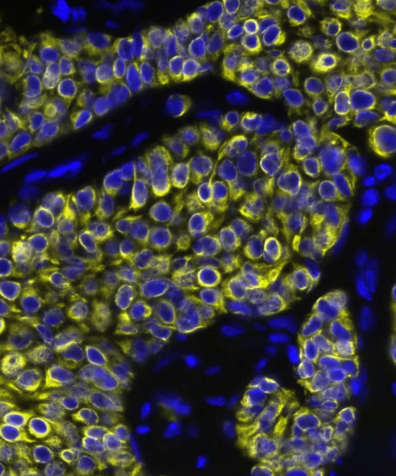
DR647MS) and counterstained with DAPI (blue; Lunaphore Catalog #
DR100). Specific staining was localized to the cytoplasm. Protocol available in
COMET™ Panel Builder." alt="CXCL12/SDF-1 was detected in immersion fixed paraffin-embedded sections of human breast tumor using Mouse Anti-Human CXCL12/SDF-1, Monoclonal Antibody (Catalog #MAB350) at 15ug/mL at 37° Celsius for 4 minutes. Before incubation with the primary antibody, tissue underwent an all-in-one dewaxing and antigen retrieval preprocessing using PreTreatment Module (PT Module) and Dewax and HIER Buffer H (pH 9; Epredia Catalog # TA-999-DHBH). Tissue was stained using the Alexa Fluor™ 647 Goat anti-Mouse IgG Secondary Antibody at 1:200 at 37 ° Celsius for 2 minutes. (Yellow; Lunaphore Catalog #
DR647MS) and counterstained with DAPI (blue; Lunaphore Catalog #
DR100). Specific staining was localized to the cytoplasm. Protocol available in
COMET™ Panel Builder." class="big_thumb" />
MAB002, open histogram), followed by Phycoerythrin-conjugated Anti-Mouse IgG Secondary Antibody (Catalog #
F0102B). To facilitate intracellular staining, cells were fixed and permeabilized with FlowX FoxP3 Fixation & Permeabilization Buffer Kit (Catalog #
FC012). View our protocol for
Staining Intracellular Molecules." class="big_lightbox">
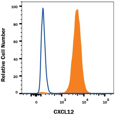
MAB002, open histogram), followed by Phycoerythrin-conjugated Anti-Mouse IgG Secondary Antibody (Catalog #
F0102B). To facilitate intracellular staining, cells were fixed and permeabilized with FlowX FoxP3 Fixation & Permeabilization Buffer Kit (Catalog #
FC012). View our protocol for
Staining Intracellular Molecules." class="big_thumb" />
VC001) and DAB chromogen (lower image, yellow-brown). Tissues were counterstained with hematoxylin (blue)." class="big_lightbox">
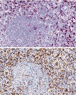
VC001) and DAB chromogen (lower image, yellow-brown). Tissues were counterstained with hematoxylin (blue)." class="big_thumb" />


![CXCR4 and CXCL12 expression in DLBCL. (a) Expression analysis of CXCR4 and CXCL12 in non-neoplastic control germinal center B cells (GC-B) and diffuse large B cell lymphoma cells (DLBCL) consisting of DLBCL-NGCB and DLCBL-GCB, by RQ-PCR. GCB-DLBCL were further subdivided into primary (DLBCL-pGCB) and transformed DLBCL (DLBCL-pGCB) originating from follicular lymphoma. (b) Expression analysis of CXCR4 and CXCL12 in DLBCL samples with early (stage 1) and advanced stage (stage 2–4) (left graphs) and DLBCL samples with and without bone marrow infiltration (right graphs) by RQ-PCR. (c) Probability of 5-year-survival in DLBCL patients (our cohort left panel and the cohort of Lenz et al. right [20]) stratified by the third quartile of CXCR4 expression, respectively. (d) Representative immunohistochemical stains of CXCR4 (I–III) and CXCL12 (IV–VI) on DLBCL samples (magnification 20×). mRNA expression levels were calculated as a relative expression in comparison to the GC-B cells. All images were captured using an Olympus BX51 microscope and an Olympus E-330 camera. Image collected and cropped by CiteAb from the following publication (//pubmed.ncbi.nlm.nih.gov/31554271), licensed under a CC-BY license. Not internally tested by R&D Systems.](//images.novusbio.com/images/mab350_human-mouse-cxcl12-sdf-1-mab-clone-79018-41202410331984.jpg)



350-NS) was serially diluted and captured by Mouse Anti-Human/Mouse CXCL12/SDF‑1 Monoclonal Antibody (Catalog # MAB350) coated on a Clear Polystyrene Microplate (Catalog #
DY990). Goat Anti-Human CXCL12/SDF‑1 Antigen Affinity-purified Polyclonal Antibody (Catalog #
AF-310-NA) was biotinylated and incubated with the protein captured on the plate. Detection of the standard curve was achieved by incubating Streptavidin-HRP (Catalog #
DY998)" class="big_lightbox">

350-NS) was serially diluted and captured by Mouse Anti-Human/Mouse CXCL12/SDF‑1 Monoclonal Antibody (Catalog # MAB350) coated on a Clear Polystyrene Microplate (Catalog #
DY990). Goat Anti-Human CXCL12/SDF‑1 Antigen Affinity-purified Polyclonal Antibody (Catalog #
AF-310-NA) was biotinylated and incubated with the protein captured on the plate. Detection of the standard curve was achieved by incubating Streptavidin-HRP (Catalog #
DY998)" class="big_thumb" />
460-SD) was serially diluted and captured by Mouse Anti-Human/Mouse CXCL12/SDF‑1 Monoclonal Antibody (Catalog # MAB350) coated on a Clear Polystyrene Microplate (Catalog #
DY990). Goat Anti-Human CXCL12/SDF‑1 Antigen Affinity-purified Polyclonal Antibody (Catalog #
AF-310-NA) was biotinylated and incubated with the protein captured on the plate. Detection of the standard curve was achieved by incubating Streptavidin-HRP (Catalog #
DY998)" class="big_lightbox">

460-SD) was serially diluted and captured by Mouse Anti-Human/Mouse CXCL12/SDF‑1 Monoclonal Antibody (Catalog # MAB350) coated on a Clear Polystyrene Microplate (Catalog #
DY990). Goat Anti-Human CXCL12/SDF‑1 Antigen Affinity-purified Polyclonal Antibody (Catalog #
AF-310-NA) was biotinylated and incubated with the protein captured on the plate. Detection of the standard curve was achieved by incubating Streptavidin-HRP (Catalog #
DY998)" class="big_thumb" />
 DR647MS) and counterstained with DAPI (blue; Lunaphore Catalog # DR100). Specific staining was localized to the cytoplasm. Protocol available in COMET™ Panel Builder." alt="CXCL12/SDF-1 was detected in immersion fixed paraffin-embedded sections of human breast tumor using Mouse Anti-Human CXCL12/SDF-1, Monoclonal Antibody (Catalog #MAB350) at 15ug/mL at 37° Celsius for 4 minutes. Before incubation with the primary antibody, tissue underwent an all-in-one dewaxing and antigen retrieval preprocessing using PreTreatment Module (PT Module) and Dewax and HIER Buffer H (pH 9; Epredia Catalog # TA-999-DHBH). Tissue was stained using the Alexa Fluor™ 647 Goat anti-Mouse IgG Secondary Antibody at 1:200 at 37 ° Celsius for 2 minutes. (Yellow; Lunaphore Catalog # DR647MS) and counterstained with DAPI (blue; Lunaphore Catalog # DR100). Specific staining was localized to the cytoplasm. Protocol available in COMET™ Panel Builder." class="big_thumb" />
 350-NS) chemoattracts the BaF3 mouse pro-B cell line transfected with human CXCR4 in a dose-dependent manner (orange line). Chemotaxis elicited by Recombinant Human/Feline/ Rhesus Macaque CXCL12/ SDF-1a (2 ng/mL) is neutralized (green line) by increasing concentrations of Mouse Anti-Human/Mouse CXCL12/SDF-1 Monoclonal Antibody (Catalog # MAB310). At 111 µg/mL, this antibody will neutralize > 50% of the chemotactic effect." alt="Recombinant Human/Feline/ Rhesus Macaque CXCL12/ SDF-1a (Catalog # 350-NS) chemoattracts the BaF3 mouse pro-B cell line transfected with human CXCR4 in a dose-dependent manner (orange line). Chemotaxis elicited by Recombinant Human/Feline/ Rhesus Macaque CXCL12/ SDF-1a (2 ng/mL) is neutralized (green line) by increasing concentrations of Mouse Anti-Human/Mouse CXCL12/SDF-1 Monoclonal Antibody (Catalog # MAB310). At 111 µg/mL, this antibody will neutralize > 50% of the chemotactic effect." class="big_thumb" />
 350-NS) chemoattracts the BaF3 mouse pro-B cell line transfected with human CXCR4 in a dose-dependent manner (orange line). The amount of cells that migrated through to the lower chemotaxis chamber was measured by Resazurin (Catalog # AR002). Chemotaxis elicited by Recombinant Human/Feline/Rhesus Macaque CXCL12/SDF-1a (2 ng/mL) is neutralized (green line) by increasing concentrations of Goat Anti-Human CXCL12/SDF-1 Antigen Affinity-purified Polyclonal Antibody (Catalog # AF-310-NA). The ND50 is typically 1.5-6.0 µg/mL." alt="Recombinant Human/Feline/Rhesus Macaque CXCL12/SDF-1a (Catalog # 350-NS) chemoattracts the BaF3 mouse pro-B cell line transfected with human CXCR4 in a dose-dependent manner (orange line). The amount of cells that migrated through to the lower chemotaxis chamber was measured by Resazurin (Catalog # AR002). Chemotaxis elicited by Recombinant Human/Feline/Rhesus Macaque CXCL12/SDF-1a (2 ng/mL) is neutralized (green line) by increasing concentrations of Goat Anti-Human CXCL12/SDF-1 Antigen Affinity-purified Polyclonal Antibody (Catalog # AF-310-NA). The ND50 is typically 1.5-6.0 µg/mL." class="big_thumb" />


 NL007) and counterstained with DAPI (blue). Specific staining was localized to cell membranes. View our protocol for
  CTS002) and counterstained with hematoxylin (blue). Specific labeling was localized to epithelial cells in cripts. View our protocol for
  CTS002) and counterstained with hematoxylin (blue). View our protocol for
  MAB002, open histogram), followed by Phycoerythrin-conjugated Anti-Mouse IgG Secondary Antibody (Catalog #
  VC001) and DAB chromogen (lower image, yellow-brown). Tissues were counterstained with hematoxylin (blue)." class="big_thumb" />


![CXCR4 and CXCL12 expression in DLBCL. (a) Expression analysis of CXCR4 and CXCL12 in non-neoplastic control germinal center B cells (GC-B) and diffuse large B cell lymphoma cells (DLBCL) consisting of DLBCL-NGCB and DLCBL-GCB, by RQ-PCR. GCB-DLBCL were further subdivided into primary (DLBCL-pGCB) and transformed DLBCL (DLBCL-pGCB) originating from follicular lymphoma. (b) Expression analysis of CXCR4 and CXCL12 in DLBCL samples with early (stage 1) and advanced stage (stage 2–4) (left graphs) and DLBCL samples with and without bone marrow infiltration (right graphs) by RQ-PCR. (c) Probability of 5-year-survival in DLBCL patients (our cohort left panel and the cohort of Lenz et al. right [20]) stratified by the third quartile of CXCR4 expression, respectively. (d) Representative immunohistochemical stains of CXCR4 (I–III) and CXCL12 (IV–VI) on DLBCL samples (magnification 20×). mRNA expression levels were calculated as a relative expression in comparison to the GC-B cells. All images were captured using an Olympus BX51 microscope and an Olympus E-330 camera. Image collected and cropped by CiteAb from the following publication (//pubmed.ncbi.nlm.nih.gov/31554271), licensed under a CC-BY license. Not internally tested by R&D Systems.](http://images.novusbio.com/fullsize/mab350_human-mouse-cxcl12-sdf-1-mab-clone-79018-41202410331984.jpg)


 350-NS) was serially diluted and captured by Mouse Anti-Human/Mouse CXCL12/SDF‑1 Monoclonal Antibody (Catalog # MAB350) coated on a Clear Polystyrene Microplate (Catalog #
  460-SD) was serially diluted and captured by Mouse Anti-Human/Mouse CXCL12/SDF‑1 Monoclonal Antibody (Catalog # MAB350) coated on a Clear Polystyrene Microplate (Catalog #
 ![N/A CXCL12/SDF-1 alpha [HRP]](http://images.novusbio.com/fullsize/elisa/DATA_CXCL12_SDF1_alpha_DSA00_ELISA_819.jpg "N/A CXCL12/SDF-1 alpha [HRP]")
![N/A CXCL12/SDF-1 alpha [HRP]](http://images.novusbio.com/fullsize/elisa/CXCL12_DSA00_ELISA_177.jpg)
![N/A CXCL12/SDF-1 alpha [HRP]](http://images.novusbio.com/fullsize/elisa/DATA_CXCL12_SDF1_alpha_MCX120_ELISA_962.jpg "N/A CXCL12/SDF-1 alpha [HRP]")
![N/A CXCL12/SDF-1 alpha [HRP]](http://images.novusbio.com/fullsize/elisa/CXCL12_MCX120_ELISA_257.jpg)


![N/A CXCL12/SDF-1 [Biotin]](http://images.novusbio.com/fullsize/elisa/DATA_CXCL12_DY350_ELISA_1951.jpg "N/A CXCL12/SDF-1 [Biotin]")
 350-NS) was serially diluted and captured by Mouse Anti-Human/Mouse CXCL12/SDF‑1 Monoclonal Antibody (Catalog #
  460-SD) was serially diluted and captured by Mouse Anti-Human/Mouse CXCL12/SDF‑1 Monoclonal Antibody (Catalog #
 ![N/A CXCL12/SDF-1 [Biotin]](http://images.novusbio.com/fullsize/elisa/DATA_CXCL12_DY460_ELISA_2043.jpg "N/A CXCL12/SDF-1 [Biotin]")
![Immunohistochemistry-Paraffin: CXCL12/SDF-1 alpha Antibody [NBP2-29480] - Formalin fixed paraffin embedded tissue section of human colorectal carcinoma stained using SDF-1 alpha antibody (NBP2-29480) with DAB detection. Sodium citrate buffer (pH6.0) based heat mediated antigen retrieval was employed in this IHC-P validation.](http://images.novusbio.com/fullsize/CXCL12-SDF-1-alpha-Antibody-Immunohistochemistry-Paraffin-NBP2-29480-img0007.jpg "Immunohistochemistry-Paraffin: CXCL12/SDF-1 alpha Antibody [NBP2-29480] - Formalin fixed paraffin embedded tissue section of human colorectal carcinoma stained using SDF-1 alpha antibody (NBP2-29480) with DAB detection. Sodium citrate buffer (pH6.0) based heat mediated antigen retrieval was employed in this IHC-P validation.")
![Immunohistochemistry-Paraffin: CXCL12/SDF-1 alpha Antibody [NBP2-29480] - Formalin fixed paraffin embedded tissue section of human colorectal carcinoma stained using SDF-1 alpha antibody (NBP2-29480) with DAB detection. Sodium citrate buffer (pH6.0) based heat mediated antigen retrieval was employed in this IHC-P validation.](http://images.novusbio.com/fullsize/CXCL12-SDF-1-alpha-Antibody-Immunohistochemistry-Paraffin-NBP2-29480-img0006.jpg)


