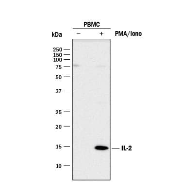
HAF017). A specific band was detected for IL-2 at approximately 14 kDa (as indicated). This experiment was conducted under reducing conditions and using
Immunoblot Buffer Group 1." class="big_lightbox">
HAF017). A specific band was detected for IL-2 at approximately 14 kDa (as indicated). This experiment was conducted under reducing conditions and using
Immunoblot Buffer Group 1." alt="Western blot shows lysates of monensin treated human peripheral blood mononuclear cells (PBMCs) with no additional treatment (-) or additionally treated (+) with 0.5 μg/mL calcium ionomycin (Iono) and 50 ng/mL PMA overnight. PVDF membrane was probed with 2 µg/mL of Goat Anti-Human/Rat IL-2 Antigen Affinity-purified Polyclonal Antibody (Catalog # AF-502-NA) followed by HRP-conjugated Anti-Goat IgG Secondary Antibody (
HAF017). A specific band was detected for IL-2 at approximately 14 kDa (as indicated). This experiment was conducted under reducing conditions and using
Immunoblot Buffer Group 1." class="big_thumb" />
502-RL) stimulates proliferation in the CTLL-2 mouse cytotoxic T cell line in a dose-dependent manner (orange line). Proliferation elicited by Recombinant Rat IL-2 (2 ng/mL) is neutralized (green line) by increasing concentrations of Goat Anti-Human/Rat IL-2 Antigen Affinity-purified Polyclonal Antibody (Catalog # AF-502-NA). The ND
50 is typically 0.15-0.75 µg/mL." class="big_lightbox">

502-RL) stimulates proliferation in the CTLL-2 mouse cytotoxic T cell line in a dose-dependent manner (orange line). Proliferation elicited by Recombinant Rat IL-2 (2 ng/mL) is neutralized (green line) by increasing concentrations of Goat Anti-Human/Rat IL-2 Antigen Affinity-purified Polyclonal Antibody (Catalog # AF-502-NA). The ND
50 is typically 0.15-0.75 µg/mL." class="big_thumb" />
VC004). Before incubation with the primary antibody, tissue was subjected to heat-induced epitope retrieval using VisUCyte Antigen Retrieval Reagent-Basic (Catalog #
VCTS021). Tissue was stained using DAB (brown) and counterstained with hematoxylin (blue). Specific staining was localized to cytoplasm in lymphocytes. View our protocol for
IHC Staining with VisUCyte HRP Polymer Detection Reagents." class="big_lightbox">

VC004). Before incubation with the primary antibody, tissue was subjected to heat-induced epitope retrieval using VisUCyte Antigen Retrieval Reagent-Basic (Catalog #
VCTS021). Tissue was stained using DAB (brown) and counterstained with hematoxylin (blue). Specific staining was localized to cytoplasm in lymphocytes. View our protocol for
IHC Staining with VisUCyte HRP Polymer Detection Reagents." class="big_thumb" />
502-RL) was serially diluted and captured by Goat Anti-Human/Rat IL‑2 Antigen Affinity-purified Polyclonal Antibody (Catalog # AF-502-NA) coated on a Clear Polystyrene Microplate (Catalog #
DY990). Goat Anti-Human/Rat IL‑2 Antigen Affinity-purified Polyclonal Antibody (Catalog #
AF-502-NA) was biotinylated and incubated with the protein captured on the plate. Detection of the standard curve was achieved by incubating Streptavidin-HRP (Catalog #
DY998)" class="big_lightbox">

502-RL) was serially diluted and captured by Goat Anti-Human/Rat IL‑2 Antigen Affinity-purified Polyclonal Antibody (Catalog # AF-502-NA) coated on a Clear Polystyrene Microplate (Catalog #
DY990). Goat Anti-Human/Rat IL‑2 Antigen Affinity-purified Polyclonal Antibody (Catalog #
AF-502-NA) was biotinylated and incubated with the protein captured on the plate. Detection of the standard curve was achieved by incubating Streptavidin-HRP (Catalog #
DY998)" class="big_thumb" />
502-RL) was serially diluted and captured by Goat Anti-Human/Rat IL‑2 Antigen Affinity-purified Polyclonal Antibody (Catalog #
AF-502-NA) coated on a Clear Polystyrene Microplate (Catalog #
DY990). Goat Anti-Human/Rat IL‑2 Antigen Affinity-purified Polyclonal Antibody (Catalog # AF-502-NA) was biotinylated and incubated with the protein captured on the plate. Detection of the standard curve was achieved by incubating Streptavidin-HRP (Catalog #
DY998)" class="big_lightbox">

502-RL) was serially diluted and captured by Goat Anti-Human/Rat IL‑2 Antigen Affinity-purified Polyclonal Antibody (Catalog #
AF-502-NA) coated on a Clear Polystyrene Microplate (Catalog #
DY990). Goat Anti-Human/Rat IL‑2 Antigen Affinity-purified Polyclonal Antibody (Catalog # AF-502-NA) was biotinylated and incubated with the protein captured on the plate. Detection of the standard curve was achieved by incubating Streptavidin-HRP (Catalog #
DY998)" class="big_thumb" />
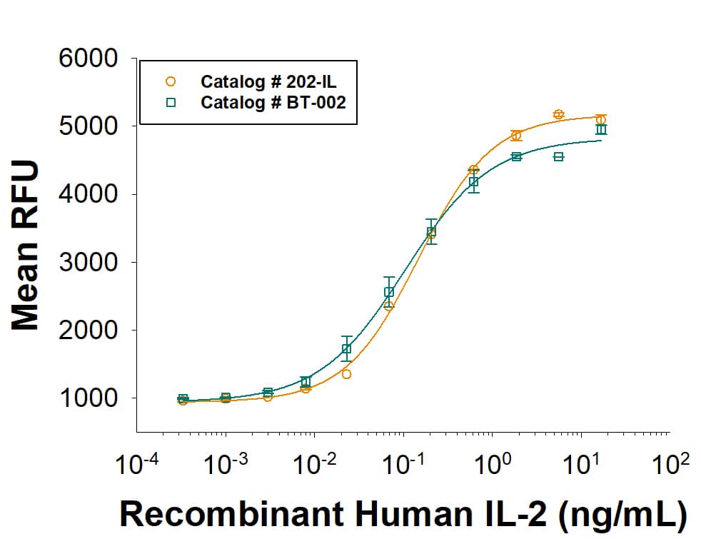 BT-002). It has equivalent bioactivity to Recombinant Human IL-2 (Catalog # 202-IL). It combines R&D Systems quality with scalability that allows for a solid supply chain. Both Recombinant Human IL-2 proteins are measured in a cell proliferation assay using CTLL 2 mouse cytotoxic T cells." alt="As an alternative, please consider our next generation Recombinant Human IL-2 (BT-002). It has equivalent bioactivity to Recombinant Human IL-2 (Catalog # 202-IL). It combines R&D Systems quality with scalability that allows for a solid supply chain. Both Recombinant Human IL-2 proteins are measured in a cell proliferation assay using CTLL 2 mouse cytotoxic T cells." class="big_thumb" />
 NL007) and counterstained with DAPI (blue). View our protocol for Fluorescent ICC Staining of Non-adherent Cells." alt="IL-2 was detected in immersion fixed human peripheral blood mononuclear cells (PBMCs) stimulated with PMA and ionomycin using 10 µg/mL Mouse Anti-Human/Primate IL-2 Monoclonal Antibody (Catalog # MAB602) for 3 hours at room temperature. Cells were stained with the NorthernLights™ 557-conjugated Anti-Mouse IgG Secondary Antibody (red; Catalog # NL007) and counterstained with DAPI (blue). View our protocol for Fluorescent ICC Staining of Non-adherent Cells." class="big_thumb" />
 FAB100A) followed by Phycoerythrin-conjugated Anti-Mouse IgG Secondary Antibody (Catalog # F0102B). To facilitate intracellular staining, cells were fixed with Flow Cytometry Fixation Buffer (Catalog # FC004) and permeabilized with Flow Cytometry Permeabilization/Wash Buffer I (Catalog # FC005). View our protocol for Staining Intracellular Molecules." alt="PBMC's treated with PMA (50ng/mL) and Ca2+ ionomycin (200ng/mL) overnight (A) vs untreated PBMC's (B) were stained with Mouse Anti-Human IL‑2 Monoclonal Antibody (Catalog # mab202) and Mouse Anti-Human CD3 epsilon APC‑conjugated Monoclonal Antibody (Catalog # FAB100A) followed by Phycoerythrin-conjugated Anti-Mouse IgG Secondary Antibody (Catalog # F0102B). To facilitate intracellular staining, cells were fixed with Flow Cytometry Fixation Buffer (Catalog # FC004) and permeabilized with Flow Cytometry Permeabilization/Wash Buffer I (Catalog # FC005). View our protocol for Staining Intracellular Molecules." class="big_thumb" />
 402-ML) stimulates proliferation in the CTLL-2 mouse cytotoxic T cell line in a dose-dependent manner (orange line), as measured by Resazurin (Catalog # AR002). Proliferation elicited by Recombinant Mouse IL-2 (1 ng/mL) is neutralized (green line) by increasing concentrations of Rat Anti-Mouse IL-2 Monoclonal Antibody (Catalog # MAB702). The ND50 is typically 0.125-0.75 µg/mL." alt="Recombinant Mouse IL-2 (Catalog # 402-ML) stimulates proliferation in the CTLL-2 mouse cytotoxic T cell line in a dose-dependent manner (orange line), as measured by Resazurin (Catalog # AR002). Proliferation elicited by Recombinant Mouse IL-2 (1 ng/mL) is neutralized (green line) by increasing concentrations of Rat Anti-Mouse IL-2 Monoclonal Antibody (Catalog # MAB702). The ND50 is typically 0.125-0.75 µg/mL." class="big_thumb" />
 HAF017). A specific band was detected for IL-2 at approximately 14 kDa (as indicated). This experiment was conducted under reducing conditions and using Immunoblot Buffer Group 1." alt="Western blot shows lysates of monensin treated human peripheral blood mononuclear cells (PBMCs) with no additional treatment (-) or additionally treated (+) with 0.5 μg/mL calcium ionomycin (Iono) and 50 ng/mL PMA overnight. PVDF membrane was probed with 2 µg/mL of Goat Anti-Human/Rat IL-2 Antigen Affinity-purified Polyclonal Antibody (Catalog # AF-502-NA) followed by HRP-conjugated Anti-Goat IgG Secondary Antibody (HAF017). A specific band was detected for IL-2 at approximately 14 kDa (as indicated). This experiment was conducted under reducing conditions and using Immunoblot Buffer Group 1." class="big_thumb" />


![N/A IL-2 [HRP]](http://images.novusbio.com/fullsize/elisa/d2050_human-il-2-quantikine-sixpak-6-plates-293202165324.jpg "N/A IL-2 [HRP]")
![N/A IL-2 [HRP]](http://images.novusbio.com/fullsize/elisa/d2050_human-il-2-quantikine-sixpak-6-plates-293202165420.jpg)
![N/A IL-2 [HRP]](http://images.novusbio.com/fullsize/elisa/d2050_human-il-2-quantikine-sixpak-6-plates-29320216567.jpg)
![N/A IL-2 [Biotin]](http://images.novusbio.com/fullsize/elisa/DATA_IL2_DY402_ELISA_1999.jpg "N/A IL-2 [Biotin]")
![N/A IL-2 [Biotin]](http://images.novusbio.com/fullsize/elisa/DATA_IL2_DY202_ELISA_1749.jpg "N/A IL-2 [Biotin]")
![N/A IL-2 [HRP]](http://images.novusbio.com/fullsize/elisa/DATA_IL2_M2000_ELISA_926.jpg "N/A IL-2 [HRP]")
![N/A IL-2 [HRP]](http://images.novusbio.com/fullsize/elisa/IL-2_M2000_ELISA_229.jpg)
 202-IL) was serially diluted and captured by Mouse Anti-Human/Primate IL‑2 Monoclonal Antibody (Catalog # MAB602) coated on a Clear Polystyrene Microplate (Catalog #
 NL007) and counterstained with DAPI (blue). Specific staining was localized to cytoplasm. View our protocol for


![N/A IL-2 [HRP]](http://images.novusbio.com/fullsize/elisa/DATA_IL2_R2000_ELISA_1082.jpg "N/A IL-2 [HRP]")
![N/A IL-2 [HRP]](http://images.novusbio.com/fullsize/elisa/IL-2_PR2000_ELISA_348.jpg)
 402-ML) was serially diluted and captured by Rat Anti-Mouse IL‑2 Monoclonal Antibody (Catalog # MAB702) coated on a Clear Polystyrene Microplate (Catalog #
  NL001) and counterstained with DAPI (blue). Specific staining was localized to cytoplasm. View our protocol for
  CTS008) and counterstained with hematoxylin (blue). Specific staining was localized to cytoplasm in lymphocytes. View our protocol for
  502-RL) stimulates proliferation in the CTLL-2 mouse cytotoxic T cell line in a dose-dependent manner (orange line). Proliferation elicited by Recombinant Rat IL-2 (2 ng/mL) is neutralized (green line) by increasing concentrations of Goat Anti-Human/Rat IL-2 Antigen Affinity-purified Polyclonal Antibody (Catalog # AF-502-NA). The ND50 is typically 0.15-0.75 µg/mL." class="big_thumb" />
 VC004). Before incubation with the primary antibody, tissue was subjected to heat-induced epitope retrieval using VisUCyte Antigen Retrieval Reagent-Basic (Catalog #
  502-RL) was serially diluted and captured by Goat Anti-Human/Rat IL‑2 Antigen Affinity-purified Polyclonal Antibody (Catalog # AF-502-NA) coated on a Clear Polystyrene Microplate (Catalog #
  502-RL) was serially diluted and captured by Goat Anti-Human/Rat IL‑2 Antigen Affinity-purified Polyclonal Antibody (Catalog #


