NL001) and counterstained with DAPI (blue). Specific staining was localized to cytoplasm. View our protocol for
Fluorescent ICC Staining of Non-adherent Cells." class="big_lightbox">
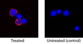
NL001) and counterstained with DAPI (blue). Specific staining was localized to cytoplasm. View our protocol for
Fluorescent ICC Staining of Non-adherent Cells." alt="IL-2 was detected in immersion fixed feline peripheral blood mononuclear cells (PBMCs) stimulated with PMA and calcium ionomycin using Goat Anti-Feline IL-2 Antigen Affinity-purified Polyclonal Antibody (Catalog # AF1890) at 15 µg/mL for 3 hours at room temperature. Cells were stained using the NorthernLights™ 557-conjugated Anti-Goat IgG Secondary Antibody (red; Catalog #
NL001) and counterstained with DAPI (blue). Specific staining was localized to cytoplasm. View our protocol for
Fluorescent ICC Staining of Non-adherent Cells." class="big_thumb" />
1890-FL) induces cell proliferation in the CTLL-2 mouse cytotoxic T cell line in a dose-dependent manner (orange line), as measured by Resazurin (Catalog #
AR002). Proliferation elicited by Recombinant Feline IL-2 (Cys146Ser) (0.2 ng/mL) is neutralized (green line) by increasing concentrations of Goat Anti-Feline IL-2 Antigen Affinity-purified Polyclonal Antibody (Catalog # AF1890). The ND
50 is typically 0.03-0.15 µg/mL." class="big_lightbox">
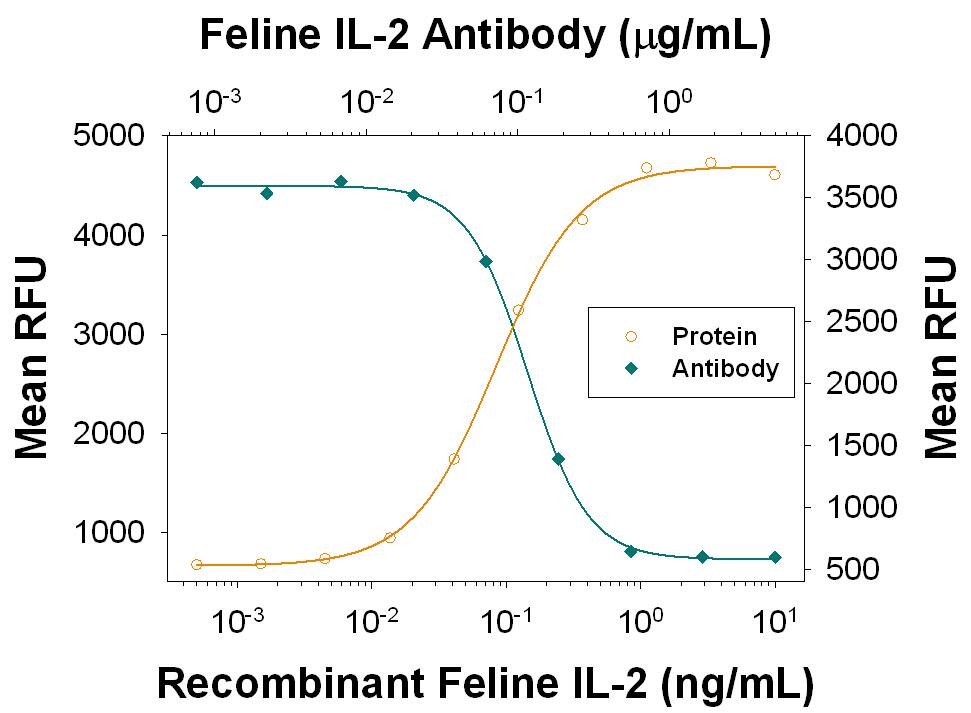
1890-FL) induces cell proliferation in the CTLL-2 mouse cytotoxic T cell line in a dose-dependent manner (orange line), as measured by Resazurin (Catalog #
AR002). Proliferation elicited by Recombinant Feline IL-2 (Cys146Ser) (0.2 ng/mL) is neutralized (green line) by increasing concentrations of Goat Anti-Feline IL-2 Antigen Affinity-purified Polyclonal Antibody (Catalog # AF1890). The ND
50 is typically 0.03-0.15 µg/mL." class="big_thumb" />
1890-FL) was serially diluted and captured by Goat Anti-Feline IL‑2 Antigen Affinity-purified Polyclonal Antibody (Catalog # AF1890) coated on a Clear Polystyrene Microplate (Catalog #
DY990). Goat Anti-Feline IL‑2 Antigen Affinity-purified Polyclonal Antibody (Catalog #
AF1890) was biotinylated and incubated with the protein captured on the plate. Detection of the standard curve was achieved by incubating Streptavidin-HRP (Catalog #
DY998)" class="big_lightbox">

1890-FL) was serially diluted and captured by Goat Anti-Feline IL‑2 Antigen Affinity-purified Polyclonal Antibody (Catalog # AF1890) coated on a Clear Polystyrene Microplate (Catalog #
DY990). Goat Anti-Feline IL‑2 Antigen Affinity-purified Polyclonal Antibody (Catalog #
AF1890) was biotinylated and incubated with the protein captured on the plate. Detection of the standard curve was achieved by incubating Streptavidin-HRP (Catalog #
DY998)" class="big_thumb" />
1890-FL) was serially diluted and captured by Goat Anti-Feline IL‑2 Antigen Affinity-purified Polyclonal Antibody (Catalog #
AF1890) coated on a Clear Polystyrene Microplate (Catalog #
DY990). Goat Anti-Feline IL‑2 Antigen Affinity-purified Polyclonal Antibody (Catalog # AF1890) was biotinylated and incubated with the protein captured on the plate. Detection of the standard curve was achieved by incubating Streptavidin-HRP (Catalog #
DY998)" class="big_lightbox">

1890-FL) was serially diluted and captured by Goat Anti-Feline IL‑2 Antigen Affinity-purified Polyclonal Antibody (Catalog #
AF1890) coated on a Clear Polystyrene Microplate (Catalog #
DY990). Goat Anti-Feline IL‑2 Antigen Affinity-purified Polyclonal Antibody (Catalog # AF1890) was biotinylated and incubated with the protein captured on the plate. Detection of the standard curve was achieved by incubating Streptavidin-HRP (Catalog #
DY998)" class="big_thumb" />
 HAF017). A specific band was detected for IL-2 at approximately 14 kDa (as indicated). This experiment was conducted under reducing conditions and using Immunoblot Buffer Group 1." alt="Western blot shows lysates of monensin treated human peripheral blood mononuclear cells (PBMCs) with no additional treatment (-) or additionally treated (+) with 0.5 μg/mL calcium ionomycin (Iono) and 50 ng/mL PMA overnight. PVDF membrane was probed with 0.5 µg/mL of Goat Anti-Human IL-2 Antigen Affinity-purified Polyclonal Antibody (Catalog # AF-202-NA) followed by HRP-conjugated Anti-Goat IgG Secondary Antibody (HAF017). A specific band was detected for IL-2 at approximately 14 kDa (as indicated). This experiment was conducted under reducing conditions and using Immunoblot Buffer Group 1." class="big_thumb" />
 NL001) and counterstained with DAPI (blue). Specific staining was localized to cytoplasm. View our protocol for Fluorescent ICC Staining of Non-adherent Cells." alt="IL-2 was detected in immersion fixed feline peripheral blood mononuclear cells (PBMCs) stimulated with PMA and calcium ionomycin using Goat Anti-Feline IL-2 Antigen Affinity-purified Polyclonal Antibody (Catalog # AF1890) at 15 µg/mL for 3 hours at room temperature. Cells were stained using the NorthernLights™ 557-conjugated Anti-Goat IgG Secondary Antibody (red; Catalog # NL001) and counterstained with DAPI (blue). Specific staining was localized to cytoplasm. View our protocol for Fluorescent ICC Staining of Non-adherent Cells." class="big_thumb" />
 NL001) and counterstained with DAPI (blue). Specific staining was localized to cytoplasm. View our protocol for Fluorescent ICC Staining of Non-adherent Cells." alt="IL-2 was detected in immersion fixed bovine peripheral blood mononuclear cells (PBMCs) using Goat Anti-Bovine IL-2 Antigen Affinity-purified Polyclonal Antibody (Catalog # AF2465) at 15 µg/mL for 3 hours at room temperature. Cells were stained using the NorthernLights™ 557-conjugated Anti-Goat IgG Secondary Antibody (red; Catalog # NL001) and counterstained with DAPI (blue). Specific staining was localized to cytoplasm. View our protocol for Fluorescent ICC Staining of Non-adherent Cells." class="big_thumb" />
 NL001) and counterstained with DAPI (blue). Specific staining was localized to cytoplasm. View our protocol for Fluorescent ICC Staining of Non-adherent Cells." alt="IL-2 was detected in immersion fixed equine peripheral blood mononuclear cells (PBMCs) using Goat Anti-Equine IL-2 Antigen Affinity-purified Polyclonal Antibody (Catalog # AF1613) at 15 µg/mL for 3 hours at room temperature. Cells were stained using the NorthernLights™ 557-conjugated Anti-Goat IgG Secondary Antibody (red; Catalog # NL001) and counterstained with DAPI (blue). Specific staining was localized to cytoplasm. View our protocol for Fluorescent ICC Staining of Non-adherent Cells." class="big_thumb" />
 402-ML) stimulates proliferation in the CTLL‑2 mouse cytotoxic T cell line in a dose-dependent manner (orange line). Proliferation elicited by Recombinant Mouse IL‑2 (2 ng/mL) is neutralized (green line) by increasing concentrations of Mouse IL‑2 Antigen Affinity-purified Polyclonal Antibody (Catalog # AF-402-NA). The ND50 is typically 0.1-0.3 µg/mL." alt="Recombinant Mouse IL‑2 (Catalog # 402-ML) stimulates proliferation in the CTLL‑2 mouse cytotoxic T cell line in a dose-dependent manner (orange line). Proliferation elicited by Recombinant Mouse IL‑2 (2 ng/mL) is neutralized (green line) by increasing concentrations of Mouse IL‑2 Antigen Affinity-purified Polyclonal Antibody (Catalog # AF-402-NA). The ND50 is typically 0.1-0.3 µg/mL." class="big_thumb" />
 652-P2)." alt="Porcine IL-2 Antibody Antigen Affinity-purified Polyclonal Goat IgG Antibody (Catalog # AF652) neutralizes IL 2-induced proliferation in the CTLL 2 mouse cytotoxic T cell line. The Neutralization Dose (ND50) is typically 0.0500-0.600 µg/mL in the presence of 2 ng/mL Recombinant Porcine IL 2 (652-P2)." class="big_thumb" />


![Immunohistochemistry-Paraffin: IL-2 Antibody [NBP2-16948] - Human tonsil. Image from verified customer review.](http://images.novusbio.com/fullsize/IL-2-Antibody-Immunohistochemistry-Paraffin-NBP2-16948-img0006.jpg "Immunohistochemistry-Paraffin: IL-2 Antibody [NBP2-16948] - Human tonsil. Image from verified customer review.")
![Flow Cytometry: IL-2 Antibody [NBP2-16948] - THP-1 cell. Black: Unlabelled sample was used as a control. Red: IL2 antibody dilution: 1:50. Acquisition of 20,000 events were collected for FACS analysis.](http://images.novusbio.com/fullsize/IL-2-Antibody-Flow-Cytometry-NBP2-16948-img0007.jpg)
![Immunohistochemistry-Paraffin: IL-2 Antibody [NBP2-16948] - Human gastric cancer, using IL2 antibody at 1:500 dilution. Antigen Retrieval: Trilogy™ (EDTA based, pH 8.0) buffer, 15min.](http://images.novusbio.com/fullsize/IL-2-Antibody-Immunohistochemistry-Paraffin-NBP2-16948-img0004.jpg)

 NL001) and counter-stained with DAPI (blue). View our protocol for
  202-IL) stimulates proliferation in the CTLL-2 mouse cytotoxic T cell line in a dose-dependent manner (orange line) as measured by Resazurin (
 202-IL) was serially diluted and captured by Mouse Anti-Human/Primate IL‑2 Monoclonal Antibody (Catalog #
 ![N/A IL-2 [Unconjugated]](http://images.novusbio.com/fullsize/elisa/IL2_HS200_ELISA_1175.jpg "N/A IL-2 [Unconjugated]")
![N/A IL-2 [Unconjugated]](http://images.novusbio.com/fullsize/elisa/IL2_HS200_ELISA_1176.jpg)
![SDS-Page: Human IL-2 Protein [NBP2-35054]](http://images.novusbio.com/fullsize/Human-IL-2-Protein-SDS-Page-NBP2-35054-img0001.jpg "SDS-Page: Human IL-2 Protein [NBP2-35054]")
 1890-FL) induces cell proliferation in the CTLL-2 mouse cytotoxic T cell line in a dose-dependent manner (orange line), as measured by Resazurin (Catalog #
  1890-FL) was serially diluted and captured by Goat Anti-Feline IL‑2 Antigen Affinity-purified Polyclonal Antibody (Catalog # AF1890) coated on a Clear Polystyrene Microplate (Catalog #
  1890-FL) was serially diluted and captured by Goat Anti-Feline IL‑2 Antigen Affinity-purified Polyclonal Antibody (Catalog #


 BT-002HS) Provides consistent CD4+/CD8+ T Cell Proliferation in Side-by-Side Testing with the Standard IL-2 Protein. CD4+ and CD8+ T cells were activated with anti-CD3- , anti-CD28- conjugated beads, and grown for 14 days in media containing 10 ng/mL Animal-free Recombinant Human IL-2 (R&D Systems, Catalog #
  BT-002-AFL) as the standard IL-2 protein or Heat Stable IL-2 (Cat #
  2465-BL) was serially diluted and captured by Goat Anti-Bovine IL‑2 Antigen Affinity-purified Polyclonal Antibody (Catalog # AF2465) coated on a Clear Polystyrene Microplate (Catalog #
  2465-BL) was serially diluted and captured by Goat Anti-Bovine IL‑2 Antigen Affinity-purified Polyclonal Antibody (Catalog #
  1613-IL) was serially diluted and captured by Goat Anti-Equine IL‑2 Antigen Affinity-purified Polyclonal Antibody (Catalog # AF1613) coated on a Clear Polystyrene Microplate (Catalog #
  1613-IL) was serially diluted and captured by Goat Anti-Equine IL‑2 Antigen Affinity-purified Polyclonal Antibody (Catalog #
 ![N/A IL-2 [HRP]](http://images.novusbio.com/fullsize/elisa/IL2_QK202_ELISA_2725.jpg "N/A IL-2 [HRP]")
![N/A IL-2 [HRP]](http://images.novusbio.com/fullsize/elisa/IL2_QK202_ELISA_2726.jpg)

 402-ML) was serially diluted and captured by Rat Anti-Mouse IL‑2 Monoclonal Antibody (Catalog #
  NL001) and counterstained with DAPI (blue). Specific staining was localized to cytoplasm. View our protocol for


