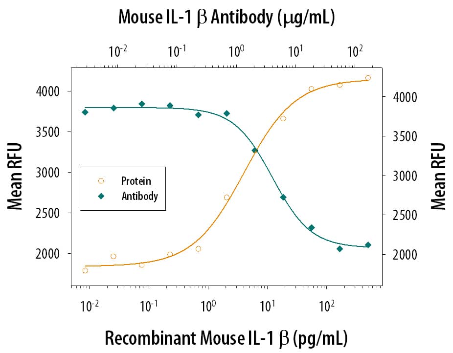
401-ML) stimulates proliferation in the the D10.G4.1 mouse helper T cell line in a dose-dependent manner (orange line). Proliferation elicited by Recombinant Mouse IL-1 beta /IL-1F2 (50 pg/mL) is neutralized (green line) by increasing concentrations of Rat Anti-Mouse IL-1 beta /IL-1F2 Monoclonal Antibody (Catalog # MAB401). The ND
50 is typically 1-5 µg/mL." class="big_lightbox">
401-ML) stimulates proliferation in the the D10.G4.1 mouse helper T cell line in a dose-dependent manner (orange line). Proliferation elicited by Recombinant Mouse IL-1 beta /IL-1F2 (50 pg/mL) is neutralized (green line) by increasing concentrations of Rat Anti-Mouse IL-1 beta /IL-1F2 Monoclonal Antibody (Catalog # MAB401). The ND
50 is typically 1-5 µg/mL." alt="Recombinant Mouse IL-1 beta /IL-1F2 (Catalog #
401-ML) stimulates proliferation in the the D10.G4.1 mouse helper T cell line in a dose-dependent manner (orange line). Proliferation elicited by Recombinant Mouse IL-1 beta /IL-1F2 (50 pg/mL) is neutralized (green line) by increasing concentrations of Rat Anti-Mouse IL-1 beta /IL-1F2 Monoclonal Antibody (Catalog # MAB401). The ND
50 is typically 1-5 µg/mL." class="big_thumb" />
401-ML) was serially diluted and captured by Rat Anti-Mouse IL‑1 beta /IL‑1F2 Monoclonal Antibody (Catalog # MAB401) coated on a Clear Polystyrene Microplate (Catalog #
DY990). Goat Anti-Mouse IL‑1 beta /IL‑1F2 Antigen Affinity-purified Polyclonal Antibody (Catalog #
AF-401-NA) was biotinylated and incubated with the protein captured on the plate. Detection of the standard curve was achieved by incubating Streptavidin-HRP (Catalog #
DY998)" class="big_lightbox">

401-ML) was serially diluted and captured by Rat Anti-Mouse IL‑1 beta /IL‑1F2 Monoclonal Antibody (Catalog # MAB401) coated on a Clear Polystyrene Microplate (Catalog #
DY990). Goat Anti-Mouse IL‑1 beta /IL‑1F2 Antigen Affinity-purified Polyclonal Antibody (Catalog #
AF-401-NA) was biotinylated and incubated with the protein captured on the plate. Detection of the standard curve was achieved by incubating Streptavidin-HRP (Catalog #
DY998)" class="big_thumb" />
401-ML) was serially diluted and captured by Rat Anti-Mouse IL‑1 beta /IL‑1F2 Monoclonal Antibody (Catalog # MAB401) coated on a Clear Polystyrene Microplate (Catalog #
DY990). Goat Anti-Mouse IL‑1 beta /IL‑1F2 Antigen Affinity-purified Polyclonal Antibody (Catalog #
AF-401-NA) was biotinylated and incubated with the protein captured on the plate. Detection of the standard curve was achieved by incubating Streptavidin-HRP (Catalog #
DY998)" class="big_lightbox">
401-ML) was serially diluted and captured by Rat Anti-Mouse IL‑1 beta /IL‑1F2 Monoclonal Antibody (Catalog # MAB401) coated on a Clear Polystyrene Microplate (Catalog #
DY990). Goat Anti-Mouse IL‑1 beta /IL‑1F2 Antigen Affinity-purified Polyclonal Antibody (Catalog #
AF-401-NA) was biotinylated and incubated with the protein captured on the plate. Detection of the standard curve was achieved by incubating Streptavidin-HRP (Catalog #
DY998)" class="big_thumb" />
401-ML) was serially diluted and captured by Rat Anti-Mouse IL‑1 beta /IL‑1F2 Monoclonal Antibody (Catalog # MAB401) coated on a Clear Polystyrene Microplate (Catalog #
DY990). Goat Anti-Mouse IL‑1 beta /IL‑1F2 Antigen Affinity-purified Polyclonal Antibody (Catalog #
AF-401-NA) was biotinylated and incubated with the protein captured on the plate. Detection of the standard curve was achieved by incubating Streptavidin-HRP (Catalog #
DY998)" class="big_lightbox">
401-ML) was serially diluted and captured by Rat Anti-Mouse IL‑1 beta /IL‑1F2 Monoclonal Antibody (Catalog # MAB401) coated on a Clear Polystyrene Microplate (Catalog #
DY990). Goat Anti-Mouse IL‑1 beta /IL‑1F2 Antigen Affinity-purified Polyclonal Antibody (Catalog #
AF-401-NA) was biotinylated and incubated with the protein captured on the plate. Detection of the standard curve was achieved by incubating Streptavidin-HRP (Catalog #
DY998)" class="big_thumb" />
401-ML) was serially diluted and captured by Rat Anti-Mouse IL‑1 beta /IL‑1F2 Monoclonal Antibody (Catalog # MAB401) coated on a Clear Polystyrene Microplate (Catalog #
DY990). Goat Anti-Mouse IL‑1 beta /IL‑1F2 Antigen Affinity-purified Polyclonal Antibody (Catalog #
AF-401-NA) was biotinylated and incubated with the protein captured on the plate. Detection of the standard curve was achieved by incubating Streptavidin-HRP (Catalog #
DY998)" class="big_lightbox">
401-ML) was serially diluted and captured by Rat Anti-Mouse IL‑1 beta /IL‑1F2 Monoclonal Antibody (Catalog # MAB401) coated on a Clear Polystyrene Microplate (Catalog #
DY990). Goat Anti-Mouse IL‑1 beta /IL‑1F2 Antigen Affinity-purified Polyclonal Antibody (Catalog #
AF-401-NA) was biotinylated and incubated with the protein captured on the plate. Detection of the standard curve was achieved by incubating Streptavidin-HRP (Catalog #
DY998)" class="big_thumb" />
401-ML) was serially diluted and captured by Rat Anti-Mouse IL‑1 beta /IL‑1F2 Monoclonal Antibody (Catalog # MAB401) coated on a Clear Polystyrene Microplate (Catalog #
DY990). Goat Anti-Mouse IL‑1 beta /IL‑1F2 Antigen Affinity-purified Polyclonal Antibody (Catalog #
AF-401-NA) was biotinylated and incubated with the protein captured on the plate. Detection of the standard curve was achieved by incubating Streptavidin-HRP (Catalog #
DY998)" class="big_lightbox">
401-ML) was serially diluted and captured by Rat Anti-Mouse IL‑1 beta /IL‑1F2 Monoclonal Antibody (Catalog # MAB401) coated on a Clear Polystyrene Microplate (Catalog #
DY990). Goat Anti-Mouse IL‑1 beta /IL‑1F2 Antigen Affinity-purified Polyclonal Antibody (Catalog #
AF-401-NA) was biotinylated and incubated with the protein captured on the plate. Detection of the standard curve was achieved by incubating Streptavidin-HRP (Catalog #
DY998)" class="big_thumb" />
 HAF018). A specific band was detected for IL-1 beta /IL-1F2 at approximately 36 kDa (as indicated). This experiment was conducted under reducing conditions and using Immunoblot Buffer Group 1." alt="Western blot shows lysates of THP-1 human acute monocytic leukemia cell line untreated (-) or treated (+) with 200 nM PMA for 24 hours and 10 µg/mL LPS and 3 hours. PVDF membrane was probed with 1 µg/mL of Mouse Anti-Human IL-1 beta /IL-1F2 Monoclonal Antibody (Catalog # MAB201) followed by HRP-conjugated Anti-Mouse IgG Secondary Antibody (Catalog # HAF018). A specific band was detected for IL-1 beta /IL-1F2 at approximately 36 kDa (as indicated). This experiment was conducted under reducing conditions and using Immunoblot Buffer Group 1." class="big_thumb" />
 HAF018). A specific band was detected for IL-1 beta /IL-1F2 at approximately 36 kDa (as indicated). This experiment was conducted under reducing conditions and using Immunoblot Buffer Group 1." alt="Western blot shows lysates of THP-1 human acute monocytic leukemia cell line untreated (-) or treated (+) with 200 nM PMA for 24 hours and 10 µg/mL LPS and 3 hours. PVDF membrane was probed with 1 µg/mL of Mouse Anti-Human IL-1 beta /IL-1F2 Monoclonal Antibody (Catalog # MAB601) followed by HRP-conjugated Anti-Mouse IgG Secondary Antibody (Catalog # HAF018). A specific band was detected for IL-1 beta /IL-1F2 at approximately 36 kDa (as indicated). This experiment was conducted under reducing conditions and using Immunoblot Buffer Group 1." class="big_thumb" />
 501-RL) stimulates proliferation in the the D10.G4.1 mouse helper T cell line in a dose-dependent manner (orange line). Proliferation elicited by Recombinant Rat IL-1 beta /IL-1F2 (10 ng/mL) is neutralized (green line) by increasing concentrations of Rat IL-1 beta /IL-1F2 Antigen Affinity-purified Polyclonal Antibody (Catalog # AF-501-NA). The ND50 is typically 2-10 µg/mL." alt="Recombinant Rat IL-1 beta /IL-1F2 (Catalog # 501-RL) stimulates proliferation in the the D10.G4.1 mouse helper T cell line in a dose-dependent manner (orange line). Proliferation elicited by Recombinant Rat IL-1 beta /IL-1F2 (10 ng/mL) is neutralized (green line) by increasing concentrations of Rat IL-1 beta /IL-1F2 Antigen Affinity-purified Polyclonal Antibody (Catalog # AF-501-NA). The ND50 is typically 2-10 µg/mL." class="big_thumb" />
 401-ML) stimulates proliferation in the the D10.G4.1 mouse helper T cell line in a dose-dependent manner (orange line). Proliferation elicited by Recombinant Mouse IL-1 beta /IL-1F2 (50 pg/mL) is neutralized (green line) by increasing concentrations of Rat Anti-Mouse IL-1 beta /IL-1F2 Monoclonal Antibody (Catalog # MAB401). The ND50 is typically 1-5 µg/mL." alt="Recombinant Mouse IL-1 beta /IL-1F2 (Catalog # 401-ML) stimulates proliferation in the the D10.G4.1 mouse helper T cell line in a dose-dependent manner (orange line). Proliferation elicited by Recombinant Mouse IL-1 beta /IL-1F2 (50 pg/mL) is neutralized (green line) by increasing concentrations of Rat Anti-Mouse IL-1 beta /IL-1F2 Monoclonal Antibody (Catalog # MAB401). The ND50 is typically 1-5 µg/mL." class="big_thumb" />
 401-ML) stimulates proliferation in the the D10.G4.1 mouse helper T cell line in a dose-dependent manner (orange line). Proliferation elicited by Recombinant Mouse IL-1 beta /IL-1F2 (10 pg/mL) is neutralized (green line) by increasing concentrations of Goat Anti-Mouse IL-1 beta /IL-1F2 Polyclonal Antibody (Catalog # AB-401-NA). The ND50 is typically 2-12 µg/mL." alt="Recombinant Mouse IL-1 beta /IL-1F2 (401-ML) stimulates proliferation in the the D10.G4.1 mouse helper T cell line in a dose-dependent manner (orange line). Proliferation elicited by Recombinant Mouse IL-1 beta /IL-1F2 (10 pg/mL) is neutralized (green line) by increasing concentrations of Goat Anti-Mouse IL-1 beta /IL-1F2 Polyclonal Antibody (Catalog # AB-401-NA). The ND50 is typically 2-12 µg/mL." class="big_thumb" />
 401-ML) stimulates proliferation in the the D10.G4.1 mouse helper T cell line in a dose-dependent manner (orange line). Proliferation elicited by Recombinant Mouse IL-1 beta /IL-1F2 (50 pg/mL) is neutralized (green line) by increasing concentrations of Mouse IL-1 beta /IL-1F2 Monoclonal Antibody (Catalog # MAB4012). The ND50 is typically 0.3-1.5 µg/mL." alt="Recombinant Mouse IL-1 beta /IL-1F2 (Catalog # 401-ML) stimulates proliferation in the the D10.G4.1 mouse helper T cell line in a dose-dependent manner (orange line). Proliferation elicited by Recombinant Mouse IL-1 beta /IL-1F2 (50 pg/mL) is neutralized (green line) by increasing concentrations of Mouse IL-1 beta /IL-1F2 Monoclonal Antibody (Catalog # MAB4012). The ND50 is typically 0.3-1.5 µg/mL." class="big_thumb" />
 HAF005). A specific band was detected for IL-1 beta /IL-1F2 at approximately 35 kDa (as indicated). This experiment was conducted under reducing conditions and using Immunoblot Buffer Group 1." alt="Western blot shows lysates of RAW 264.7 mouse monocyte/macrophage cell line untreated (-) or treated (+) with LPS. PVDF membrane was probed with 1 µg/mL of Rat Anti-Mouse IL-1 beta /IL-1F2 Monoclonal Antibody (Catalog # MAB4011) followed by HRP-conjugated Anti-Rat IgG Secondary Antibody (Catalog # HAF005). A specific band was detected for IL-1 beta /IL-1F2 at approximately 35 kDa (as indicated). This experiment was conducted under reducing conditions and using Immunoblot Buffer Group 1." class="big_thumb" />
 501-RL) stimulates proliferation in the the D10.G4.1 mouse helper T cell line in a dose-dependent manner (orange line). Proliferation elicited by Recombinant Rat IL-1 beta /IL-1F2 (4 ng/mL) is neutralized (green line) by increasing concentrations of Mouse Anti-Rat IL-1 beta /IL-1F2 Monoclonal Antibody (Catalog # MAB501). The ND50 is typically 10-30 µg/mL." alt="Recombinant Rat IL-1 beta /IL-1F2 (Catalog # 501-RL) stimulates proliferation in the the D10.G4.1 mouse helper T cell line in a dose-dependent manner (orange line). Proliferation elicited by Recombinant Rat IL-1 beta /IL-1F2 (4 ng/mL) is neutralized (green line) by increasing concentrations of Mouse Anti-Rat IL-1 beta /IL-1F2 Monoclonal Antibody (Catalog # MAB501). The ND50 is typically 10-30 µg/mL." class="big_thumb" />


 NL007) and counterstained with DAPI (blue). View our protocol for
  NL007) and counterstained with DAPI (blue). View our protocol for
 201-LB) stimulates proliferation in the the D10.G4.1 mouse helper T cell line in a dose-dependent manner (orange line). Proliferation elicited by Recombinant Human IL-1 beta /IL-1F2 (50 pg/mL) is neutralized (green line) by increasing concentrations of Mouse Anti-Human IL-1 beta /IL-1F2 Monoclonal Antibody (Catalog # MAB201). The ND50 is typically 0.001-0.003 µg/mL." class="big_thumb" />


 NL007) and counterstained with DAPI (blue). Specific staining was localized to the cytoplasm of THP-1 cells treated with 200nM PMA for 24 hours then 10ug/mL LPS for 24 hours. View our protocol for
  NL007) and counterstained with DAPI (blue). Specific staining was localized to cytoplasm. View our protocol for
 201-LB) stimulates proliferation in the the D10.G4.1 mouse helper T cell line in a dose-dependent manner (orange line). Proliferation elicited by Recombinant Human IL-1 beta /IL-1F2 (0.05 ng/mL) is neutralized (green line) by increasing concentrations of Mouse Anti-Human IL-1 beta /IL-1F2 Monoclonal Antibody (Catalog # MAB601). The ND50 is typically 0.05-0.2 µg/mL." class="big_thumb" />


 201-LB) was serially diluted and captured by Mouse Anti-Human IL‑1 beta /IL‑1F2 Monoclonal Antibody (Catalog # MAB601) coated on a Clear Polystyrene Microplate (Catalog #
  NL001) and counterstained (green). View our protocol for


 501-RL) was serially diluted and captured by Goat Anti-Rat IL‑1 beta /IL‑1F2 Antigen Affinity-purified Polyclonal Antibody (Catalog # AF-501-NA) coated on a Clear Polystyrene Microplate (Catalog #
  501-RL) was serially diluted and captured by Goat Anti-Rat IL‑1 beta /IL‑1F2 Antigen Affinity-purified Polyclonal Antibody (Catalog #

 401-ML) was serially diluted and captured by Rat Anti-Mouse IL‑1 beta /IL‑1F2 Monoclonal Antibody (Catalog # MAB401) coated on a Clear Polystyrene Microplate (Catalog #
  401-ML) was serially diluted and captured by Rat Anti-Mouse IL‑1 beta /IL‑1F2 Monoclonal Antibody (Catalog # MAB401) coated on a Clear Polystyrene Microplate (Catalog #
  401-ML) was serially diluted and captured by Rat Anti-Mouse IL‑1 beta /IL‑1F2 Monoclonal Antibody (Catalog # MAB401) coated on a Clear Polystyrene Microplate (Catalog #
  401-ML) was serially diluted and captured by Rat Anti-Mouse IL‑1 beta /IL‑1F2 Monoclonal Antibody (Catalog # MAB401) coated on a Clear Polystyrene Microplate (Catalog #
  401-ML) was serially diluted and captured by Rat Anti-Mouse IL‑1 beta /IL‑1F2 Monoclonal Antibody (Catalog # MAB401) coated on a Clear Polystyrene Microplate (Catalog #
  HAF017). A specific band was detected for IL-1 beta /IL-1F2 at approximately 35 kDa (as indicated). This experiment was conducted under reducing conditions and using


![Western Blot: IL-1 beta/IL-1F2 Antibody [NBP1-19775] - Analysis of IL1 beta Antibody in extracts from HeLa cells.](http://images.novusbio.com/fullsize/IL-1-beta-IL-1F2-Antibody-Western-Blot-NBP1-19775-img0004.jpg "Western Blot: IL-1 beta/IL-1F2 Antibody [NBP1-19775] - Analysis of IL1 beta Antibody in extracts from HeLa cells.")

![Immunohistochemistry: IL-1 beta/IL-1F2 Antibody [NBP1-19775] - Staining in the cytoplasm of villi and in the crypt cells of human lung carcinoma tissue.](http://images.novusbio.com/fullsize/IL-1-beta-IL-1F2-Antibody-Immunohistochemistry-NBP1-19775-img0005.jpg)
![Western Blot: IL-1 beta/IL-1F2 Antibody [NBP1-42767] - Analysis using the HRP conjugate of NBP1-42767. Detection of Lane 1: Mouse IL-1 Beta Load: 50 ug per Detection of Lane Secondary antibody: IL1 beta Antibody Peroxidase conjugated at 1:1,000 for 30 min at RT Block for30 min RT Predicted/Observed size: 18 kDa, 18 kDa.](http://images.novusbio.com/fullsize/IL-1-beta-IL-1F2-Antibody-Western-Blot-NBP1-42767-img0012.jpg "Western Blot: IL-1 beta/IL-1F2 Antibody [NBP1-42767] - Analysis using the HRP conjugate of NBP1-42767. Detection of Lane 1: Mouse IL-1 Beta Load: 50 ug per Detection of Lane Secondary antibody: IL1 beta Antibody Peroxidase conjugated at 1:1,000 for 30 min at RT Block for30 min RT Predicted/Observed size: 18 kDa, 18 kDa.")
![Immunocytochemistry/Immunofluorescence: IL-1 beta/IL-1F2 Antibody [NBP1-42767] - Staining of mouse carotid artery. A) No Ab staining of WT uninjured Ms. B) IL-1 beta staining of cells after surgical injury of tissue. C) No Ab staining of injured tissue from an IL-1 beta KO.](http://images.novusbio.com/fullsize/IL-1-beta-IL-1F2-Antibody-Immunocytochemistry-Immunofluorescence-NBP1-42767-img0004.jpg)
![Immunohistochemistry: IL-1 beta/IL-1F2 Antibody [NBP1-42767] - Analysis of Mouse Embryonic Kidney Tissue: Mouse Embryonic Kidney Fixation: FFPE buffered formalin 10% conc Ag Retrieval: Heat, Citrate pH 6.2. Pressure Cooker Primary antibody: 2ug/ml 1.5 hour @ room T Secondary Ab: MACH 1 HRP POLYMER 1:50 45C RT](http://images.novusbio.com/fullsize/IL-1-beta-IL-1F2-Antibody-Immunohistochemistry-NBP1-42767-img0010.jpg)
![Western Blot: IL-1 beta/IL-1F2 Antibody [NBP1-42767] - Analysis using the Biotin conjugate of NBP1-42767. Detection of Lane 1: Mouse IL-1beta Recombinant Protein. Load: 50 ng per lane. Used at a dilution of 1:1,000 overnight at 4C.](http://images.novusbio.com/fullsize/IL-1-beta-IL-1F2-Antibody-Western-Blot-NBP1-42767-img0008.jpg)
![Western Blot: IL-1 beta/IL-1F2 Antibody [NBP1-42767] - Detects 10% of the non-denatured (native) precursor 31,000 MW mouse IL-1Beta containing samples but will primarily detect all of the 17,000 MW mature molecule. However, in western blot analysis, the usual procedure of heating the sample in SDS with or without reducing agents will facilitate denaturing of the 31,000 MW IL- 1Beta precursor molecule. Denatured IL-1Beta will have a 18 kDa band.](http://images.novusbio.com/fullsize/IL-1-beta-IL-1F2-Antibody-Western-Blot-NBP1-42767-img0011.jpg)


![N/A IL-1 beta/IL-1F2 [Biotin]](http://images.novusbio.com/fullsize/elisa/DATA_IL1_beta_DY681_ELISA_2177.jpg "N/A IL-1 beta/IL-1F2 [Biotin]")


 NL007) and counterstained with DAPI (blue). Specific staining was localized to the cytoplasm. View our protocol for
  VCTS021). Tissue was stained using the HRP-conjugated Anti-Mouse IgG Secondary Antibody (Catalog #
  VCTS021). Tissue was stained using the HRP-conjugated Anti-Mouse IgG Secondary Antibody (Catalog #
  VCTS021). Tissue was stained using the HRP-conjugated Anti-Mouse IgG Secondary Antibody (Catalog #


