VC001). Before incubation with the primary antibody, tissue was subjected to heat-induced epitope retrieval using Antigen Retrieval Reagent-Basic (
CTS013). Tissue was stained using DAB (brown) and counterstained with hematoxylin (blue). Specific staining was localized to cytoplasm. View our protocol for
IHC Staining with VisUCyte HRP Polymer Detection Reagents." class="big_lightbox">
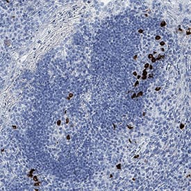
VC001). Before incubation with the primary antibody, tissue was subjected to heat-induced epitope retrieval using Antigen Retrieval Reagent-Basic (
CTS013). Tissue was stained using DAB (brown) and counterstained with hematoxylin (blue). Specific staining was localized to cytoplasm. View our protocol for
IHC Staining with VisUCyte HRP Polymer Detection Reagents." alt="CXCL13/BLC/BCA-1 was detected in immersion fixed paraffin-embedded sections of human tonsil using Mouse Anti-Human CXCL13/BLC/BCA-1 Monoclonal Antibody (Catalog # MAB8012) at 1.7 µg/mL for 1 hour at room temperature followed by incubation with the Anti-Mouse IgG VisUCyte™ HRP Polymer Antibody
VC001). Before incubation with the primary antibody, tissue was subjected to heat-induced epitope retrieval using Antigen Retrieval Reagent-Basic (
CTS013). Tissue was stained using DAB (brown) and counterstained with hematoxylin (blue). Specific staining was localized to cytoplasm. View our protocol for
IHC Staining with VisUCyte HRP Polymer Detection Reagents." class="big_thumb" />
801-CX) chemoattracts the BaF3 mouse pro-B cell line transfected with human CXCR5 in a dose-dependent manner (orange line). The amount of cells that migrated through to the lower chemotaxis chamber was measured by Resazurin (
AR002). Chemotaxis elicited by Recombinant Human CXCL13/BLC/BCA-1 (0.05 µg/mL) is neutralized (green line) by increasing concentrations of Mouse Anti-Human CXCL13/BLC/BCA-1 Monoclonal Antibody (Catalog # MAB8012). The ND
50 is typically 0.5‑2.5 µg/mL." class="big_lightbox">
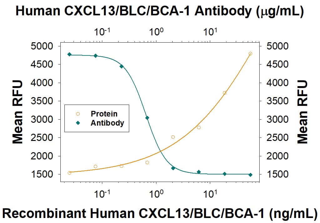
801-CX) chemoattracts the BaF3 mouse pro-B cell line transfected with human CXCR5 in a dose-dependent manner (orange line). The amount of cells that migrated through to the lower chemotaxis chamber was measured by Resazurin (
AR002). Chemotaxis elicited by Recombinant Human CXCL13/BLC/BCA-1 (0.05 µg/mL) is neutralized (green line) by increasing concentrations of Mouse Anti-Human CXCL13/BLC/BCA-1 Monoclonal Antibody (Catalog # MAB8012). The ND
50 is typically 0.5‑2.5 µg/mL." class="big_thumb" />
204-IL) and 50 ng/mL Recombinant Human GM-CSF Protein (
215-GM) for 6 days) were stained with Mouse Anti-Human CXCL13/BLC/BCA‑1 Monoclonal Antibody (Catalog # MAB8012, filled histogram) or isotype control antibody (Catalog #
MAB002, open histogram), followed by Allophycocyanin-conjugated Anti-Mouse IgG Secondary Antibody (Catalog #
F0101B). To facilitate intracellular staining, cells were fixed and permeabilized with FlowX FoxP3 Fixation & Permeabilization Buffer Kit (
FC012). View our protocol for
Staining Intracellular Molecules." class="big_lightbox">
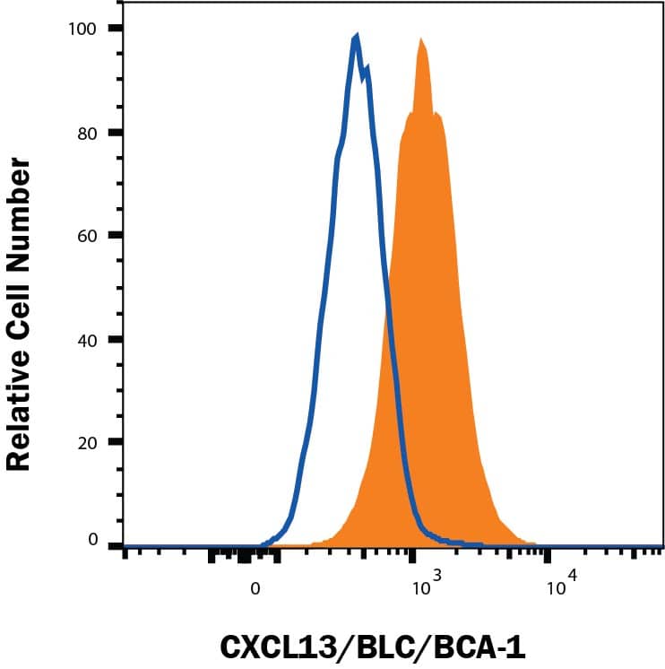
204-IL) and 50 ng/mL Recombinant Human GM-CSF Protein (
215-GM) for 6 days) were stained with Mouse Anti-Human CXCL13/BLC/BCA‑1 Monoclonal Antibody (Catalog # MAB8012, filled histogram) or isotype control antibody (Catalog #
MAB002, open histogram), followed by Allophycocyanin-conjugated Anti-Mouse IgG Secondary Antibody (Catalog #
F0101B). To facilitate intracellular staining, cells were fixed and permeabilized with FlowX FoxP3 Fixation & Permeabilization Buffer Kit (
FC012). View our protocol for
Staining Intracellular Molecules." class="big_thumb" />
204-IL) and 50 ng/mL Recombinant Human GM-CSF Protein (
215-GM) for 6 days) were stained with Mouse Anti-Human CD11c PE‑conjugated Monoclonal Antibody (Catalog #
FAB1777P) and either (A) Mouse Anti-Human CXCL13/BLC/BCA‑1 Monoclonal Antibody (Catalog # MAB8012) or (B) Mouse IgG
1 Isotype Control (Catalog #
MAB002) followed by Allophycocyanin-conjugated Anti-Mouse IgG Secondary Antibody (Catalog #
F0101B). To facilitate intracellular staining, cells were fixed and permeabilized with FlowX FoxP3 Fixation & Permeabilization Buffer Kit (Catalog #
FC012). View our protocol for
Staining Intracellular Molecules." class="big_lightbox">

204-IL) and 50 ng/mL Recombinant Human GM-CSF Protein (
215-GM) for 6 days) were stained with Mouse Anti-Human CD11c PE‑conjugated Monoclonal Antibody (Catalog #
FAB1777P) and either (A) Mouse Anti-Human CXCL13/BLC/BCA‑1 Monoclonal Antibody (Catalog # MAB8012) or (B) Mouse IgG
1 Isotype Control (Catalog #
MAB002) followed by Allophycocyanin-conjugated Anti-Mouse IgG Secondary Antibody (Catalog #
F0101B). To facilitate intracellular staining, cells were fixed and permeabilized with FlowX FoxP3 Fixation & Permeabilization Buffer Kit (Catalog #
FC012). View our protocol for
Staining Intracellular Molecules." class="big_thumb" />
MAB002, open histogram), followed by Allophycocyanin-conjugated Anti-Mouse IgG Secondary Antibody (Catalog #
F0101B). To facilitate intracellular staining, cells were fixed and permeabilized with FlowX FoxP3 Fixation & Permeabilization Buffer Kit (Catalog #
FC012). View our protocol for
Staining Intracellular Molecules." class="big_lightbox">

MAB002, open histogram), followed by Allophycocyanin-conjugated Anti-Mouse IgG Secondary Antibody (Catalog #
F0101B). To facilitate intracellular staining, cells were fixed and permeabilized with FlowX FoxP3 Fixation & Permeabilization Buffer Kit (Catalog #
FC012). View our protocol for
Staining Intracellular Molecules." class="big_thumb" />
 470-BC) chemoattracts the BaF3 mouse pro-B cell line transfected with human CXCR5 in a dose-dependent manner (orange line). The amount of cells that migrated through to the lower chemotaxis chamber was measured by Resazurin staining. Chemotaxis elicited by Recombinant Mouse CXCL13/BLC/BCA-1 (4 µg/mL) is neutralized (green line) by increasing concentrations of Goat Anti-Mouse CXCL13/ BLC/BCA-1 Antigen Affinity-purified Polyclonal Antibody (Catalog # AF470). The ND50 is typically 20-50 µg/mL." alt="Recombinant Mouse CXCL13/BLC/BCA-1 (Catalog # 470-BC) chemoattracts the BaF3 mouse pro-B cell line transfected with human CXCR5 in a dose-dependent manner (orange line). The amount of cells that migrated through to the lower chemotaxis chamber was measured by Resazurin staining. Chemotaxis elicited by Recombinant Mouse CXCL13/BLC/BCA-1 (4 µg/mL) is neutralized (green line) by increasing concentrations of Goat Anti-Mouse CXCL13/ BLC/BCA-1 Antigen Affinity-purified Polyclonal Antibody (Catalog # AF470). The ND50 is typically 20-50 µg/mL." class="big_thumb" />
 801-CX) chemoattracts the BaF3 mouse pro-B cell line transfected with human CXCR5 in a dose-dependent manner (orange line). The amount of cells that migrated through to the lower chemotaxis chamber was measured by Resazurin. Chemotaxis elicited by Recombinant Human CXCL13/BLC/BCA-1 (50 ng/mL) is neutralized (green line) by increasing concentrations of Goat Anti-Human CXCL13/BLC/BCA-1 Antigen Affinity-purified Polyclonal Antibody (Catalog # AF801). The ND50 is typically 1-4 µg/mL." alt="Recombinant Human CXCL13/BLC/BCA-1 (Catalog # 801-CX) chemoattracts the BaF3 mouse pro-B cell line transfected with human CXCR5 in a dose-dependent manner (orange line). The amount of cells that migrated through to the lower chemotaxis chamber was measured by Resazurin. Chemotaxis elicited by Recombinant Human CXCL13/BLC/BCA-1 (50 ng/mL) is neutralized (green line) by increasing concentrations of Goat Anti-Human CXCL13/BLC/BCA-1 Antigen Affinity-purified Polyclonal Antibody (Catalog # AF801). The ND50 is typically 1-4 µg/mL." class="big_thumb" />
 801-CX) and Recombinant Mouse CXCL13/BLC/BCA-1 (Catalog # 470-BC). PVDF Membrane was probed with 1 µg/mL of Mouse Anti-Human CXCL13/BLC/BCA-1 Monoclonal Antibody (Catalog # MAB801) followed by HRP-conjugated Anti-Mouse IgG Secondary Antibody (Catalog # HAF007). A specific band was detected for CXCL13/BLC/BCA-1 at approximately 12 kDa (as indicated). This experiment was conducted under reducing conditions and using Immunoblot Buffer Group 3." alt="Western blot shows 100 ng of Recombinant Human CXCL13/BLC/BCA-1 (Catalog # 801-CX) and Recombinant Mouse CXCL13/BLC/BCA-1 (Catalog # 470-BC). PVDF Membrane was probed with 1 µg/mL of Mouse Anti-Human CXCL13/BLC/BCA-1 Monoclonal Antibody (Catalog # MAB801) followed by HRP-conjugated Anti-Mouse IgG Secondary Antibody (Catalog # HAF007). A specific band was detected for CXCL13/BLC/BCA-1 at approximately 12 kDa (as indicated). This experiment was conducted under reducing conditions and using Immunoblot Buffer Group 3." class="big_thumb" />
 470-BC) chemoattracts the BaF3 mouse pro-B cell line transfected with human CXCR5 in a dose-dependent manner (orange line). The amount of cells that migrated through to the lower chemotaxis chamber was measured by Resazurin (Catalog # AR002). Chemotaxis elicited by Recombinant Mouse CXCL13/BLC/BCA-1 (4 µg/mL) is neutralized (green line) by increasing concentrations of Rat Anti-Mouse CXCL13/BLC/BCA-1 Monoclonal Antibody (Catalog # MAB470). The ND50 is typically 12-30 µg/mL." alt="Recombinant Mouse CXCL13/BLC/BCA-1 (Catalog # 470-BC) chemoattracts the BaF3 mouse pro-B cell line transfected with human CXCR5 in a dose-dependent manner (orange line). The amount of cells that migrated through to the lower chemotaxis chamber was measured by Resazurin (Catalog # AR002). Chemotaxis elicited by Recombinant Mouse CXCL13/BLC/BCA-1 (4 µg/mL) is neutralized (green line) by increasing concentrations of Rat Anti-Mouse CXCL13/BLC/BCA-1 Monoclonal Antibody (Catalog # MAB470). The ND50 is typically 12-30 µg/mL." class="big_thumb" />
 VC001). Before incubation with the primary antibody, tissue was subjected to heat-induced epitope retrieval using Antigen Retrieval Reagent-Basic (CTS013). Tissue was stained using DAB (brown) and counterstained with hematoxylin (blue). Specific staining was localized to cytoplasm. View our protocol for IHC Staining with VisUCyte HRP Polymer Detection Reagents." alt="CXCL13/BLC/BCA-1 was detected in immersion fixed paraffin-embedded sections of human tonsil using Mouse Anti-Human CXCL13/BLC/BCA-1 Monoclonal Antibody (Catalog # MAB8012) at 1.7 µg/mL for 1 hour at room temperature followed by incubation with the Anti-Mouse IgG VisUCyte™ HRP Polymer Antibody VC001). Before incubation with the primary antibody, tissue was subjected to heat-induced epitope retrieval using Antigen Retrieval Reagent-Basic (CTS013). Tissue was stained using DAB (brown) and counterstained with hematoxylin (blue). Specific staining was localized to cytoplasm. View our protocol for IHC Staining with VisUCyte HRP Polymer Detection Reagents." class="big_thumb" />


 NL001) and counterstained with DAPI (blue). Specific staining was localized to endothelial cells. View our protocol for


 470-BC) was serially diluted and captured by Rat Anti-Mouse CXCL13/BLC/BCA‑1 Monoclonal Antibody (Catalog #
 ![N/A CXCL13/BLC/BCA-1 [HRP]](http://images.novusbio.com/fullsize/elisa/DATA_CXCL13_DCX130_ELISA_642.jpg "N/A CXCL13/BLC/BCA-1 [HRP]")
![N/A CXCL13/BLC/BCA-1 [HRP]](http://images.novusbio.com/fullsize/elisa/CXCL13_DCX130_ELISA_72.jpg)

 CTS013). Tissue was stained using the Anti-Goat HRP-DAB Cell & Tissue Staining Kit (brown; Catalog #
  AF801) at 5ug/mL with 1-hour incubation at room temperature followed by incubation with anti-goat IgG VisUCyte HRP Polymer Antibody (Catalog #
 801-CX) was serially diluted and captured by Mouse Anti-Human CXCL13/BLC/BCA‑1 Monoclonal Antibody (Catalog #
 ![Western Blot: CXCL13/BLC/BCA-1 Antibody [NBP2-16041] - Non-transfected (-) and transfected (+) 293T whole cell extracts (30 ug) were separated by 15% SDS-PAGE, and the membrane was blotted with BCA1 antibody [N3C3] diluted at 1:1000. The HRP-conjugated anti-rabbit IgG antibody (NBP2-19301) was used to detect the primary antibody.](http://images.novusbio.com/fullsize/CXCL13-BLC-BCA-1-Antibody-Western-Blot-NBP2-16041-img0017.jpg "Western Blot: CXCL13/BLC/BCA-1 Antibody [NBP2-16041] - Non-transfected (-) and transfected (+) 293T whole cell extracts (30 ug) were separated by 15% SDS-PAGE, and the membrane was blotted with BCA1 antibody [N3C3] diluted at 1:1000. The HRP-conjugated anti-rabbit IgG antibody (NBP2-19301) was used to detect the primary antibody.")
![Immunocytochemistry/Immunofluorescence: CXCL13/BLC/BCA-1 Antibody [NBP2-16041] - HepG2 cells were fixed for 10 minutes using 10% formalin and then permeabilized for 5 minutes using 1X PBS + 0.05% Triton X-100. The cells were incubated with anti-CXCL13 at 5 ug/ml overnight at 4C and detected with an anti-rabbit Dylight 488 (Green) at a 1:500 dilution. Nuclei were counterstained with DAPI (Blue). Cells were imaged using a 40X objective.](http://images.novusbio.com/fullsize/CXCL13-BLC-BCA-1-Antibody-Immunocytochemistry-Immunofluorescence-NBP2-16041-img0019.jpg)
![Immunohistochemistry-Paraffin: CXCL13/BLC/BCA-1 Antibody - BSA Free [NBP2-16041] - Human breast cancer tissue stained for CXCL13/BLC/BCA-1 (green) and counterstained with DAPI (blue). Alexa Fluor 488 version of antibody used (NBP2-16041AF488). Image from verified customer review.](http://images.novusbio.com/fullsize/CXCL13-BLC-BCA-1-Antibody---BSA-Free-Immunohistochemistry-Paraffin-NBP2-16041-img0020.jpg)
![Western Blot: CXCL13/BLC/BCA-1 Antibody [NBP2-16041] - Non-transfected (-) and transfected (+) 293T whole cell extracts (60 ug) were separated by 15% SDS-PAGE, and the membrane was blotted with BCA1 antibody [N3C3] diluted at 1:5000. The HRP-conjugated anti-rabbit IgG antibody (NBP2-19301) was used to detect the primary antibody.](http://images.novusbio.com/fullsize/CXCL13-BLC-BCA-1-Antibody-Western-Blot-NBP2-16041-img0015.jpg)
![Immunohistochemistry-Paraffin: CXCL13/BLC/BCA-1 Antibody [NBP2-16041] - Analysis of a FFPE tissue section of human esophageal carcinoma using 1:300 dilution of CXCL13 antibody (NBP2-16041). The signal was developed using HRP-DAB method which followed counterstaining of the cells with hematoxylin.](http://images.novusbio.com/fullsize/CXCL13-BLC-BCA-1-Antibody-Immunohistochemistry-Paraffin-NBP2-16041-img0018.jpg)
 801-CX) chemoattracts the BaF3 mouse pro-B cell line transfected with human CXCR5 in a dose-dependent manner (orange line). The amount of cells that migrated through to the lower chemotaxis chamber was measured by Resazurin (Catalog #
 801-CX) was serially diluted and captured by Mouse Anti-Human CXCL13/BLC/BCA‑1 Monoclonal Antibody (Catalog # MAB801) coated on a Clear Polystyrene Microplate (Catalog #
 ![N/A CXCL13/BLC/BCA-1 [HRP]](http://images.novusbio.com/fullsize/elisa/DATA_CXCL13_MCX130_ELISA_963.jpg "N/A CXCL13/BLC/BCA-1 [HRP]")
![N/A CXCL13/BLC/BCA-1 [HRP]](http://images.novusbio.com/fullsize/elisa/CXCL13_MCX130_ELISA_258.jpg)
![N/A CXCL13/BLC/BCA-1 [Biotin]](http://images.novusbio.com/fullsize/elisa/DATA_CXCL13_DY801_ELISA_2235.jpg "N/A CXCL13/BLC/BCA-1 [Biotin]")
![N/A CXCL13/BLC/BCA-1 [Biotin]](http://images.novusbio.com/fullsize/elisa/DATA_CXCL13_DY470_ELISA_2051.jpg "N/A CXCL13/BLC/BCA-1 [Biotin]")
 470-BC) was serially diluted and captured by Rat Anti-Mouse CXCL13/BLC/BCA‑1 Monoclonal Antibody (Catalog # MAB470) coated on a Clear Polystyrene Microplate (Catalog #
  801-CX) chemoattracts the BaF3 mouse pro-B cell line transfected with human CXCR5 in a dose-dependent manner (orange line). The amount of cells that migrated through to the lower chemotaxis chamber was measured by Resazurin (
 204-IL) and 50 ng/mL Recombinant Human GM-CSF Protein (
 204-IL) and 50 ng/mL Recombinant Human GM-CSF Protein (
 MAB002, open histogram), followed by Allophycocyanin-conjugated Anti-Mouse IgG Secondary Antibody (Catalog #


