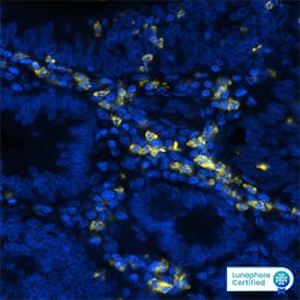
MAB16992) at 15ug/mL at 37 ° Celsius for 4 minutes. Before incubation with the primary antibody, tissue underwent an all-in-one dewaxing and antigen retrieval preprocessing using PreTreatment Module (PT Module) and Dewax and HIER Buffer H (pH 9; Epredia Catalog # TA-999-DHBH). Tissue was stained using the Alexa Fluor™ 647 Goat anti-Mouse IgG Secondary Antibody at 1:200 at 37 ° Celsius for 2 minutes. (Yellow; Lunaphore Catalog #
DR647MS) and counterstained with DAPI (blue; Lunaphore Catalog #
DR100). Specific staining was localized to the membrane. Protocol available in
COMET™ Panel Builder." class="big_lightbox">
MAB16992) at 15ug/mL at 37 ° Celsius for 4 minutes. Before incubation with the primary antibody, tissue underwent an all-in-one dewaxing and antigen retrieval preprocessing using PreTreatment Module (PT Module) and Dewax and HIER Buffer H (pH 9; Epredia Catalog # TA-999-DHBH). Tissue was stained using the Alexa Fluor™ 647 Goat anti-Mouse IgG Secondary Antibody at 1:200 at 37 ° Celsius for 2 minutes. (Yellow; Lunaphore Catalog #
DR647MS) and counterstained with DAPI (blue; Lunaphore Catalog #
DR100). Specific staining was localized to the membrane. Protocol available in
COMET™ Panel Builder." alt="CD11b/Integrin alpha M was detected in immersion fixed paraffin-embedded sections of human colon cancer using Mouse Anti-Human CD11b/Integrin alpha M Monoclonal Antibody (Catalog #
MAB16992) at 15ug/mL at 37 ° Celsius for 4 minutes. Before incubation with the primary antibody, tissue underwent an all-in-one dewaxing and antigen retrieval preprocessing using PreTreatment Module (PT Module) and Dewax and HIER Buffer H (pH 9; Epredia Catalog # TA-999-DHBH). Tissue was stained using the Alexa Fluor™ 647 Goat anti-Mouse IgG Secondary Antibody at 1:200 at 37 ° Celsius for 2 minutes. (Yellow; Lunaphore Catalog #
DR647MS) and counterstained with DAPI (blue; Lunaphore Catalog #
DR100). Specific staining was localized to the membrane. Protocol available in
COMET™ Panel Builder." class="big_thumb" />
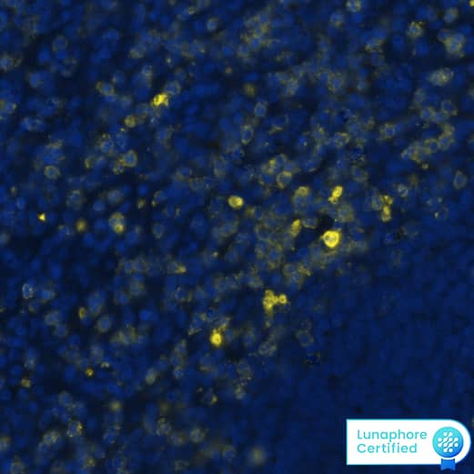
MAB16992) at 15 μg/mL at 37° Celsius for 4 minutes. Before incubation with the primary antibody, tissue underwent an all-in-one dewaxing and antigen retrieval preprocessing using PreTreatment Module (PT Module) and Dewax and HIER Buffer H (pH 9). Tissue was stained using the Alexa Fluor™ 647 Goat anti-Mouse IgG Secondary Antibody at 1:200 at 37° Celsius for 2 minutes. (Yellow; Lunaphore Catalog #
DR647MS) and counterstained with DAPI (blue; Lunaphore Catalog #
DR100). Specific staining was localized to the membrane. Protocol available in
COMET™ Panel Builder." class="big_lightbox">
MAB16992) at 15 μg/mL at 37° Celsius for 4 minutes. Before incubation with the primary antibody, tissue underwent an all-in-one dewaxing and antigen retrieval preprocessing using PreTreatment Module (PT Module) and Dewax and HIER Buffer H (pH 9). Tissue was stained using the Alexa Fluor™ 647 Goat anti-Mouse IgG Secondary Antibody at 1:200 at 37° Celsius for 2 minutes. (Yellow; Lunaphore Catalog #
DR647MS) and counterstained with DAPI (blue; Lunaphore Catalog #
DR100). Specific staining was localized to the membrane. Protocol available in
COMET™ Panel Builder." class="big_thumb" />
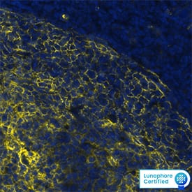
MAB16992) at 15ug/mL at 37 ° Celsius for 4 minutes. Before incubation with the primary antibody, tissue underwent an all-in-one dewaxing and antigen retrieval preprocessing using PreTreatment Module (PT Module) and Dewax and HIER Buffer H (pH 9; Epredia Catalog # TA-999-DHBH). Tissue was stained using the Alexa Fluor™ 647 Goat anti-Mouse IgG Secondary Antibody at 1:200 at 37 ° Celsius for 2 minutes. (Yellow; Lunaphore Catalog #
DR647MS) and counterstained with DAPI (blue; Lunaphore Catalog #
DR100). Specific staining was localized to the membrane. Protocol available in
COMET™ Panel Builder." class="big_lightbox">
MAB16992) at 15ug/mL at 37 ° Celsius for 4 minutes. Before incubation with the primary antibody, tissue underwent an all-in-one dewaxing and antigen retrieval preprocessing using PreTreatment Module (PT Module) and Dewax and HIER Buffer H (pH 9; Epredia Catalog # TA-999-DHBH). Tissue was stained using the Alexa Fluor™ 647 Goat anti-Mouse IgG Secondary Antibody at 1:200 at 37 ° Celsius for 2 minutes. (Yellow; Lunaphore Catalog #
DR647MS) and counterstained with DAPI (blue; Lunaphore Catalog #
DR100). Specific staining was localized to the membrane. Protocol available in
COMET™ Panel Builder." class="big_thumb" />
VC001). Before incubation with the primary antibody, tissue was subjected to heat-induced epitope retrieval using VisUCyte Antigen Retrieval Reagent-Basic (Catalog #
VCTS021). Tissue was stained using DAB (brown) and counterstained with Hematoxylin (blue). Specific staining was localized to cell surface and cytoplasm of lymphocytes. View our protocol for
Chromogenic IHC Staining of Paraffin-embedded Tissue Sections." class="big_lightbox">

VC001). Before incubation with the primary antibody, tissue was subjected to heat-induced epitope retrieval using VisUCyte Antigen Retrieval Reagent-Basic (Catalog #
VCTS021). Tissue was stained using DAB (brown) and counterstained with Hematoxylin (blue). Specific staining was localized to cell surface and cytoplasm of lymphocytes. View our protocol for
Chromogenic IHC Staining of Paraffin-embedded Tissue Sections." class="big_thumb" />
 MAB16992) at 15ug/mL at 37 ° Celsius for 4 minutes. Before incubation with the primary antibody, tissue underwent an all-in-one dewaxing and antigen retrieval preprocessing using PreTreatment Module (PT Module) and Dewax and HIER Buffer H (pH 9; Epredia Catalog # TA-999-DHBH). Tissue was stained using the Alexa Fluor™ 647 Goat anti-Mouse IgG Secondary Antibody at 1:200 at 37 ° Celsius for 2 minutes. (Yellow; Lunaphore Catalog # DR647MS) and counterstained with DAPI (blue; Lunaphore Catalog # DR100). Specific staining was localized to the membrane. Protocol available in COMET™ Panel Builder." alt="CD11b/Integrin alpha M was detected in immersion fixed paraffin-embedded sections of human colon cancer using Mouse Anti-Human CD11b/Integrin alpha M Monoclonal Antibody (Catalog # MAB16992) at 15ug/mL at 37 ° Celsius for 4 minutes. Before incubation with the primary antibody, tissue underwent an all-in-one dewaxing and antigen retrieval preprocessing using PreTreatment Module (PT Module) and Dewax and HIER Buffer H (pH 9; Epredia Catalog # TA-999-DHBH). Tissue was stained using the Alexa Fluor™ 647 Goat anti-Mouse IgG Secondary Antibody at 1:200 at 37 ° Celsius for 2 minutes. (Yellow; Lunaphore Catalog # DR647MS) and counterstained with DAPI (blue; Lunaphore Catalog # DR100). Specific staining was localized to the membrane. Protocol available in COMET™ Panel Builder." class="big_thumb" />
 AB-108-C, open histogram), followed by Phycoerythrin-conjugated Anti-Goat IgG Secondary Antibody (Catalog # F0107)." alt="Canine peripheral blood mononuclear cells (PBMCs) were stained with Goat Anti-Human/Canine CD11b/Integrin aM Antigen Affinity-purified Polyclonal Antibody (Catalog # AF1699, filled histogram) or isotype control antibody (Catalog # AB-108-C, open histogram), followed by Phycoerythrin-conjugated Anti-Goat IgG Secondary Antibody (Catalog # F0107)." class="big_thumb" />
 MAB002, open histogram), followed by Allophycocyanin-conjugated Anti-Mouse IgG Secondary Antibody (Catalog # F0101B). View our protocol for Staining Membrane-associated Proteins." alt="Human peripheral blood monocytes were stained with Mouse Anti-Human CD11b/Integrin alpha M Monoclonal Antibody (Catalog # MAB1699, filled histogram) or isotype control antibody (Catalog # MAB002, open histogram), followed by Allophycocyanin-conjugated Anti-Mouse IgG Secondary Antibody (Catalog # F0101B). View our protocol for Staining Membrane-associated Proteins." class="big_thumb" />
 HAF005). A specific band was detected for CD11b/Integrin alpha M at approximately 160 kDa (as indicated). This experiment was conducted under reducing conditions and using Immunoblot Buffer Group 1." alt="Western blot shows lysates of RAW 264.7 mouse monocyte/macrophage cell line. PVDF membrane was probed with 1 µg/mL of Mouse Anti-Mouse CD11b/Integrin alpha M Monoclonal Antibody (Catalog # MAB11241) followed by HRP-conjugated Anti-Rat IgG Secondary Antibody (Catalog # HAF005). A specific band was detected for CD11b/Integrin alpha M at approximately 160 kDa (as indicated). This experiment was conducted under reducing conditions and using Immunoblot Buffer Group 1." class="big_thumb" />
 MAB002, open histogram), followed by Phycoerythrin-conjugated Anti-Mouse IgG Secondary Antibody (Catalog # F0102B). View our protocol for Staining Membrane-associated Proteins. " alt="PBMC monocytes were stained with Mouse Anti-Human/Equine CD11b/Integrin alpha M Monoclonal Antibody (Catalog # MAB16991, filled histogram) or isotype control antibody (Catalog # MAB002, open histogram), followed by Phycoerythrin-conjugated Anti-Mouse IgG Secondary Antibody (Catalog # F0102B). View our protocol for Staining Membrane-associated Proteins. " class="big_thumb" />
 DR647RB) and counterstained with DAPI (blue; Lunaphore Catalog # DR100). Specific staining was localized to the cytoplasm. Protocol available in COMET™ Panel Builder." alt="CD11b was detected in immersion fixed paraffin-embedded sections of mouse Spleen using Rabbit Anti-Mouse CD11b, Monoclonal Antibody (Catalog #MAB11242) at 3ug/mL dilution at 37 ° Celsius for 4 minutes. Before incubation with the primary antibody, tissue underwent an all-in-one dewaxing and antigen retrieval preprocessing using PreTreatment Module (PT Module) and Dewax and HIER Buffer H (pH 9; Epredia Catalog # TA-999-DHBH). Tissue was stained using the Alexa Fluor™ Plus 647 Goat anti-Rabbit IgG Secondary Antibody at 1:200 at 37 ° Celsius for 2 minutes. (Yellow; Lunaphore Catalog # DR647RB) and counterstained with DAPI (blue; Lunaphore Catalog # DR100). Specific staining was localized to the cytoplasm. Protocol available in COMET™ Panel Builder." class="big_thumb" />


![Immunocytochemistry/Immunofluorescence: CD11b Antibody - BSA Free [NB110-89474] - Raw264.7 cells were fixed in 4% paraformaldehyde for 10 minutes and permeabilized in 0.05% Triton X-100 in PBS for 5 minutes. The cells were incubated with CD11b Antibody (NB110-89474) at 1ug/ml overnight at 4C and detected with an anti-rabbit DyLight 488 (Green) at a 1:1000 dilution for 60 minutes. Nuclei were counterstained with DAPI (Blue). Cells were imaged using a 100X objective and digitally deconvolved.](http://images.novusbio.com/fullsize/CD11b-Antibody---BSA-Free-Immunocytochemistry-Immunofluorescence-NB110-89474-img0031.jpg "Immunocytochemistry/Immunofluorescence: CD11b Antibody - BSA Free [NB110-89474] - Raw264.7 cells were fixed in 4% paraformaldehyde for 10 minutes and permeabilized in 0.05% Triton X-100 in PBS for 5 minutes. The cells were incubated with CD11b Antibody (NB110-89474) at 1ug/ml overnight at 4C and detected with an anti-rabbit DyLight 488 (Green) at a 1:1000 dilution for 60 minutes. Nuclei were counterstained with DAPI (Blue). Cells were imaged using a 100X objective and digitally deconvolved.")
![Immunocytochemistry/Immunofluorescence: CD11b Antibody - BSA Free [NB110-89474] - Raw264.7 cells were fixed in 4% paraformaldehyde for 10 minutes and permeabilized in 0.05% Triton X-100 in PBS for 5 minutes. The cells were incubated with CD11b Antibody conjugated to Alexa Fluor 488 (NB110-89474AF488) at 5 ug/ml for 1 hour at room temperature. Nuclei were counterstained with DAPI (Blue). Cells were imaged using a 100X objective and digitally deconvolved.](http://images.novusbio.com/fullsize/CD11b-Antibody---BSA-Free-Immunocytochemistry-Immunofluorescence-NB110-89474-img0028.jpg)
![Western Blot: CD11b Antibody - BSA Free [NB110-89474] - Western Blot detection of CD11b in RAW 264.7 whole cell lysates using [NB110-89474].](http://images.novusbio.com/fullsize/CD11b-Antibody---BSA-Free-Western-Blot-NB110-89474-img0013.jpg)
![Immunocytochemistry/Immunofluorescence: CD11b Antibody - BSA Free [NB110-89474] - Staining of mouse pancreas using [NB110-89474] at 1:400 dilution. Nuclei counterstained with 4',6-diamidino-2-phenylindole (DAPI) (blue). ICC/IF image submitted by a verified customer review.](http://images.novusbio.com/fullsize/CD11b-Antibody---BSA-Free-Immunocytochemistry-Immunofluorescence-NB110-89474-img0010.jpg)

![Flow (Cell Surface): CD11b Antibody - BSA Free [NB110-89474] - Surface staining of CD11b in CT26 colorectal carcinoma tumor model. Using Alexa Fluor 405 conjugated version of the antibody (NB110-89474AF405). Image from verified customer review.](http://images.novusbio.com/fullsize/CD11b-Antibody-BSA-Free-Flow-Cell-Surface-NB110-89474-img0033.jpg)
![Immunocytochemistry/Immunofluorescence: CD11b Antibody - BSA Free [NB110-89474] - Raw264.7 cells were fixed in 4% paraformaldehyde for 10 minutes and permeabilized in 0.05% Triton X-100 in PBS for 5 minutes. The cells were incubated with CD11b Antibody conjugated to Alexa Fluor 647 (NB110-89474AF647) at 5 ug/ml for 1 hour at room temperature. Nuclei were counterstained with DAPI (Blue). Cells were imaged using a 100X objective and digitally deconvolved.](http://images.novusbio.com/fullsize/CD11b-Antibody---BSA-Free-Immunocytochemistry-Immunofluorescence-NB110-89474-img0029.jpg)
![Immunocytochemistry/Immunofluorescence: CD11b Antibody - BSA Free [NB110-89474] - Raw264.7 cells were fixed in 4% paraformaldehyde for 10 minutes and permeabilized in 0.05% Triton X-100 in PBS for 5 minutes. The cells were incubated with CD11b Antibody conjugated to DyLight 650 (NB110-8947C) at 5 ug/ml for 1 hour at room temperature. Nuclei were counterstained with DAPI (Blue). Cells were imaged using a 100X objective and digitally deconvolved.](http://images.novusbio.com/fullsize/CD11b-Antibody---BSA-Free-Immunocytochemistry-Immunofluorescence-NB110-89474-img0030.jpg)
![Immunocytochemistry/Immunofluorescence: CD11b Antibody - BSA Free [NB110-89474] - CD11b Antibody [NB110-89474] - CD11b antibody [NB110-89474] was tested in Raw264.7 cells with DyLight 488 (green). Nuclei and alpha-tubulin were counterstained with 4',6-diamidino-2-phenylindole (DAPI) (blue) and DyLight 550 (red).](http://images.novusbio.com/fullsize/CD11b-Antibody---BSA-Free-Immunocytochemistry-Immunofluorescence-NB110-89474-img0009.jpg)

![Immunohistochemistry: CD11b Antibody - BSA Free [NB110-89474] - Immunohistochemical analysis of CD11b in human renal cancer with [NB110-89474] using 3,3'-Diaminobenzidine (DAB) with hematoxylin counterstain.](http://images.novusbio.com/fullsize/CD11b-Antibody---BSA-Free-Immunohistochemistry-NB110-89474-img0011.jpg)
![Immunohistochemistry-Frozen: CD11b Antibody - BSA Free [NB110-89474] - Immunohistochemical analysis of frozen sections of mouse spleen using [NB110-89474]. IHC-Fr image submitted by a verified customer review.](http://images.novusbio.com/fullsize/CD11b-Antibody---BSA-Free-Immunohistochemistry-Frozen-NB110-89474-img0021.jpg)

![Flow (Cell Surface): CD11b Antibody - BSA Free [NB110-89474] - CD11b Antibody [NB110-89474] - A surface stain was performed on Raw264.7 cells with DyLight 550-conjugated [NB110-89474R] (blue) and a matched isotype control (orange). Cells were incubated in an antibody dilution of 10 ug/mL for 20 minutes at room temperature. Both antibodies were conjugated to DyLight 550.](http://images.novusbio.com/fullsize/CD11b-Antibody-BSA-Free-Flow-Cell-Surface-NB110-89474-img0022.jpg)
![Flow (Cell Surface): CD11b Antibody - BSA Free [NB110-89474] - An intracellular stain was performed on THP-1 cells with CD11b NB110-89474 (blue) and a matched isotype control NBP2-24891 (orange). Cells were incubated in an antibody dilution of 2.5 ug/mL for 30 minutes at room temperature, followed by Rabbit IgG (H+L) Cross-Adsorbed Secondary Antibody, Dylight 550 (SA5-10033, Thermo Fisher).](http://images.novusbio.com/fullsize/CD11b-Antibody-BSA-Free-Flow-Cell-Surface-NB110-89474-img0032.jpg)
![Simple Western: CD11b Antibody - BSA Free [NB110-89474] - Specific band for Cd11b in 1.0 mg/mL of Dentate Gyrus from Rat Brain. [NB110-89474] was used at a dilution of 1:50. This experiment was performed under reducing conditions using the 12-230 kDa separation system. Simple Western image submitted by a verified customer review.](http://images.novusbio.com/fullsize/CD11b-Antibody---BSA-Free-Simple-Western-NB110-89474-img0012.jpg)
![Dual RNAscope ISH-IHC: CD11b Antibody - BSA Free [NB110-89474] - Formalin-fixed paraffin-embedded tissue sections of human lymph node were probed for CD11b mRNA (ACD RNAScope Probe, catalog #555098; Fast Red chromogen, ACD catalog # 322750). Adjacent tissue section was processed for immunohistochemistry using Rabbit Polyclonal (Novus Biologicals catalog #NB110-89474) at 1:50 dilution with 1 hour incubation at room temperature followed by incubation with anti-rabbit IgG VisUCyte HRP Polymer Antibody (Catalog # VC003) and DAB chromogen (yellow-brown). Tissue was counterstained with hematoxylin (blue). Specific staining was localized to lymphocytes.](http://images.novusbio.com/fullsize/CD11b-Antibody---BSA-Free-Dual-RNAscope-ISH-IHC-NB110-89474-img0025.jpg)
![Dual RNAscope ISH-IHC: CD11b Antibody - BSA Free [NB110-89474] - CD14 mRNA (red) and CD11b protein (green) were detected in formalin-fixed paraffin-embedded tissue sections of human malignant lymph node. ACD's Integrated Co-Detection Workflow was performed using ACD RNAScope Probe Hs-CD14 and CD11b antibody at 1:200 dilution. Tissue was stained on Leica Bond RX using RNAscope (TM) 2.5 LS Reagent Kit-RED, BOND Polymer Refine Detection (DAB) and Hematoxylin, BOND Polymer Refine Red Detection and Hematoxylin and RNA scope (TM) 2.5 LS Green Accessory Pack. Tissue was counterstained with 50% hematoxylin (blue).](http://images.novusbio.com/fullsize/CD11b-Antibody---BSA-Free-Dual-RNAscope-ISH-IHC-NB110-89474-img0026.jpg)

![Western Blot: CD11b Antibody - BSA Free [NB110-89474] - Immunodensities of (a) CD11b, (b) GFAP & (c) NF-kappa B (p65) proteins with representative immunoblots in striatum from Munc18-OE (n = 5) & wild-type (n = 5) mice. Bar graphs are ratios of optical densities of our proteins of interest to beta -actin (42 kDa band), expressed as immunoreactivity in percentage of mean value of the WT group (100%). No differences were detected between groups for any of the analyzed proteins. Right panels are representative immunoblots for target proteins & beta -actin which included Munc18-OE (OE) & wild-type (WT) mice samples. The molecular masses were estimated from referenced standards. Image collected & cropped by CiteAb from the following publication (//pubmed.ncbi.nlm.nih.gov/25069615), licensed under a CC-BY license. Not internally tested by Novus Biologicals.](http://images.novusbio.com/fullsize/nb110-89474_rabbit-polyclonal-cd11b-antibody-31020241535670.jpg)
![Immunohistochemistry: CD11b Antibody - BSA Free [NB110-89474] - Comparison of Ki-67 or CD11b immunoreactivity of Aldara-treated dorsal skin samples of C57BL/6 mice using OP or MP. Nuclear proliferation marker Ki-67 immunostaining on OP (a) & MP (b) dorsal skin tissue samples at 400x magnification. Dermal dendritic cells were labelled with anti-CD11b antibody on OP (c) & MP samples (d) at 400x magnification. Image collected & cropped by CiteAb from the following publication (//pubmed.ncbi.nlm.nih.gov/30842501), licensed under a CC-BY license. Not internally tested by Novus Biologicals.](http://images.novusbio.com/fullsize/nb110-89474_rabbit-polyclonal-cd11b-antibody-310202415384158.jpg)
![Immunohistochemistry: CD11b Antibody - BSA Free [NB110-89474] - Comparison of Ki-67 or CD11b immunoreactivity of Aldara-treated dorsal skin samples of C57BL/6 mice using OP or MP. Nuclear proliferation marker Ki-67 immunostaining on OP (a) & MP (b) dorsal skin tissue samples at 400x magnification. Dermal dendritic cells were labelled with anti-CD11b antibody on OP (c) & MP samples (d) at 400x magnification. Image collected & cropped by CiteAb from the following publication (//pubmed.ncbi.nlm.nih.gov/30842501), licensed under a CC-BY license. Not internally tested by Novus Biologicals.](http://images.novusbio.com/fullsize/nb110-89474_rabbit-polyclonal-cd11b-antibody-31020241539450.jpg)
![Western Blot: CD11b Antibody - BSA Free [NB110-89474] - Immunodensities of (a) CD11b, (b) GFAP & (c) NF-kappa B (p65) proteins with representative immunoblots in cerebral cortex from Munc18-OE (n = 5) & wild-type (n = 5) mice. Bar graphs are ratios of optical densities of our proteins of interest to beta -actin (42 kDa band), expressed as immunoreactivity in percentage of the mean value of the WT group (100%). CD11b was significantly decreased in Munc18-OE mice compared to the WT (t = 3.01; P <0.05). No differences between groups were observed in the levels of GFAP or NF-kappa B. Right panels are representative immunoblots for target proteins & beta -actin which included Munc18-OE (OE) & wild-type (WT) mice samples. The molecular masses were estimated from referenced standards. Image collected & cropped by CiteAb from the following publication (//pubmed.ncbi.nlm.nih.gov/25069615), licensed under a CC-BY license. Not internally tested by Novus Biologicals.](http://images.novusbio.com/fullsize/nb110-89474_rabbit-polyclonal-cd11b-antibody-310202415394511.jpg)
![Immunocytochemistry/ Immunofluorescence: CD11b Antibody - BSA Free [NB110-89474] - CD11b staining in IMhu. Cells were stained with antibodies against the CD11b surface antigen. (A) Panel A shows positive staining for CD11b & (B) panel B shows nuclear DAPI staining. Magnitude 40×. Image collected & cropped by CiteAb from the following publication (//pubmed.ncbi.nlm.nih.gov/31096716), licensed under a CC-BY license. Not internally tested by Novus Biologicals.](http://images.novusbio.com/fullsize/nb110-89474_rabbit-polyclonal-cd11b-antibody-310202415291998.jpg)

![Immunohistochemistry: CD11b Antibody (M1/70.15) [NB600-1327] - Immunohistochemical analysis of frozen lymph node section using CD11b Antibody (M1/70.15) [NB600-1327] followed by staining with HRP-conjugated secondary antibody.](http://images.novusbio.com/fullsize/CD11b-Antibody-M1-70-15-Immunohistochemistry-NB600-1327-img0012.jpg "Immunohistochemistry: CD11b Antibody (M1/70.15) [NB600-1327] - Immunohistochemical analysis of frozen lymph node section using CD11b Antibody (M1/70.15) [NB600-1327] followed by staining with HRP-conjugated secondary antibody.")
![Immunocytochemistry/Immunofluorescence: CD11b Antibody (M1/70.15) [NB600-1327] - Characterization of microglia cultures by multiple staining with CD11b Antibody (M1/70.15) [NB600-1327] (panels a. & c.) and anti GFAP (panels b & d). Nuclei appear blue due to 4',6-diamidino-2-phenylindole (DAPI) counter staining.](http://images.novusbio.com/fullsize/CD11b-Antibody-M1-70-15-Immunocytochemistry-Immunofluorescence-NB600-1327-img0013.jpg)
![Flow Cytometry: CD11b Antibody (M1/70.15) [NB600-1327] - A surface stain was performed on Raw264.7 cells with CD11b (M1/70.15) Antibody [NB600-1327R] (blue) and a matched isotype control (orange), both conjugated to DyLight 550. Cells were incubated in an antibody dilution of 5 ug/mL for 20 minutes at room temperature.](http://images.novusbio.com/fullsize/CD11b-Antibody-M1-70-15-Flow-Cytometry-NB600-1327-img0017.jpg)
![Immunocytochemistry/Immunofluorescence: CD11b Antibody (M1/70.15) - BSA Free [NB600-1327] - Raw264.7 cells were fixed in 4% paraformaldehyde for 10 minutes and permeabilized in 0.05% Triton X-100 in PBS for 5 minutes. The cells were incubated with CD11b Antibody [M1/70.15] (NB600-1327) at 1ug/ml overnight at 4C and detected with an anti-rat DyLight 488 (Green) at a 1:1000 dilution for 60 minutes. Nuclei were counterstained with DAPI (Blue). Cells were imaged using a 100X objective and digitally deconvolved.](http://images.novusbio.com/fullsize/CD11b-Antibody-M1-70-15-BSA-Free-Immunocytochemistry-Immunofluorescence-NB600-1327-img0020.jpg)
![Immunocytochemistry/Immunofluorescence: CD11b Antibody (M1/70.15) - BSA Free [NB600-1327] - Raw264.7 cells were fixed in 4% paraformaldehyde for 10 minutes and permeabilized in 0.05% Triton X-100 in PBS for 5 minutes. The cells were incubated with CD11b Antibody [M1/70.15] conjugated to Alexa Fluor 488 (NB600-1327AF488) at 5 ug/ml for 1 hour at room temperature. Nuclei were counterstained with DAPI (Blue). Cells were imaged using a 100X objective and digitally deconvolved.](http://images.novusbio.com/fullsize/CD11b-Antibody-M1-70-15-BSA-Free-Immunocytochemistry-Immunofluorescence-NB600-1327-img0018.jpg)
![Immunocytochemistry/Immunofluorescence: CD11b Antibody (M1/70.15) [NB600-1327] - Immunocytochemistry/Immunofluorescence (ICC/IF) analysis of RAW264.7 cells using CD11b antibody (M1/70.15) [NB600-1327] with Dylight 488 conjugated secondary (green). Nuclei and tubulin were stained with 4',6-diamidino-2-phenylindole (DAPI) (blue) and Dylight 550 (red).](http://images.novusbio.com/fullsize/CD11b-Antibody-M1-70-15-Immunocytochemistry-Immunofluorescence-NB600-1327-img0010.jpg)
![<b>Biological Strategies Validation. </b>Immunocytochemistry/Immunofluorescence: CD11b Antibody (M1/70.15) [NB600-1327] - Immunocytochemical/Immunofluorescent staining in untreated and treated mouse microglia cells using CD11b Antibody (M1/70.15) [NB600-1327]. Image from a verified customer review.](http://images.novusbio.com/fullsize/CD11b-Antibody-M1-70-15-Immunocytochemistry-Immunofluorescence-NB600-1327-img0015.jpg)
![<b>Biological Strategies Validation. </b>Immunocytochemistry/Immunofluorescence: CD11b Antibody (M1/70.15) [NB600-1327] - Immunocytochemical/Immunofluorescent analysis in mouse primary microglia. Cells were treated with lysophosphatidic acid (LPA) or LPA and an LPAR5 inhibitor in order to investigate the expression of iNOS after 24 hours. CD11b Antibody (M1/70.15) [NB600-1327] was used in a dilution 1:100 in order to stain primary murine microglia. Cy2 (green) and Cy3 (red) were used as secondary antibodies.](http://images.novusbio.com/fullsize/CD11b-Antibody-M1-70-15-Immunocytochemistry-Immunofluorescence-NB600-1327-img0016.jpg)
![Immunocytochemistry/Immunofluorescence: CD11b Antibody (M1/70.15) - BSA Free [NB600-1327] - Raw264.7 cells were fixed in 4% paraformaldehyde for 10 minutes and permeabilized in 0.05% Triton X-100 in PBS for 5 minutes. The cells were incubated with CD11b Antibody [M1/70.15] conjugated to Alexa Fluor 647 (NB600-1327AF647) at 5 ug/ml for 1 hour at room temperature. Nuclei were counterstained with DAPI (Blue). Cells were imaged using a 100X objective and digitally deconvolved.](http://images.novusbio.com/fullsize/CD11b-Antibody-M1-70-15-BSA-Free-Immunocytochemistry-Immunofluorescence-NB600-1327-img0019.jpg)
![Flow Cytometry: CD11b Antibody (M1/70.15) [NB600-1327] - Staining of mouse peritoneal macrophages with Rat anti Mouse CD11b Antibody (M1/70.15) [NB600-1327].](http://images.novusbio.com/fullsize/CD11b-Antibody-M1-70-15-Flow-Cytometry-NB600-1327-img0004.jpg)
![Flow Cytometry: CD11b Antibody (M1/70.15) [NB600-1327] - Staining of mouse peritoneal macrophages with Rat anti Mouse CD11b Antibody (M1/70.15) [NB600-1327]: RPE - Alexa Fluor 750](http://images.novusbio.com/fullsize/CD11b-Antibody-M1-70-15-Flow-Cytometry-NB600-1327-img0007.jpg)
![Flow Cytometry: CD11b Antibody (M1/70.15) [NB600-1327] - Staining of mouse peritoneal macrophages using the Alexa Fluor (R) 488 conjugate of CD11b Antibody (M1/70.15) [NB600-1327AF488].](http://images.novusbio.com/fullsize/CD11b-Antibody-M1-70-15-Flow-Cytometry-NB600-1327-img0014.jpg)

![Immunohistochemistry-Paraffin: CD11b Antibody (CL1719) [NBP2-34490] - Immunohistochemical staining in paraffin-embedded sections of human tonsil and skeletal muscle tissues using [NBP2-34490]. Corresponding ITGAM RNA-seq data are presented for the same tissues.](http://images.novusbio.com/fullsize/CD11b-Antibody-CL1719-Immunohistochemistry-Paraffin-NBP2-34490-img0017.jpg "<b>Orthogonal Strategies Validation. </b>Immunohistochemistry-Paraffin: CD11b Antibody (CL1719) [NBP2-34490] - Immunohistochemical staining in paraffin-embedded sections of human tonsil and skeletal muscle tissues using [NBP2-34490]. Corresponding ITGAM RNA-seq data are presented for the same tissues.")
![<b>Orthogonal Strategies Validation. </b>Western Blot: CD11b Antibody (CL1719) [NBP2-34490] - Western Blot analysis using CD11b Antibody (CL1719) [NBP2-34490]. Lane 1: Marker [kDa] Lane 2: Human tonsil. Detection at ~190 kDa.](http://images.novusbio.com/fullsize/CD11b-Antibody-CL1719-Western-Blot-NBP2-34490-img0008.jpg)
![Immunohistochemistry-Paraffin: CD11b Antibody (CL1719) [NBP2-34490] - Immunohistochemical staining in paraffin-embedded sections of human cerebral cortex using CD11b Antibody (CL1719) [NBP2-34490] shows moderate membranous positivity in microglia.](http://images.novusbio.com/fullsize/CD11b-Antibody-CL1719-Immunohistochemistry-Paraffin-NBP2-34490-img0018.jpg)
![Immunohistochemistry-Paraffin: CD11b Antibody (CL1719) [NBP2-34490] - Immunohistochemical staining in paraffin-embedded sections of human tonsil using CD11b Antibody (CL1719) [NBP2-34490] shows strong membranous positivity in germinal center cells.](http://images.novusbio.com/fullsize/CD11b-Antibody-CL1719-Immunohistochemistry-Paraffin-NBP2-34490-img0012.jpg)
![Immunohistochemistry-Paraffin: CD11b Antibody (CL1719) [NBP2-34490] - Immunohistochemical staining of human colon using CD11b Antibody (CL1719) [NBP2-34490] shows moderate membranous positivity in lymphoid cells.](http://images.novusbio.com/fullsize/CD11b-Antibody-CL1719-Immunohistochemistry-Paraffin-NBP2-34490-img0013.jpg)
![Immunohistochemistry-Paraffin: CD11b Antibody (CL1719) [NBP2-34490] - Immunohistochemical staining of Paraffin-embedded sections of human skeletal muscle using CD11b Antibody (CL1719) [NBP2-34490] shows no positivity in striated muscle fibers as expected.](http://images.novusbio.com/fullsize/CD11b-Antibody-CL1719-Immunohistochemistry-Paraffin-NBP2-34490-img0014.jpg)
 MAB16992) at 15 μg/mL at 37° Celsius for 4 minutes. Before incubation with the primary antibody, tissue underwent an all-in-one dewaxing and antigen retrieval preprocessing using PreTreatment Module (PT Module) and Dewax and HIER Buffer H (pH 9). Tissue was stained using the Alexa Fluor™ 647 Goat anti-Mouse IgG Secondary Antibody at 1:200 at 37° Celsius for 2 minutes. (Yellow; Lunaphore Catalog #
  MAB16992) at 15ug/mL at 37 ° Celsius for 4 minutes. Before incubation with the primary antibody, tissue underwent an all-in-one dewaxing and antigen retrieval preprocessing using PreTreatment Module (PT Module) and Dewax and HIER Buffer H (pH 9; Epredia Catalog # TA-999-DHBH). Tissue was stained using the Alexa Fluor™ 647 Goat anti-Mouse IgG Secondary Antibody at 1:200 at 37 ° Celsius for 2 minutes. (Yellow; Lunaphore Catalog #
  VC001). Before incubation with the primary antibody, tissue was subjected to heat-induced epitope retrieval using VisUCyte Antigen Retrieval Reagent-Basic (Catalog #
 ![Western Blot: CD11b Antibody [NBP2-92978] - Analysis of extracts of various cell lines, using CD11b antibody (NBP2-92978) at 1:500 dilution. Secondary antibody: HRP Goat Anti-Rabbit IgG (H+L) at 1:10000 dilution. Lysates/proteins: 25ug per lane. Blocking buffer: 3% nonfat dry milk in TBST. Detection: ECL Enhanced Kit. Exposure time: 90s.](http://images.novusbio.com/fullsize/CD11b Antibody-Western Blot-NBP2-92978-img0005.jpg "Western Blot: CD11b Antibody [NBP2-92978] - Analysis of extracts of various cell lines, using CD11b antibody (NBP2-92978) at 1:500 dilution. Secondary antibody: HRP Goat Anti-Rabbit IgG (H+L) at 1:10000 dilution. Lysates/proteins: 25ug per lane. Blocking buffer: 3% nonfat dry milk in TBST. Detection: ECL Enhanced Kit. Exposure time: 90s.")
![Immunohistochemistry-Paraffin: CD11b Antibody [NBP2-92978] - Mouse spleen using CD11b antibody (NBP2-92978) at dilution of 1:100 (40x lens). Perform high pressure antigen retrieval with 10 mM citrate buffer pH 6.0 before commencing with IHC staining protocol.](http://images.novusbio.com/fullsize/CD11b-Antibody-Immunohistochemistry-Paraffin-NBP2-92978-img0004.jpg)
![Immunohistochemistry-Paraffin: CD11b Antibody [NBP2-92978] - Human tonsil using CD11b antibody (NBP2-92978) at dilution of 1:100 (40x lens). Perform high pressure antigen retrieval with 10 mM citrate buffer pH 6.0 before commencing with IHC staining protocol.](http://images.novusbio.com/fullsize/CD11b-Antibody-Immunohistochemistry-Paraffin-NBP2-92978-img0003.jpg)
![Immunocytochemistry/Immunofluorescence: CD11b Antibody [NBP2-92978] - Immunofluorescence analysis of TF-1 cells using CD11b Rabbit pAb (NBP2-92978) at dilution of 1:100 (40x lens). Blue: DAPI for nuclear staining.](http://images.novusbio.com/fullsize/CD11b-Antibody-Immunocytochemistry-Immunofluorescence-NBP2-92978-img0001.jpg)
![Immunocytochemistry/ Immunofluorescence: CD11b Antibody - Azide and BSA Free [NBP2-92978] - Immunofluorescence analysis of TF-1 cells using CD11b Rabbit pAb at dilution of 1:100 (40x lens). Secondary antibody: Cy3-conjugated Goat anti-Rabbit IgG (H+L) at 1:500 dilution. Blue: DAPI for nuclear staining.](http://images.novusbio.com/fullsize/nbp2-92978_rabbit-polyclonal-cd11b-antibody-4220251429043.jpg)
![Immunocytochemistry/ Immunofluorescence: CD11b Antibody - Azide and BSA Free [NBP2-92978] - Immunofluorescence analysis of THP-1 cells using CD11b Rabbit pAb at dilution of 1:100 (40x lens). Secondary antibody: Cy3-conjugated Goat anti-Rabbit IgG (H+L) at 1:500 dilution. Blue: DAPI for nuclear staining.](http://images.novusbio.com/fullsize/nbp2-92978_rabbit-polyclonal-cd11b-antibody-4220251430438.jpg)
![Immunocytochemistry/ Immunofluorescence: CD11b Antibody - Azide and BSA Free [NBP2-92978] - Immunofluorescence analysis of THP-1 cells using CD11b Rabbit pAb at dilution of 1:100 (40x lens). Secondary antibody: Cy3-conjugated Goat anti-Rabbit IgG (H+L) at 1:500 dilution. Blue: DAPI for nuclear staining.](http://images.novusbio.com/fullsize/nbp2-92978_rabbit-polyclonal-cd11b-antibody-4220251429086.jpg)
![Immunocytochemistry/ Immunofluorescence: CD11b Antibody - Azide and BSA Free [NBP2-92978] - Immunofluorescence analysis of THP-1 cells using CD11b Rabbit pAb at dilution of 1:100 (40x lens). Secondary antibody: Cy3-conjugated Goat anti-Rabbit IgG (H+L) at 1:500 dilution. Blue: DAPI for nuclear staining.](http://images.novusbio.com/fullsize/nbp2-92978_rabbit-polyclonal-cd11b-antibody-4220251427526.jpg)
![Immunocytochemistry/ Immunofluorescence: CD11b Antibody - Azide and BSA Free [NBP2-92978] - Immunofluorescence analysis of TF-1 cells using CD11b Rabbit pAb at dilution of 1:100 (40x lens). Secondary antibody: Cy3-conjugated Goat anti-Rabbit IgG (H+L) at 1:500 dilution. Blue: DAPI for nuclear staining.](http://images.novusbio.com/fullsize/nbp2-92978_rabbit-polyclonal-cd11b-antibody-42202514251860.jpg)
 AB-108-C, open histogram), followed by Phycoerythrin-conjugated Anti-Goat IgG Secondary Antibody (Catalog #
 ![Flow Cytometry: CD11b Antibody (OX-42) [NB100-65284] - Detection of CD11b in Rat Splenocytes by Flow Cytometry. Rat splenocytes were stained with Mouse Anti-Rat CD11b Monoclonal Antibody (OX-42) [NB100-65284, filled histogram] or Mouse IgG2A Isotype Control Antibody [MAB003, open histogram] followed by anti-Mouse IgG PE-conjugated secondary antibody [F0102B] and Mouse Anti-Rat CD161 Alexa Fluor 647-conjugated Monoclonal Antibody [FAB9766R]. Splenocytes were gated on CD161+ cells.](http://images.novusbio.com/fullsize/CD11b-Antibody-OX-42-Flow-Cytometry-NB100-65284-img0001.jpg "Flow Cytometry: CD11b Antibody (OX-42) [NB100-65284] - Detection of CD11b in Rat Splenocytes by Flow Cytometry. Rat splenocytes were stained with Mouse Anti-Rat CD11b Monoclonal Antibody (OX-42) [NB100-65284, filled histogram] or Mouse IgG2A Isotype Control Antibody [MAB003, open histogram] followed by anti-Mouse IgG PE-conjugated secondary antibody [F0102B] and Mouse Anti-Rat CD161 Alexa Fluor 647-conjugated Monoclonal Antibody [FAB9766R]. Splenocytes were gated on CD161+ cells.")
![Flow Cytometry: CD11b Antibody (OX-42) [NB100-65284] - Detection of CD11b in Rat Bone Marrow cells by Flow Cytometry. Rat cells were stained with Mouse Anti-Rat CD11b Monoclonal Antibody (OX-42) [NB100-65284, filled histogram] or Mouse IgG2A Isotype Control Antibody [MAB003, open histogram] followed by anti-Mouse IgG APC-conjugated secondary antibody [F0101B] .](http://images.novusbio.com/fullsize/antibody/nb100-65284_mouse-monoclonal-cd11b-antibody-ox-42-flow-cell-surface--31102023115850..jpg)
 NL007) and counterstained with DAPI (blue). View our protocol for
  MAB0061, open histogram), followed by Phycoerythrin-conjugated Anti-Rat IgG Secondary Antibody (Catalog #
  HAF008). A specific band was detected for CD11b/Integrin alpha M at approximately 150 kDa (as indicated). This experiment was conducted under reducing conditions and using Western Blot Buffer Group 1." class="big_thumb" />

![Western Blot: CD11b/c Antibody - BSA Free [NB110-40766] - Analysis of CD11b/c in Raw 264.7 whole cell lysate.](http://images.novusbio.com/fullsize/CD11b-c-Antibody---BSA-Free-Western-Blot-NB110-40766-img0002.jpg "Western Blot: CD11b/c Antibody - BSA Free [NB110-40766] - Analysis of CD11b/c in Raw 264.7 whole cell lysate.")
![Immunocytochemistry/Immunofluorescence: CD11b/c Antibody - BSA Free [NB110-40766] - CD11 antibody was tested in Raw264.7 cells with FITC (green). Nuclei were counterstained with DAPI (blue).](http://images.novusbio.com/fullsize/CD11b-c-Antibody---BSA-Free-Immunocytochemistry-Immunofluorescence-NB110-40766-img0006.jpg)
![Immunohistochemistry-Frozen: CD11b/c Antibody - BSA Free [NB110-40766] - 10 um Cryosection of murine acute brain slices stained with CD11b/c antibody (1:300 = 3.3 ug/ml) and Alexa Fluor 555 donkey anti rabbit IgG. Antibody shows specific staining, but the background is quite heavy. Image from verified customer review.](http://images.novusbio.com/fullsize/CD11b-c-Antibody---BSA-Free-Immunohistochemistry-Frozen-NB110-40766-img0009.jpg)
![Flow Cytometry: CD11b/c Antibody - BSA Free [NB110-40766] - A surface stain was performed on RAW 264.7 with NB110-40766 and a matched isotype control. Cells were incubated in an antibody dilution of 2.5 ug/mL for 30 minutes at room temperature, followed by Rabbit IgG (H+L) Cross-Adsorbed Secondary Antibody, DyLight 550.](http://images.novusbio.com/fullsize/CD11b-c-Antibody---BSA-Free-Flow-Cytometry-NB110-40766-img0008.jpg)


![Immunohistochemistry: CD11b/c Antibody - BSA Free [NB110-40766] - Staining of human bone marrow, myeloid precursors.](http://images.novusbio.com/fullsize/CD11b-c-Antibody---BSA-Free-Immunohistochemistry-NB110-40766-img0007.jpg)
![Western Blot: CD11b/c Antibody - BSA Free [NB110-40766] - Effects of oltipraz on the expression of CD11b/c, phosphoinositide 3-kinase (PI3K), phosphorylated protein kinase B (p-Akt), & phosphorylated inhibitor of kappa B alpha (p-IkB alpha ) in the spinal cord of the CCI-injured mice. The relative protein levels of (A) CD11b/c, (B) PI3K, (D) p-Akt, & (E) p-IKB alpha on the ipsilateral side of the spinal cord in the CCI-injured mice treated with oltipraz (OLT) or vehicle are represented. The sham-operated mice (SHAM) treated with vehicle were used as controls. (C) Representative examples of blots for CD11b/c (160 kDa), PI3K (130 kDa), & GAPDH (37 kDa), & (F) for p-Akt (60 kDa), Akt (60 kDa), p-IKB alpha (40 kDa) & IKB alpha (40 kDa). CD11b/c & PI3K are expressed relative to GAPDH levels whereas phosphorylated proteins are expressed relative to their corresponding total proteins. In all panels, * denotes significant differences vs. sham-operated mice treated with vehicle (p < 0.05; one-way ANOVA followed by the SNK test). Results are presented as the mean ± SEM; n = 5 samples per experimental group. Image collected & cropped by CiteAb from the following publication (//pubmed.ncbi.nlm.nih.gov/31234342), licensed under a CC-BY license. Not internally tested by Novus Biologicals.](http://images.novusbio.com/fullsize/nb110-40766_rabbit-polyclonal-cd11b-c-antibody-310202416235533.jpg)
![Western Blot: CD11b/c Antibody - BSA Free [NB110-40766] - Effects of oltipraz on the expression of CD11b/c, PI3K, p-Akt, & p-IkB alpha in the prefrontal cortex of the CCI-injured mice. The relative protein levels of (A) CD11b/c, (B) PI3K, (D) p-Akt, & (E) p-IKB alpha in the prefrontal cortex of the CCI-injured mice treated with oltipraz (OLT) or vehicle. The sham-operated mice (SHAM) treated with vehicle were used as controls. Representative examples of blots for (C) CD11b/c (160 kDa), PI3K (130 kDa) & GAPDH (37 kDa), & for (F) p-Akt (60 kDa), Akt (60 kDa), p-IKB alpha (40 kDa) & IKB alpha (40 kDa). CD11b/c & PI3K are expressed relative to GAPDH levels whereas phosphorylated proteins are expressed relative to their corresponding total proteins. In all panels, * denotes significant differences vs. sham-operated mice treated with vehicle (p < 0.05; one-way ANOVA followed by the SNK test). Results are presented as the mean ± SEM; n = 4-–5 samples per experimental group. Image collected & cropped by CiteAb from the following publication (//pubmed.ncbi.nlm.nih.gov/31234342), licensed under a CC-BY license. Not internally tested by Novus Biologicals.](http://images.novusbio.com/fullsize/nb110-40766_rabbit-polyclonal-cd11b-c-antibody-310202416235518.jpg)
![Western Blot: CD11b/c Antibody - BSA Free [NB110-40766] - Effects of oltipraz on the expression of CD11b/c, PI3K, p-Akt, & p-IkB alpha in the hippocampus of the CCI-injured mice. The relative protein levels of (A) CD11b/c, (B) PI3K, (D) p-Akt, & (E) p-IKB alpha in the hippocampus of the CCI-injured mice treated with oltipraz (OLT) or vehicle. The sham-operated mice (SHAM) treated with vehicle were used as controls. Representative examples of blots for (C) CD11b/c (160 kDa), PI3K (130 kDa), & GAPDH (37 kDa); & (F) for p-Akt (60 kDa), Akt (60 kDa), p-IKB alpha (40 kDa), & IKB alpha (40 kDa). CD11b/c & PI3K are expressed relative to GAPDH levels whereas phosphorylated proteins are expressed relative to their corresponding total proteins. In all panels, * denotes significant differences vs. sham-operated mice treated with vehicle (p < 0.05; one-way ANOVA followed by the SNK test). Results are presented as the mean ± SEM; n = 5 samples per experimental group. Image collected & cropped by CiteAb from the following publication (//pubmed.ncbi.nlm.nih.gov/31234342), licensed under a CC-BY license. Not internally tested by Novus Biologicals.](http://images.novusbio.com/fullsize/nb110-40766_rabbit-polyclonal-cd11b-c-antibody-310202416235538.jpg)
![Immunocytochemistry/ Immunofluorescence: CD11b/c Antibody - BSA Free [NB110-40766] - Phase contrast microscopy images of HFA & brain tumor cells & immunofluorescence images of cultured primary brain tumor cells, AA & HFA.HFA were derived from three different foetal brains. (A) 18-week foetus (10× magnification); (B) 19-week foetus, first week in culture (10× magnification); (C) 19-week foetus, day 5 in culture (20× magnification); (D) Primary GBM after 2 trypsinisations (20x); (E) Recurrent GBM (20x); (F) Secondary GBM after 3 trypsinisations (10x); (G) OII 14 days in culture (10x); (H) AII 13 days in culture (10x); (I) Negative control for NGS merged with DAPI; (J) Negative control for secondary antibodies merged with DAPI; (K) GBM cells derived from one patient – GFAP (DAKO) merged with DAPI & CD68 (zoomed: 2.4× magnification); (L) primary brain tumor cells from one patient (male, 40 years old) (passaged three times) - GFAP (Novocastra) merged with DAPI; (K & L) GFAP (green) was used as a GBM marker & CD68 (red) was used as a marker for microglia. (M) AA from one Female, 61 years old; (passaged twice) - GFAP (Novocastra) merged with DAPI; (N) AA from one male 40 years old; (passaged once) - GFAP (Sigma) merged with DAPI & CD68 (Abcam); (M & N) GFAP (green) was used as an astrocyte marker & CD68 (red) & CD11b (green) were used as markers for microglia; (O) HFA from one 17-week- old foetus (passaged once) - GFAP (Novocastra) merged with DAPI & CD11b (Novus); (P) HFA from an 18-week- old foetus (passaged once) - GFAP (DAKO) merged with DAPI & CD68 (zoomed: 2.1× magnification); (O & P) GFAP (green & red) was used as an astrocyte marker & CD68 (red) & CD11b (green) were used as markers for microglia. Nuclei indicated by DAPI (blue) in all images. Image collected & cropped by CiteAb from the following publication (//dx.plos.org/10.1371/journal.pone.0112945), licensed under a CC-BY license. Not internally tested by Novus Biologicals.](http://images.novusbio.com/fullsize/nb110-40766_rabbit-polyclonal-cd11b-c-antibody-310202416235543.jpg)


