Recombinant Human Muscle Glycogen Phosphorylase His-tag, CF Summary
| Details of Functionality |
Measured by its ability to hydrolyze alpha -D-Glucose 1-phosphate. The specific activity is >3000 pmol/min/μg, as measured under the described conditions. |
| Source |
Human embryonic kidney cell, HEK293-derived human Glycogen phosphorylase, muscle form protein
Ser2-Ile842, with an N-terminal Met and 6-His tag |
| Accession # |
|
| N-terminal Sequence |
Protein identity confirmed by mass spectrometry. |
| Protein/Peptide Type |
Recombinant Enzymes |
| Purity |
>95%, by SDS-PAGE visualized with Silver Staining and quantitative densitometry by Coomassie® Blue Staining |
| Endotoxin Note |
<0.10 EU per 1 μg of the protein by the LAL method. |
Applications/Dilutions
| Dilutions |
|
| Theoretical MW |
98 kDa.
Disclaimer note: The observed molecular weight of the protein may vary from the listed predicted molecular weight due to post translational modifications, post translation cleavages, relative charges, and other experimental factors. |
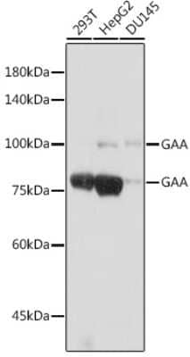
| SDS-PAGE |
86-96 kDa, under reducing conditions |
Packaging, Storage & Formulations
| Storage |
Use a manual defrost freezer and avoid repeated freeze-thaw cycles.- 6 months from date of receipt, -20 to -70 °C as supplied.
- 3 months, -20 to -70 °C under sterile conditions after opening.
|
| Buffer |
Supplied as a 0.2 μm filtered solution in Tris, NaCl, TCEP and Glycerol. |
| Purity |
>95%, by SDS-PAGE visualized with Silver Staining and quantitative densitometry by Coomassie® Blue Staining |
| Assay Procedure |
- Assay Buffer: 50 mM Tris, pH 7.0
- Recombinant Human Muscle Glycogen Phosphorylase His-tag (rhPYGM) (Catalog # 11786-PM)
- Adenosine monophosphate (AMP), 5 mM stock in deionized water
- Substrate: alpha -D-Glucose-1-Phosphate, 10 mM stock in deionized water
- Substrate: Glycogen, 20 mg/mL stock in deionized water
- Malachite Green Phosphate Detection Kit
(Catalog #
DY996)
- Clear 96-well Plate (Catalog #
DY990)
- Plate Reader with Absorbance Read Capability
- Dilute 1 M Phosphate Standard (supplied in kit) by adding 10 µL of the 1 M Phosphate Standard to 990 µL of Assay Buffer for a 10 mM stock. Continue by adding 10 µL of the 10 mM Phosphate stock to 990 µL of Assay Buffer for a 100 µM stock. This is the first point of the standard curve.
- Complete the standard curve by performing six one-half serial dilutions of the 100 µM Phosphate stock in Assay Buffer. The standard curve has a range of 0.078 to 5 nmol per well.
- Load 50 µL of each dilution of the standard curve into a plate. Include a curve blank containing 50 μL of Assay Buffer.
- Dilute rhPYGM to 0.5 µg/mL in Assay Buffer.
- Load 25 µL of 0.5 µg/mL rhPYGM into empty wells of the same plate as the curve. Include a Control containing 25 µL of Assay Buffer.
- Create a reaction mixture containing 1 mM AMP, 1 mM alpha -D-Glucose-1-Phosphate and 2 mg/mL Glycogen in Assay Buffer.
- Add 25 µL of reaction mixture to the wells, excluding the standard curve.
- Seal plate and incubate at room temperature for 20 minutes.
- Add 30 µL of the Malachite Green Reagent A to all wells. Mix briefly.
- Add 100 µL of deionized water to all wells. Mix briefly.
- Add 30 µL of the Malachite Green Reagent B to all wells. Mix and incubate for 20 minutes at room temperature.
- Read plate at 620 nm (absorbance) in endpoint mode.
- Calculate specific activity:
Specific Activity (pmol/min/µg) = | Phosphate released* (nmol) x (1000 pmol/nmol) | | Incubation time (min) x amount of enzyme (µg) |
*Derived from the phosphate standard curve using linear or 4-parameter fitting and adjusted for Control. Per Reaction: - rhPYGM: 0.0125 µg
- AMP: 0.5 mM
- alpha -D-Glucose-1-Phosphate: 0.5 mM
- Glycogen: 50 µg
|
Notes
This product is produced by and ships from R&D Systems, Inc., a Bio-Techne brand.
Alternate Names for Recombinant Human Muscle Glycogen Phosphorylase His-tag, CF
Background
Recombinant Human Muscle Glycogen Phosphorylase (PYGM), also known as myophosphorylase, is a cytoplasmic muscle isoform of glycogen phosphorylase (GP) that catalyzes the cleavage of alpha ‑1,4‑glycosidic bonds in glycogen to release glucose‑1‑phosphate. PYGM is one of three mammalian isoforms known as liver, muscle, or brain GP that differ in sequence, activation regulation, kinetics, and physiological roles (1-4). PYGM is biologically active as a homodimer where each 842-residue monomer comprises N‑ and C‑terminal domains (1,3). The C-terminal domain contains a cofactor binding site for covalently bound pyridoxal cofactor. The N-terminal domain contains a key phosphorylation site that determines whether the protein is in an active or inactive state; each state is further regulated by binding of AMP, ATP, and glucose-6-phosphate in an allosteric site within the N-terminal domain. The N-terminal domain also contains a glycogen storage site. The muscle isoform PYGM is uniquely responsive to glucose for transition to an inactive state and binds AMP in a cooperative manner unique from the other isoforms. PYGM must be able to quickly respond to acute demands of energy through production of ATP for many biological processes in cells including contraction in muscles (1,3,4) and is responsive to extracellular control through neural and hormonal signals (2). PYGM is expressed in skeletal muscle but also is expressed as a predominant form of glycogen phosphorylase in the nervous system (5-7) and T lymphocytes where it binds the active form of proto-oncogenic RAC1 and leads to T-cell migration and proliferation (4,8,9). Based on regulation through extracellular signaling pathways, PYGM influences cellular processes such as signal transduction, transcription, protein stability, and cell viability through its involvement in insulin and glucagon signaling, the insulin resistance pathway, the hexosamine biosynthetic pathway where it plays a role in dynamic post-translational protein O-GlcNAcylation (7) in addition to glycogen metabolism. PYGM is also implicated and targeted as a metabolism-related oncogenic biomarker that is mis-regulated in many types of cancer including sarcoma, head and neck squamous cell carcinoma, and rectal cancer where it is often correlated with poor survival rate (4, 10-12). Finally, pathogenic dominant and recessive PYGM mutations result in glycogen accumulation and aggregates in a dominant mutation and over 200 described mutations result in autosomal recessive metabolic disease results from deficiencies of functional active enzyme known collectively as Glycogen storage disorder type V (GSDV) or McArdle disease (4,13,14). Pharmaceutical research on PYGM has focused on targeting the enzyme via gene and replacement therapy as well as small molecule treatments in McArdle disease, cardiac dysfunction, and osteosarcoma (15-18).
- Browner, M.F. and R.J. Fletterick. (1992) Trends. Biochem. Sci. 17:66.
- Crerar, M.M. et al. (1995) J. Biol. Chem. 270:13748.
- Lukacs, C.M. et al. (2006) Proteins. 63:1123.
- Migocka-Patrzalek, M. and M. Elias. (2021) Cells. 10:883.
- Hernández, C. et al. (2014) Acta Diabetol. 51:543.
- Jakobsen, E. et al. (2017) Neurochem. Res. 42:2490.
- Llavero, F. and J.L. Zugaza. (2024) Biochem. Soc. Trans. 52:1265.
- Arrizabalaga, O. et al. (2012) J. Biol. Chem. 287:11878.
- Llavero, F. et al. (2016) Cell Signal. 28:1713.
- Tang, Z. et al. (2017) Nucleic Acids Res. 45:W98.
- Jin, Y. and Y. Yang. (2019) Biosci. Rep. 39:BSR20191612.
- Xu, C. et al. (2025) Front. Immunol. 16:1639303.
- Llavero, F. et al. (2019) Int. J. Mol. Sci. 20:5919.
- Echaniz-Laguna, A. et al. (2020) Ann. Neurol. 88:274.
- McNamara, E.L. et al. (2020) Hum. Mol. Genet. 29:20.
- Villarreal-Salazar, M. et al. (2021) Genes (Basel). 13:74.
- Luo, L. et al. (2023) Mol. Med. 29:36.
- Gan, J. et al. (2025) Circulation. 152:1146.
Customers Who Viewed This Item Also Viewed...
Species: Hu, Mu
Applications: IHC, IHC-P, IP, Simple Western, WB
Species: Hu
Applications: BA
Species: Bv, Ca, Hu, Mu, Ma-Op, Pm, Rt
Applications: IHC, IHC-P, WB
Species: Hu
Applications: IHC, IHC-P, WB
Species: Hu
Applications: ICC/IF, IHC, IHC-P
Species: Hu
Applications: ELISA, WB
Species: Hu
Applications: ChIP-EXO-SEQ, IHC, IHC-P, WB
Species: Hu
Applications: ICC, WB
Species: Hu, Mu
Applications: ELISA, ICC/IF, IHC, IHC-P, IP, WB
Species: Mu
Applications: CyTOF-ready, Flow, IHC, IP, Simple Western, WB
Species: Hu
Applications: ICC/IF, IHC, IHC-P
Species: Mu
Applications: IHC, WB
Species: Hu, Pm, Mu, Rt
Applications: Flow, ICC/IF, IHC, IHC-P, WB
Species: Hu
Applications: IHC, IHC-P, WB
Species: Hu, Pm, Mu, Pm, Rt
Applications: Flow, ICC/IF, IHC, IHC-P, WB
Species: Hu
Applications: ICC/IF, IHC, IHC-P
Species: Hu, Mu, Pm
Applications: ICC/IF, IHC, IHC-P, WB
Publications for Glycogen phosphorylase, muscle form (11786-PM) (0)
There are no publications for Glycogen phosphorylase, muscle form (11786-PM).
By submitting your publication information earn gift cards and discounts for future purchases.
Reviews for Glycogen phosphorylase, muscle form (11786-PM) (0)
There are no reviews for Glycogen phosphorylase, muscle form (11786-PM).
By submitting a review you will receive an Amazon e-Gift Card or Novus Product Discount.
- Review with no image -- $10/€7/£6/$10 CAD/¥70 Yuan/¥1110 Yen
- Review with an image -- $25/€18/£15/$25 CAD/¥150 Yuan/¥2500 Yen
FAQs for Glycogen phosphorylase, muscle form (11786-PM) (0)
Additional Glycogen phosphorylase, muscle form Products
Blogs on Glycogen phosphorylase, muscle form
 is measured by its ability to hydrolyze alpha -D-Glucose 1-phosphate.")


![Immunohistochemistry ACE/CD143 Antibody [Unconjugated]](https://images.novusbio.com/images/antibody/ACE_AF1513_Immunohistochemistry_6703.jpg)
![Western Blot ACE/CD143 Antibody [Unconjugated]](https://images.novusbio.com/images/antibody/ACE_AF1513_Flow_Cytometry_20028.jpg)
![Simple Western ACE/CD143 Antibody [Unconjugated]](https://images.novusbio.com/images/antibody/ACE_AF1513_Simple_Western_20109.jpg)


 was resolved with SDS-PAGE under reducing (R) and non-reducing (NR) conditions and visualized by Coomassie® Blue staining, showing bands at 86-96 kDa, under reducing conditions.")


![Immunocytochemistry FUBI/MNSF beta/FAU Antibody (861504) [Unconjugated]](https://images.novusbio.com/images/antibody/FUBI_MAB9036_Immunocytochemistry__Immunofluorescence_19164.jpg)
![Western Blot FUBI/MNSF beta/FAU Antibody (861504) [Unconjugated]](https://images.novusbio.com/images/antibody/FUBI_MAB9036_Western_Blot_19167.jpg)
![Western Blot Ret Antibody [Unconjugated]](https://images.novusbio.com/images/af482_mouse-ret-affinity-purified-polyclonal-ab-8120255541732.jpg)
![Western Blot Ret Antibody [Unconjugated]](https://images.novusbio.com/images/af482_mouse-ret-affinity-purified-polyclonal-ab-8120255541771.jpg)
![Western Blot Ret Antibody [Unconjugated]](https://images.novusbio.com/images/af482_mouse-ret-affinity-purified-polyclonal-ab-8120255534722.jpg)