| Reactivity | HuSpecies Glossary |
| Applications | Bioactivity |
| Format | Carrier-Free |
| Details of Functionality | Measured by its ability to inhibit IL-2 secretion by HuT 78 human cutaneous T cell lymphoma cells Chen, C.J. and J.E. Shively (2004) J. Immunol. 172:3544. The ED50 for rhCEACAM-1 inhibition of anti-CD3 induced IL-2 production is 50.0-200 ng/mL. |
| Source | Mouse myeloma cell line, NS0-derived human CEACAM-1/CD66a protein Gln35-Gly428, with a C-terminal 10-His tag |
| Accession # | |
| N-terminal Sequence | Gln35 |
| Protein/Peptide Type | Recombinant Proteins |
| Gene | CEACAM1 |
| Purity | >90%, by SDS-PAGE visualized with Silver Staining and quantitative densitometry by Coomassie® Blue Staining |
| Endotoxin Note | <0.10 EU per 1 μg of the protein by the LAL method. |
| Dilutions |
|
|
| Theoretical MW | 44.6 kDa. Disclaimer note: The observed molecular weight of the protein may vary from the listed predicted molecular weight due to post translational modifications, post translation cleavages, relative charges, and other experimental factors. |
|
| SDS-PAGE | 100-130 kDa, reducing conditions |
|
| Publications |
|
| Storage | Use a manual defrost freezer and avoid repeated freeze-thaw cycles.
|
| Buffer | Lyophilized from a 0.2 μm filtered solution in PBS. |
| Purity | >90%, by SDS-PAGE visualized with Silver Staining and quantitative densitometry by Coomassie® Blue Staining |
| Reconstitution Instructions | Reconstitute at 100 μg/mL in sterile PBS. |
Carcinoembryonic antigen (CEA)-related cell adhesion molecule 1 (CEACAM-1; also BGP) is a 160 kDa member of the CEACAM branch of the CEA gene family of the immunoglobulin superfamily (1 - 3). It is one of seven human CEACAM subfamily genes that are essentially divided equally between type I transmembrane proteins (CEACAM-1, 3 & 4) and GPI-linked molecules (CEACAM-5-8). There is no CEACAM-2 in human. The gene for human CEACAM-1 codes for a 526 amino acid (aa) type I transmembrane protein that contains a 34 aa signal sequence, a 394 aa extracellular domain (ECD), a 24 aa transmembrane segment, and a 74 aa cytoplasmic region (4, 5). The ECD contains one N-terminal V-type Ig-like domain, followed by three C2-type Ig-like domains. It shows considerable glycosylation, including high mannose residues and (sialyl) LewisX (1). The cytoplasmic region shows one ITIM motif and a calmodulin binding site (1 - 3). In addition to the full length form, ten alternate splice forms have been reported (1, 4, 6, 7, 8). There are three soluble and seven transmembrane isoforms, with variations occurring in both the ECD and cytoplasmic region. All ten alternate splice forms contain the V-type Ig-like domain (aa’s 35 - 142). The three soluble forms also contain the first two C2-type Ig-like domains (aa’s 145 - 317), with differences coming in the third C2-type Ig-like domain (6). The seven transmembrane isoforms are highly divergent. Five of the seven contain the V-type plus the first two C2-type domains and then diverge considerably both in the ECD and cytoplasmic region. The remaining two contain only the V-type Ig-like domain, the transmembrane region, and either a full-length or truncated cytoplasmic tail (1, 8). The actual functions of the isoforms are unclear.
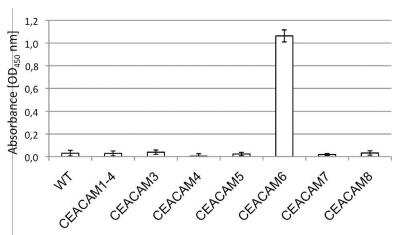
Full-length mouse and rat CEACAM-1 are approximately 57% aa identical to human CEACAM-1; in the V-type Ig-like domain, they are 58% and 56% aa identical, respectively. The full-length molecule is found on neutrophils, bile duct epithelium, activated NK cells, colonic columnar epithelium and endothelium. It is known to act as an intercellular adhesion molecule, forming both homotypic, and heterotypic bonds with CEA and CEACAM-6/NCA (3, 9). On neutrophils, CEACAM-1 also binds to dendritic cell CD-SIGN via its LeX moiety, inducing dendritic cell maturation and a subsequent Th1-type response (10,11).


The concentration calculator allows you to quickly calculate the volume, mass or concentration of your vial. Simply enter your mass, volume, or concentration values for your reagent and the calculator will determine the rest.


![Bioactivity CTLA-4 [Unconjugated]](https://images.novusbio.com/images/protein/CTLA4_7268CT_2293.jpg)


![Western Blot CEACAM7 Antibody [Unconjugated]](https://images.novusbio.com/images/antibody/CEACAM-7_AF4478_Western_Blot_5475.jpg)
![Immunohistochemistry CEACAM7 Antibody [Unconjugated]](https://images.novusbio.com/images/antibody/CEACAM7_AF4478_Immunohistochemistry_20616.jpg)


![N/A SLPI [HRP]](https://images.novusbio.com/images/elisa/SLPI_DPI00_ELISA_160.jpg)
![N/A SLPI [HRP]](https://images.novusbio.com/images/elisa/DATA_SLPI_DPI00_ELISA_792.jpg)
![N/A CCL27/CTACK [HRP]](https://images.novusbio.com/images/elisa/DATA_CCL27_DCC270_ELISA_615.jpg)
![N/A CCL27/CTACK [HRP]](https://images.novusbio.com/images/elisa/CCL27_DCC270_ELISA_54.jpg)


