Can't find what you are looking for? Use our Antibody Concierge Service & we will help you locate your antibody!
Or feel free to contact us for alternative products.
Datasheet
Reviews & Publications
Protocols & FAQs
Support & Research
IP3R1 Antibody (2B6) Summary
Description
Quality control test: Antibody Reactive Against Recombinant Protein.
Immunogen
ITPR1 (NP_002213, 2470 a.a. ~ 2577 a.a) partial recombinant protein with GST tag. MW of the GST tag alone is 26 KDa. EHTCETLLMCIVTVLSHGLRSGGGVGDVLRKPSKEEPLFAARVIYDLLFFFMVIIIVLNLIFGVIIDTFADLRSEKQKKEEILKTTCFICGLERDKFDNKTVTFEEHI
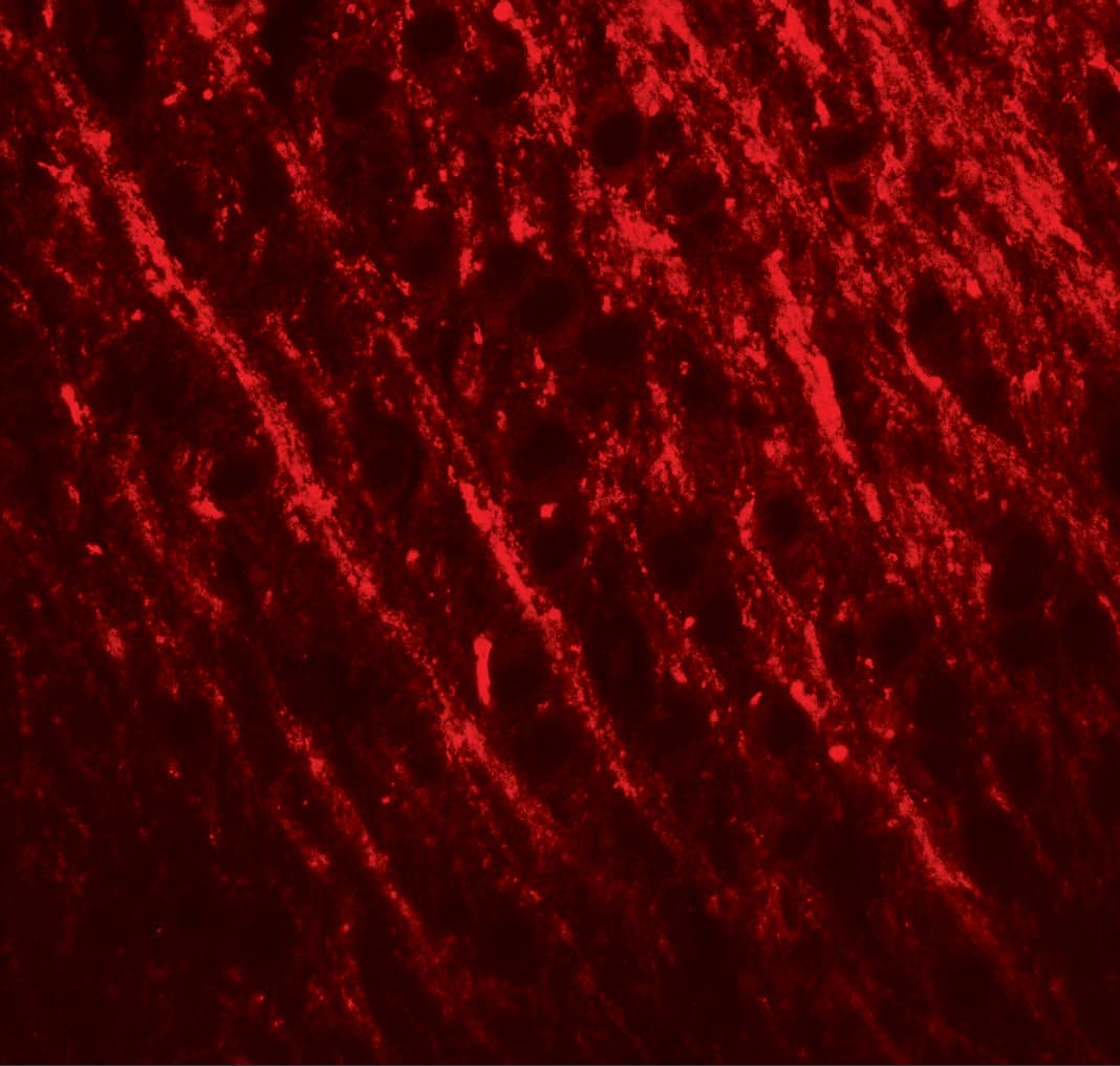
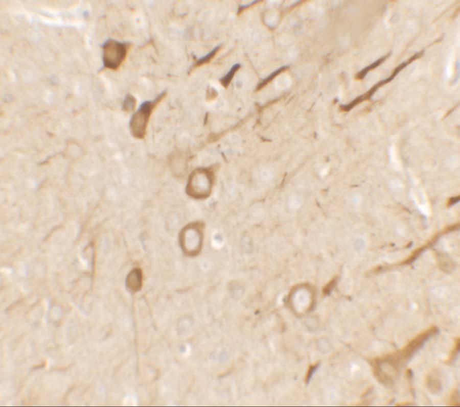
Specificity
ITPR1 - inositol 1,4,5-triphosphate receptor, type 1
Isotype
IgG2a Kappa
Clonality
Monoclonal
Host
Mouse
Gene
ITPR1
Purity
IgG purified
Innovator's Reward
Test in a species/application not listed above to receive a full credit towards a future purchase.
Porcine reactivity reported in scientific literature (PMID: 22946060).
Packaging, Storage & Formulations
Storage
Aliquot and store at -20C or -80C. Avoid freeze-thaw cycles.
Buffer
In 1x PBS, pH 7.4
Preservative
No Preservative
Purity
IgG purified
Notes
This product is produced by and distributed for Abnova, a company based in Taiwan.
Alternate Names for IP3R1 Antibody (2B6)
DKFZp313E1334
DKFZp313N1434
inositol 14,5-triphosphate receptor, type 1
inositol 14,5-trisphosphate receptor type 1
InsP3R1
IP3 receptor isoform 1
IP3 receptor
IP3R 1
IP3R
IP3R1
ITPR1
SCA15
SCA16
spinocerebellar ataxia 15
spinocerebellar ataxia 16
Type 1 inositol 14,5-trisphosphate receptor
Type 1 InsP3 receptor
Background
Inositol 1,4,5-trisphosphate (IP3) is a second messenger for many growth factors, hormones, and neurotransmitters. Upon binding to the IP3 receptor (IP3R), IP3 triggers the release of intracellular, luminal calcium to the cytosol. Functional IP3R is a homo- or heterotetramer of ~240 kDa glycoprotein subunits, that is structurally reminiscent of members of the voltage-gated ion channel superfamily. IP3R protein is structurally and functionally related to one other important intracellular calcium-release channel, the ryanodine receptor. The similarity between the two receptors continues at the physiological level owing to a physical association each receptor can have with the immunophilin protein, FKBP12. Cytosolic calcium levels appear to be regulated by a feedback loop that starts with calcium activation of protein kinase C (PKC) and calcineurin, a protein phosphatase. Phosphorylation of the IP3R by PKC causes an increase in IP3-mediated calcium release. Concomitantly, the phosphatase activity of calcineurin is stimulated upon its association with the FKBP12-IP3R complex. Calcium release is reduced when the PKC target site on the IP3R is dephosphorylated by calcineurin resulting in calcium oscillations. Mammalian IP3R subunits are the product of three distinct genes that are widely expressed and differentially regulated. IP3R type I (IP3R-I) has been detected in heart, liver, kidney, ovary, and Purkinje neurons of the cerebellum. IP3R-II is found predominantly in the brain. IP3R-III is known to be expressed in pancreatic islets, kidney, and the gastrointestinal tract. Inositol 1,4,5-trisphosphate (IP3) is a second messenger for many growth factors, hormones, and neuro-transmitters. Upon binding to the IP3 receptor (IP3R), IP3 triggers the release of intracellular, luminal calcium to the cytosol. Functional IP3R is a homo- or heterotetramer of ~240 kDa glycoprotein subunits, that is structurally reminiscent of members of the voltage-gated ion channel superfamily. IP3R protein is structurally and functionally related to one other important intracellular calcium-release channel, the ryanodine receptor. The similarity between the two receptors continues at the physiological level owing to a physical association each receptor can have with the immunophilin protein, FKBP12. Cytosolic calcium levels appear to be regulated by a feedback loop that starts with calcium activation of protein kinase C (PKC) and calcineurin, a protein phosphatase. Phosphorylation of the IP3R by PKC causes an increase in IP3-mediated calcium release. Concomitantly, the phosphatase activity of calcineurin is stimulated upon its association with the FKBP12-IP3R complex. Calcium release is reduced when the PKC target site on the IP3R is dephosphorylated by calcineurin resulting in calcium oscillations. Mammalian IP3R subunits are the product of three distinct genes that are widely expressed and differentially regulated. Type-I IP3R has been detected in heart, liver, kidney, ovary, and Purkinje neurons of the cerebellum. IP3R-II protein is found predominantly in the brain. Isoform III expression has been shown to occur in pancreatic islets, kidney and the gastrointestinal tract.
Limitations
This product is for research use only and is not approved for use in humans or in clinical diagnosis. Primary Antibodies are guaranteed for 1 year from date of receipt.
The concentration calculator allows you to quickly calculate the volume, mass or concentration of your vial. Simply enter your mass, volume, or concentration values for your reagent and the calculator will determine the rest.
=
÷
Review this Product
Be the first to review our IP3R1 Antibody (2B6) and receive a gift card or discount.