 HAF008). Specific bands were detected for GSK-3a/ beta at approximately 46 and 51 kDa (as indicated). This experiment was conducted under reducing conditions and using Immunoblot Buffer Group 1." title="Western blot shows lysates of HeLa human cervical epithelial carcinoma cell line, HT-29 human colon adenocarcinoma cell line, MDA-MB-468 and MCF-7 human breast cancer cell line. PVDF membrane was probed with 0.1 µg/mL of Rabbit Anti-Human/Mouse/ Rat GSK-3a/ beta Antigen Affinity-purified Polyclonal Antibody (Catalog # AF2157) followed by HRP-conjugated Anti-Rabbit IgG Secondary Antibody (Catalog # HAF008). Specific bands were detected for GSK-3a/ beta at approximately 46 and 51 kDa (as indicated). This experiment was conducted under reducing conditions and using Immunoblot Buffer Group 1." />
| Reactivity | Hu, Mu, RtSpecies Glossary |
| Applications | WB, CyTOF-ready, Flow, ICC/IF |
| Clonality | Polyclonal |
| Host | Rabbit |
| Conjugate | Unconjugated |
| Concentration | LYOPH |
| Immunogen | E. coli-derived recombinant human GSK-3 beta Accession # P49841 |
| Specificity | Detects human, mouse, and rat GSK-3 beta and the closely related GSK-3 alpha . |
| Source | N/A |
| Isotype | IgG |
| Clonality | Polyclonal |
| Host | Rabbit |
| Gene | GSK3A |
| Purity Statement | Antigen Affinity-purified |
| Innovator's Reward | Test in a species/application not listed above to receive a full credit towards a future purchase. |
| Dilutions |
|
|
| Publications |
|
| Storage | Use a manual defrost freezer and avoid repeated freeze-thaw cycles.
|
| Buffer | Lyophilized from a 0.2 μm filtered solution in PBS with Trehalose. *Small pack size (SP) is supplied either lyophilized or as a 0.2 µm filtered solution in PBS. |
| Preservative | No Preservative |
| Concentration | LYOPH |
| Reconstitution Instructions | Reconstitute at 0.2 mg/mL in sterile PBS. |
GSK-3 is a Ser/Thr kinase first identified as an inactivator of Glycogen Synthase. GSK-3 acts as a multifunctional downstream switch that determines the output of numerous signaling pathways. There are two mammalian GSK-3 isoforms encoded by distinct genes, GSK-3 alpha and GSK-3 beta, which are structurally similar, but functionally non-identical. GSK-3a is inhibited by phosphorylation at S21 by Akt and other kinases. GSK-3 alpha and GSK-3 beta share 85% amino acid identity. Dysregulated GSK-3 has been implicated in several diseases including type II diabetes, Alzheimer's disease, bipolar disorder, and cancer.


![Intracellular Staining by Flow Cytometry AKT [p Ser473] Antibody [Unconjugated] - Pan Specific](https://images.novusbio.com/images/antibody/Akt3_AF887_Flow_Cytometry_8283.jpg)
![Western Blot AKT [p Ser473] Antibody [Unconjugated] - Pan Specific](https://images.novusbio.com/images/af887_phospho-akt-s473-pan-specific-affinity-purified-pab-41202410485440.jpg)
![Western Blot AKT [p Ser473] Antibody [Unconjugated] - Pan Specific](https://images.novusbio.com/images/af887_phospho-akt-s473-pan-specific-affinity-purified-pab-8120255552843.jpg)
![Immunohistochemistry-Paraffin Tau [p Thr181] Antibody - BSA Free](https://images.novusbio.com/images/Tau-[p-Thr181]-Antibody-Immunohistochemistry-Paraffin-NB100-82245-img0005.jpg)
![Simple Western Tau [p Thr181] Antibody - BSA Free](https://images.novusbio.com/images/Tau-[p-Thr181]-Antibody-Simple-Western-NB100-82245-img0004.jpg)
![Western Blot Tau [p Thr181] Antibody - BSA Free](https://images.novusbio.com/images/Tau-[p-Thr181]-Antibody-Western-Blot-NB100-82245-img0001.jpg)


![Western Blot beta-Catenin Antibody [Unconjugated]](https://images.novusbio.com/images/af1329_human-mouse-rat-beta-catenin-affinity-purified-polyclonal-ab-western-blot-12122025922481.jpg)
![Western Blot beta-Catenin Antibody [Unconjugated]](https://images.novusbio.com/images/af2649_mouse-chitinase-3-like-1-affinity-purified-pab-western-blot-12122025848165.jpg)
![Western Blot beta-Catenin Antibody [Unconjugated]](https://images.novusbio.com/images/af1329_human-mouse-rat-beta-catenin-affinity-purified-polyclonal-ab-western-blot-121220258282812.jpg)



![GSK-3 alpha/beta Antibody [Unconjugated]](/sites/all/modules/enterprise-tech/et_datasheets/images/novus_guarantee.png "GSK-3 alpha/beta Antibody [Unconjugated]")
Secondary Antibodies |
Isotype Controls |
The concentration calculator allows you to quickly calculate the volume, mass or concentration of your vial. Simply enter your mass, volume, or concentration values for your reagent and the calculator will determine the rest.


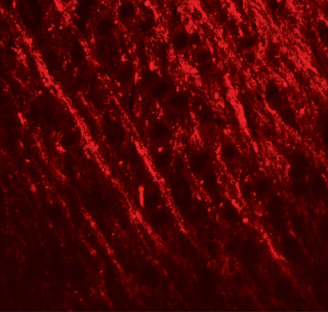
 NL004) and counterstained with DAPI (blue). Specific staining was localized to cytoplasm. View our protocol for
  AB-105-C, open histogram), followed by Allophycocyanin-conjugated Anti-Rabbit IgG Secondary Antibody (Catalog #

![Western Blot PYK2/FAK2 [p Tyr402] Antibody (592918) [Unconjugated]](https://images.novusbio.com/images/mab6210_human-phospho-pyk2-y402-mab-clone-592918-western-blot-1212202585785.jpg)
![Western Blot PYK2/FAK2 [p Tyr402] Antibody (592918) [Unconjugated]](https://images.novusbio.com/images/mab6210_human-phospho-pyk2-y402-mab-clone-592918-western-blot-121220259163819.jpg)
![Western Blot PYK2/FAK2 [p Tyr402] Antibody (592918) [Unconjugated]](https://images.novusbio.com/images/mab6210_human-phospho-pyk2-y402-mab-clone-592918-western-blot-1212202591401.jpg)


![Western Blot ERK2 Antibody [Unconjugated]](https://images.novusbio.com/images/antibody/ERK2_AF1230_Western_Blot_5097.jpg)
![Knockout Validated ERK2 Antibody [Unconjugated]](https://images.novusbio.com/images/antibody/ERK2_AF1230_Knockout_Validated_22864.jpg)
![Immunohistochemistry ERK2 Antibody [Unconjugated]](https://images.novusbio.com/images/antibody/ERK2_AF1230_Immunohistochemistry_20696.jpg)
![Immunohistochemistry TOR/mTOR [p Ser2448] Antibody - BSA Free](https://images.novusbio.com/images/nb600-607_rabbit-polyclonal-tor-mtor-p-ser2448-antibody-235202318143142.jpg)
![Western Blot TOR/mTOR [p Ser2448] Antibody - BSA Free](https://images.novusbio.com/images/nb600-607_rabbit-polyclonal-tor-mtor-p-ser2448-antibody-31020241535647.jpg)
![Data TOR/mTOR [p Ser2448] Antibody - BSA Free](https://images.novusbio.com/images/TOR-mTOR-[p-Ser2448]-Antibody-N-A-NB600-607-img0008.jpg)
![Immunocytochemistry/ Immunofluorescence c-jun [p Ser63] Antibody (SY0297)](https://images.novusbio.com/images/c-jun-p-Ser63-Antibody-SY0297-Immunocytochemistry-Immunofluorescence-NBP2-67471-img0006.jpg)
![Western Blot c-jun [p Ser63] Antibody (SY0297)](https://images.novusbio.com/images/c-jun-p-Ser63-Antibody-SY0297-Western-Blot-NBP2-67471-img0008.jpg)
![Western Blot c-jun [p Ser63] Antibody (SY0297)](https://images.novusbio.com/images/c-jun-p-Ser63-Antibody-SY0297-Western-Blot-NBP2-67471-img0009.jpg)
![Immunohistochemistry Insulin Antibody (182410) [Unconjugated]](https://images.novusbio.com/images/antibody/mab1417_human-bovine-mouse-insulin-mab-clone-182410-immunohistochemistry-308202115145.jpg)
![Immunocytochemistry Insulin Antibody (182410) [Unconjugated]](https://images.novusbio.com/images/antibody/Insulin_MAB1417_Immunocytochemistry_9376.jpg)


