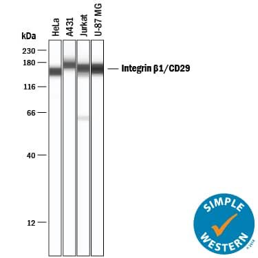
HAF017). Specific bands were detected for Integrin beta 1/CD29 at approximately 130-140 kDa (as indicated). This experiment was conducted under reducing conditions and using
Immunoblot Buffer Group 1." class="big_lightbox">
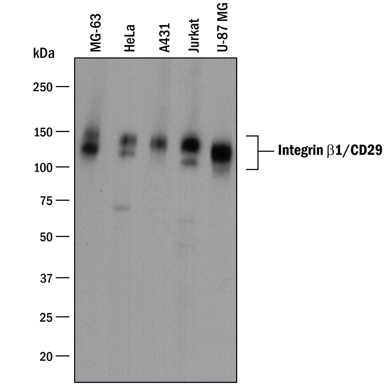
HAF017). Specific bands were detected for Integrin beta 1/CD29 at approximately 130-140 kDa (as indicated). This experiment was conducted under reducing conditions and using
Immunoblot Buffer Group 1." alt="Western blot shows lysates of MG-63 human osteosarcoma cell line, HeLa human cervical epithelial carcinoma cell line, A431 human epithelial carcinoma cell line, Jurkat human acute T cell leukemia cell line, and U-87 MG human glioblastoma/astrocytoma cell line. PVDF membrane was probed with 1 µg/mL of Goat Anti-Human/Canine Integrin beta 1/CD29 Antigen Affinity-purified Polyclonal Antibody (Catalog # AF1778) followed by HRP-conjugated Anti-Goat IgG Secondary Antibody (Catalog #
HAF017). Specific bands were detected for Integrin beta 1/CD29 at approximately 130-140 kDa (as indicated). This experiment was conducted under reducing conditions and using
Immunoblot Buffer Group 1." class="big_thumb" />

AB-108-C, open histogram), followed by Phycoerythrin-conjugated Anti-Goat IgG Secondary Antibody (Catalog #
F0107)." class="big_lightbox">
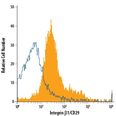
AB-108-C, open histogram), followed by Phycoerythrin-conjugated Anti-Goat IgG Secondary Antibody (Catalog #
F0107)." class="big_thumb" />
AB-108-C, open histogram), followed by Phycoerythrin-conjugated Anti-Goat IgG Secondary Antibody (Catalog #
F0107)." class="big_lightbox">
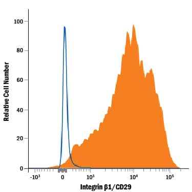
AB-108-C, open histogram), followed by Phycoerythrin-conjugated Anti-Goat IgG Secondary Antibody (Catalog #
F0107)." class="big_thumb" />
AB-108-C, open histogram), followed by Phycoerythrin-conjugated Anti-Goat IgG Secondary Antibody (Catalog #
F0107)." class="big_lightbox">
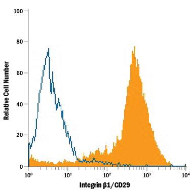
AB-108-C, open histogram), followed by Phycoerythrin-conjugated Anti-Goat IgG Secondary Antibody (Catalog #
F0107)." class="big_thumb" />
HAF109). This experiment was conducted under reducing conditions and using the 12-230 kDa separation system." class="big_lightbox">
HAF109). This experiment was conducted under reducing conditions and using the 12-230 kDa separation system." class="big_thumb" />
NL001) and counterstained with DAPI (blue). Specific staining was localized to cytoplasm. Staining was performed using our protocol for Fluorescent ICC Staining of Non-adherent Cells." class="big_lightbox">
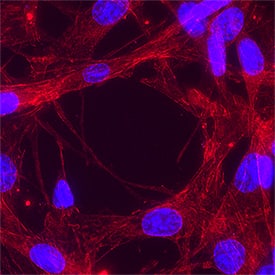
NL001) and counterstained with DAPI (blue). Specific staining was localized to cytoplasm. Staining was performed using our protocol for Fluorescent ICC Staining of Non-adherent Cells." class="big_thumb" />
HAF017). Specific bands were detected for Integrin beta 1/CD29 at approximately 105 kDa and 120 kDa (as indicated) in the parental A549 cell line, but is not detectable in knockout A549 cell line. GAPDH (Catalog #
MAB5718) is shown as a loading control. This experiment was conducted under reducing conditions and using
Immunoblot Buffer Group 1." class="big_lightbox">
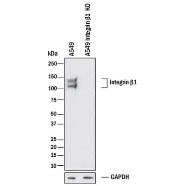
HAF017). Specific bands were detected for Integrin beta 1/CD29 at approximately 105 kDa and 120 kDa (as indicated) in the parental A549 cell line, but is not detectable in knockout A549 cell line. GAPDH (Catalog #
MAB5718) is shown as a loading control. This experiment was conducted under reducing conditions and using
Immunoblot Buffer Group 1." class="big_thumb" />
 MAB002, open histogram), followed by Phycoerythrin-conjugated Anti-Mouse IgG Secondary Antibody (Catalog # F0102B)." alt="Human peripheral blood mononuclear cells (PBMCs) were stained with Mouse Anti-Human Integrin beta 1/CD29 Monoclonal Antibody (Catalog # MAB17781, filled histogram) or isotype control antibody (Catalog # MAB002, open histogram), followed by Phycoerythrin-conjugated Anti-Mouse IgG Secondary Antibody (Catalog # F0102B)." class="big_thumb" />
 HAF017). A specific band was detected for Integrin beta 1/CD29 at approximately 110-140 kDa (as indicated). This experiment was conducted under reducing conditions and using Immunoblot Buffer Group 1." alt="Western blot shows lysates of L-929 mouse fibroblast cell line, NIH-3T3 mouse embryonic fibroblast cell line, PC-3 human prostate cancer cell line, Rat-2 rat embryonic fibroblast cell line, and RAW 264.7 mouse monocyte/macrophage cell line. PVDF membrane was probed with 1 µg/mL of Goat Anti-Mouse/Rat Integrin beta 1/CD29 Antigen Affinity-purified Polyclonal Antibody (Catalog # AF2405) followed by HRP-conjugated Anti-Goat IgG Secondary Antibody (Catalog # HAF017). A specific band was detected for Integrin beta 1/CD29 at approximately 110-140 kDa (as indicated). This experiment was conducted under reducing conditions and using Immunoblot Buffer Group 1." class="big_thumb" />
 HAF017). Specific bands were detected for Integrin beta 1/CD29 at approximately 130-140 kDa (as indicated). This experiment was conducted under reducing conditions and using Immunoblot Buffer Group 1." alt="Western blot shows lysates of MG-63 human osteosarcoma cell line, HeLa human cervical epithelial carcinoma cell line, A431 human epithelial carcinoma cell line, Jurkat human acute T cell leukemia cell line, and U-87 MG human glioblastoma/astrocytoma cell line. PVDF membrane was probed with 1 µg/mL of Goat Anti-Human/Canine Integrin beta 1/CD29 Antigen Affinity-purified Polyclonal Antibody (Catalog # AF1778) followed by HRP-conjugated Anti-Goat IgG Secondary Antibody (Catalog # HAF017). Specific bands were detected for Integrin beta 1/CD29 at approximately 130-140 kDa (as indicated). This experiment was conducted under reducing conditions and using Immunoblot Buffer Group 1." class="big_thumb" />
 MAB002, open histogram), followed by Phycoerythrin-conjugated Anti-Mouse IgG Secondary Antibody (F0102B)." alt="Human peripheral blood mononuclear cells (PBMCs) were stained with Mouse Anti-Human Integrin beta 1/CD29 Monoclonal Antibody (Catalog # MAB1778, filled histogram) or isotype control antibody (MAB002, open histogram), followed by Phycoerythrin-conjugated Anti-Mouse IgG Secondary Antibody (F0102B)." class="big_thumb" />
 MAB006, open histogram), followed by Phycoerythrin-conjugated Anti-Rat IgG Secondary Antibody (Catalog # F0105B)." alt="B16-F1 mouse melanoma cell line was stained with Rat Anti-Mouse Integrin beta 1/CD29 Monoclonal Antibody (Catalog # MAB2405, filled histogram) or isotype control antibody (Catalog # MAB006, open histogram), followed by Phycoerythrin-conjugated Anti-Rat IgG Secondary Antibody (Catalog # F0105B)." class="big_thumb" />
 MAB004), open histogram), followed by Phycoerythrin-conjugated Anti-Mouse IgG Secondary Antibody (F0102B)." alt="Human peripheral blood mononuclear cells (PBMCs) were stained with Mouse Anti-Human/Porcine/Equine Integrin beta 1/CD29 Monoclonal Antibody (Catalog # MAB17783, filled histogram) or isotype control antibody (MAB004), open histogram), followed by Phycoerythrin-conjugated Anti-Mouse IgG Secondary Antibody (F0102B)." class="big_thumb" />


 MAB002, open histogram), followed by Phycoerythrin-conjugated Anti-Mouse IgG Secondary Antibody (Catalog #
  NL007) and counterstained with DAPI (blue). Specific staining was localized to plasma membrane. View our protocol for
  MAB002, open histogram) followed by anti-Mouse IgG PE-conjugated secondary antibody (Catalog#
![Immunocytochemistry/Immunofluorescence: Integrin beta 1/CD29 Antibody (P4C10) [NBP2-36561] - Integrin beta 1 (P4C10) antibody was tested in -20 degree MeOH fixed HeLa cells at a 1:200 dilution against DyLight 488 (green). Actin and nuclei were counterstained against Phalloidin 568 (red) and DAPI (blue), respectively.](http://images.novusbio.com/fullsize/Integrin-beta-1-CD29-Antibody-P4C10-Immunocytochemistry-Immunofluorescence-NBP2-36561-img0001.jpg "Immunocytochemistry/Immunofluorescence: Integrin beta 1/CD29 Antibody (P4C10) [NBP2-36561] - Integrin beta 1 (P4C10) antibody was tested in -20 degree MeOH fixed HeLa cells at a 1:200 dilution against DyLight 488 (green). Actin and nuclei were counterstained against Phalloidin 568 (red) and DAPI (blue), respectively.")
![Flow Cytometry: Integrin beta 1/CD29 Antibody (P4C10) [NBP2-36561] - An intracellular stain was performed on A431 cells with Integrin beta 1/CD29 [P4C10] Antibody NBP2-36561B (blue) and a matched isotype control (orange). Both antibodies were conjugated to Biotin. Cells were fixed with 4% PFA and then permeabilized with 0.1% saponin. Cells were incubated in an antibody dilution of 2.5 ug/mL for 30 minutes at room temperature, followed by Streptavidin - R-Phycoerythrin Protein (2012-1000, Novus Biologicals).](http://images.novusbio.com/fullsize/Integrin-beta-1-CD29-Antibody-P4C10-Flow-Cytometry-NBP2-36561-img0005.jpg)
![Flow Cytometry: Integrin beta 1/CD29 Antibody (P4C10) [NBP2-36561]](http://images.novusbio.com/fullsize/Integrin-beta-1-CD29-Antibody-P4C10-Flow-Cytometry-NBP2-36561-img0002.jpg)
![Flow Cytometry: Integrin beta 1/CD29 Antibody (P4C10) [NBP2-36561] - A surface stain was performed on HeLa cells with Integrin beta 1/CD29 [P4C10] Antibody NBP2-36561PE (blue) and a matched isotype control (orange). Cells were incubated in an antibody dilution of 2.5 ug/mL for 20 minutes at room temperature. Both antibodies were conjugated to Phycoerythrin.](http://images.novusbio.com/fullsize/Integrin-beta-1-CD29-Antibody-P4C10-Flow-Cytometry-NBP2-36561-img0003.jpg)
![Flow Cytometry: Integrin beta 1/CD29 Antibody (P4C10) [NBP2-36561] - A surface stain was performed on HeLa cells with Integrin beta 1/CD29 [P4C10] Antibody NBP2-36561AF488 (blue) and a matched isotype control (orange). Cells were incubated in an antibody dilution of 5 ug/mL for 20 minutes at room temperature. Both antibodies were conjugated to Alexa Fluor 488.](http://images.novusbio.com/fullsize/Integrin-beta-1-CD29-Antibody-P4C10-Flow-Cytometry-NBP2-36561-img0004.jpg)
![Immunocytochemistry/Immunofluorescence: Integrin beta 1/CD29 Antibody (P4C10) [NBP2-36561] - Overnight incubation with 1:50 Integrin beta 1/CD29 primary antibody in PBS/BSA 1%, at 4ºC. Green: Integrin beta 1/CD29 Antibody (P4C10), Red: Phalloidin, Blue: DAPI. Image from verified customer review.](http://images.novusbio.com/fullsize/antibody/nbp2-36561_mouse-monoclonal-integrin-beta-1-cd29-antibody-p4c10-immunocytochemistry-immunofluorescence-1462023134549..jpg)

 MAB734), Human/Mouse/Rat p38a Antigen Affinity-purified Polyclonal Antibody (green; Catalog #
  HAF109). This experiment was conducted under reducing conditions and using the 12-230 kDa separation system." class="big_thumb" />

 AB-108-C, open histogram), followed by Phycoerythrin-conjugated Anti-Goat IgG Secondary Antibody (Catalog #
  AB-108-C, open histogram), followed by Phycoerythrin-conjugated Anti-Goat IgG Secondary Antibody (Catalog #
  AB-108-C, open histogram), followed by Phycoerythrin-conjugated Anti-Goat IgG Secondary Antibody (Catalog #
  NL001) and counterstained with DAPI (blue). Specific staining was localized to cell surfaces. View our protocol for
  NL001) and counterstained with DAPI (blue). Specific staining was localized to cell surfaces. View our protocol for
  HAF109). This experiment was conducted under reducing conditions and using the 12-230 kDa separation system." class="big_thumb" />
 NL001) and counterstained with DAPI (blue). Specific staining was localized to cytoplasm. Staining was performed using our protocol for Fluorescent ICC Staining of Non-adherent Cells." class="big_thumb" />
 HAF017). Specific bands were detected for Integrin beta 1/CD29 at approximately 105 kDa and 120 kDa (as indicated) in the parental A549 cell line, but is not detectable in knockout A549 cell line. GAPDH (Catalog #
  NL007) and counter-stained with DAPI (blue). View our protocol for
  NL007) and counterstained with DAPI (blue). Specific staining was localized to plasma membrane." class="big_thumb" />
![Immunocytochemistry/Immunofluorescence: Integrin beta 1/CD29 Antibody [NBP2-16974] - Analysis of methanol-fixed HeLa, using antibody at 1:500 dilution.](http://images.novusbio.com/fullsize/Integrin-beta-1-Antibody-Immunocytochemistry-Immunofluorescence-NBP2-16974-img0003.jpg "Immunocytochemistry/Immunofluorescence: Integrin beta 1/CD29 Antibody [NBP2-16974] - Analysis of methanol-fixed HeLa, using antibody at 1:500 dilution.")
![Immunohistochemistry-Paraffin: Integrin beta 1/CD29 Antibody [NBP2-16974] - Human colon carcinoma, using Integrin beta 1 / CD29 antibody at 1:500 dilution. Antigen Retrieval: Trilogy™ (EDTA based, pH 8.0) buffer, 15minof paraffin-embedded Colon ca, using antibody at 1:500 dilution.](http://images.novusbio.com/fullsize/Integrin-beta-1-Antibody-Immunohistochemistry-Paraffin-NBP2-16974-img0002.jpg)
![Western Blot: Integrin beta 1/CD29 Antibody [NBP2-16974] - Whole cell extract (30 ug) was separated by 7.5% SDS-PAGE, and the membrane was blotted with Integrin beta 1/CD29 antibody (NBP2-16974) diluted at 1:5000. The HRP-conjugated anti-rabbit IgG antibody was used to detect the primary antibody, and the signal was developed with Trident ECL plus-Enhanced.](http://images.novusbio.com/fullsize/nbp2-16974_rabbit-polyclonal-integrin-beta-1-cd29-antibody-91020242011496.jpg)
![<b>Genetic Strategies Validation. </b>Western Blot: Integrin beta 1/CD29 Antibody [NBP2-16974] - Non-transfected (–) and transfected (+) HeLa whole cell extracts (15 ug) were separated by 7.5% SDS-PAGE, and the membrane was blotted with Integrin beta 1 / CD29 antibody diluted at 1:5000. The HRP-conjugated anti-rabbit IgG antibody was used to detect the primary antibody.](http://images.novusbio.com/fullsize/nbp2-16974_rabbit-polyclonal-integrin-beta-1-cd29-antibody-1610202419414566.jpg)


 NL013) and counterstained with DAPI (blue). View our protocol for
 ![Western Blot: Integrin beta 1/CD29 Antibody (MEM-101A) [NB500-383] - Western Blotting analysis (non-reducing conditions) of isolated peripheral blood lymphocytes of various species using anti-Integrin beta 1 Antibody (MEM-101A). Lane 1: lysate of human PBL Lane 2: lysate of canine PBL Lane 3: lysate of porcine PBL.](http://images.novusbio.com/fullsize/Integrin-beta-1-CD29-Antibody-MEM-101A-Western-Blot-NB500-383-img0003.jpg "Western Blot: Integrin beta 1/CD29 Antibody (MEM-101A) [NB500-383] - Western Blotting analysis (non-reducing conditions) of isolated peripheral blood lymphocytes of various species using anti-Integrin beta 1 Antibody (MEM-101A). Lane 1: lysate of human PBL Lane 2: lysate of canine PBL Lane 3: lysate of porcine PBL.")
![Flow Cytometry: Integrin beta 1/CD29 Antibody (MEM-101A) [NB500-383] - Staining of human peripheral blood with anti-human CD29 (MEM-101A) PE.](http://images.novusbio.com/fullsize/Integrin-beta-1-CD29-Antibody-MEM-101A-Flow-Cytometry-NB500-383-img0004.jpg)
![Flow Cytometry: Integrin beta 1/CD29 Antibody (MEM-101A) [NB500-383] - Surface staining of human peripheral blood with anti-human CD29 (MEM-101A) APC conjugated.](http://images.novusbio.com/fullsize/Integrin-beta-1-CD29-Antibody-MEM-101A-Flow-Cytometry-NB500-383-img0002.jpg)
![Western Blot: Integrin beta 1/CD29 Antibody (3B6) [NBP2-22191] - Western blot analysis using Integrin beta 1 mouse mAb against Hela (1), HepG2 (2), A549 (3), Jurkat(4), L1210 (5) and Cos7 (6) cell lysate.](http://images.novusbio.com/fullsize/Integrin-beta-1-CD29-Antibody-3B6-Western-Blot-NBP2-22191-img0006.jpg "Western Blot: Integrin beta 1/CD29 Antibody (3B6) [NBP2-22191] - Western blot analysis using Integrin beta 1 mouse mAb against Hela (1), HepG2 (2), A549 (3), Jurkat(4), L1210 (5) and Cos7 (6) cell lysate.")
![Immunohistochemistry-Paraffin: Integrin beta 1/CD29 Antibody (3B6) [NBP2-22191] - Immunohistochemical analysis of paraffin-embedded ovarian cancer tissues using Integrin beta 1 mouse mAb with DAB staining.](http://images.novusbio.com/fullsize/Integrin-beta-1-CD29-Antibody-3B6-Immunohistochemistry-Paraffin-NBP2-22191-img0003.jpg)
![Flow Cytometry: Integrin beta 1/CD29 Antibody (3B6) [NBP2-22191] - Flow cytometric analysis of Hela cells using Integrin beta 1 mouse mAb (green) and negative control (red).](http://images.novusbio.com/fullsize/Integrin-beta-1-CD29-Antibody-3B6-Flow-Cytometry-NBP2-22191-img0001.jpg)
![Western Blot: Integrin beta 1/CD29 Antibody (3B6) [NBP2-22191] - Western blot analysis using Integrin beta 1 mAb against human ITGB1 (AA: 50-270) recombinant protein. (Expected MW is 50.6 kDa)](http://images.novusbio.com/fullsize/Integrin-beta-1-CD29-Antibody-3B6-Western-Blot-NBP2-22191-img0005.jpg)
![Immunohistochemistry-Paraffin: Integrin beta 1/CD29 Antibody (3B6) [NBP2-22191] - Immunohistochemical analysis of paraffin-embedded cervical cancer tissues using Integrin beta 1 mouse mAb with DAB staining.](http://images.novusbio.com/fullsize/Integrin-beta-1-CD29-Antibody-3B6-Immunohistochemistry-Paraffin-NBP2-22191-img0002.jpg)
![ELISA: Integrin beta 1/CD29 Antibody (3B6) [NBP2-22191] - Red: Control Antigen (100ng); Purple: Antigen (10ng); Green: Antigen (50ng); Blue: Antigen (100ng)](http://images.novusbio.com/fullsize/Integrin-beta-1-CD29-Antibody-3B6-ELISA-NBP2-22191-img0004.jpg)
![CyTOF-ready: Integrin beta 1/CD29 Antibody (3B6) [NBP2-22191] - Analysis of Integrin beta 1/CD29 antibody on human tissue. Integrin beta 1/CD29 (green). Scale, 50um. Image from verified customer review.](http://images.novusbio.com/fullsize/Integrin-beta-1-CD29-Antibody-3B6-CyTOF-ready-NBP2-22191-img0007.jpg)

![<b>Genetic Strategies Validation. </b>Flow Cytometry: Integrin beta 1/CD29 Antibody (12G10) [NB100-63255] - Flow Cytometry: Integrin beta 1/CD29 Antibody (12G10) [Alexa Fluor 488] [NB100-63255AF488] - A cell surface stain was performed on A549 cells with Integrin beta 1/CD29 (12G10) antibody NB100-63255AF488 (blue) and a matched isotype control NBP1-97005AF488 (orange). Cells were incubated in an antibody dilution of 5 ug/mL for 20 minutes at room temperature. Both antibodies were conjugated to Alexa Fluor 488. Image using the Alexa Fluor 488 form of this antibody.](http://images.novusbio.com/fullsize/Integrin-beta-1-CD29-Antibody-12G10-Flow-Cytometry-NB100-63255-img0003.jpg)
![Immunocytochemistry/Immunofluorescence: Integrin beta 1/CD29 Antibody (12G10) [NB100-63255] - Human salivary gland progenitor cells express integrin beta 1 by day 1 in three-dimensional hyaluronan-based hydrogels. Image from verified customer review.](http://images.novusbio.com/fullsize/Integrin-beta-1-CD29-Antibody-12G10-Immunocytochemistry-Immunofluorescence-NB100-63255-img0004.jpg)
![Flow Cytometry: Integrin beta 1/CD29 Antibody (12G10) [NB100-63255] - Staining of human peripheral blood monocytes with MOUSE ANTI HUMAN CD29.](http://images.novusbio.com/fullsize/Integrin-beta-1-CD29-Antibody-12G10-Flow-Cytometry-NB100-63255-img0002.jpg)

 MAB004, open histogram), followed by PE-conjugated Anti-Mouse IgG Secondary Antibody (Catalog #
  MAB004, open histogram), followed by PE-conjugated Anti-Mouse IgG Secondary Antibody (Catalog #
  NL007) and counterstained with DAPI (blue). Specific staining was localized to cell surfaces. View our protocol for
  NL007) and counterstained with DAPI (blue). Specific staining was localized to plasma membrane." class="big_thumb" />
![Flow Cytometry: Integrin beta 1/CD29 Antibody (K20) [NBP2-52708] - Flow-cytometry using anti-CD29 antibody K20 (NBP2-52708) Human leukocytes were stained with an isotype control (panel A) or the rabbit-chimeric version of K20 (panel B) at a concentration of 1 ug/ml for 30 mins at RT. After washing, bound antibody was detected using a AF488 conjugated donkey anti-rabbit antibody and cells analysed on a FACSCanto flow-cytometer.](http://images.novusbio.com/fullsize/Integrin-beta-1-CD29-Antibody-K20-NBP2-52708-img0001.jpg "Flow Cytometry: Integrin beta 1/CD29 Antibody (K20) [NBP2-52708] - Flow-cytometry using anti-CD29 antibody K20 (NBP2-52708) Human leukocytes were stained with an isotype control (panel A) or the rabbit-chimeric version of K20 (panel B) at a concentration of 1 ug/ml for 30 mins at RT. After washing, bound antibody was detected using a AF488 conjugated donkey anti-rabbit antibody and cells analysed on a FACSCanto flow-cytometer.")


