HAF016). A specific band was detected for Fibroblast Activation Protein a/FAP at approximately 97 kDa (as indicated). This experiment was conducted under reducing conditions and using
Immunoblot Buffer Group 1." class="big_lightbox">
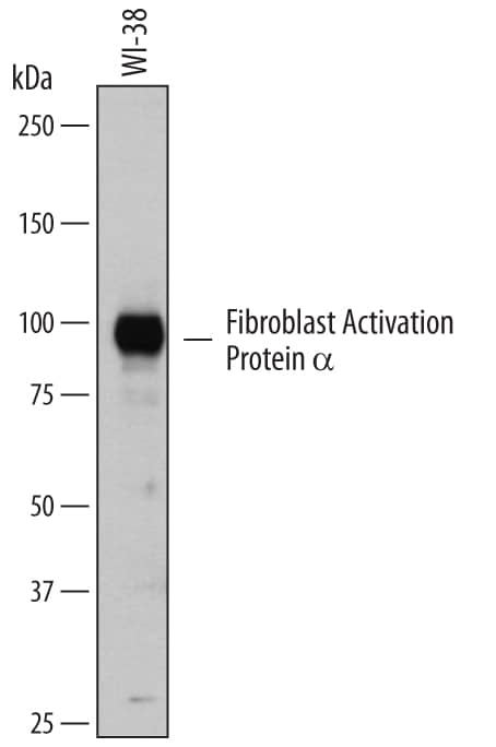
HAF016). A specific band was detected for Fibroblast Activation Protein a/FAP at approximately 97 kDa (as indicated). This experiment was conducted under reducing conditions and using
Immunoblot Buffer Group 1." alt="Western blot shows lysates of WI-38 human lung fibroblast cell line. PVDF membrane was probed with 0.5 µg/mL of Sheep Anti-Human Fibroblast Activation Protein a/FAP Antigen Affinity-purified Polyclonal Antibody (Catalog # AF3715) followed by HRP-conjugated Anti-Sheep IgG Secondary Antibody (Catalog #
HAF016). A specific band was detected for Fibroblast Activation Protein a/FAP at approximately 97 kDa (as indicated). This experiment was conducted under reducing conditions and using
Immunoblot Buffer Group 1." class="big_thumb" />
HAF016). This experiment was conducted under reducing conditions and using the 12-230 kDa separation system." class="big_lightbox">
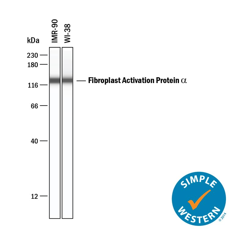
HAF016). This experiment was conducted under reducing conditions and using the 12-230 kDa separation system." class="big_thumb" />
HAF016). A specific band was detected for Fibroblast Activation Protein alpha /FAP at approximately 97 kDa (as indicated) in the parental WI‑38 human lung fibroblast cell line, but is not detectable in knockout WI‑38 human lung fibroblast cell line. GAPDH (
AF5718) is shown as a loading control. This experiment was conducted under reducing conditions and using Western Blot Buffer Group 1." class="big_lightbox">
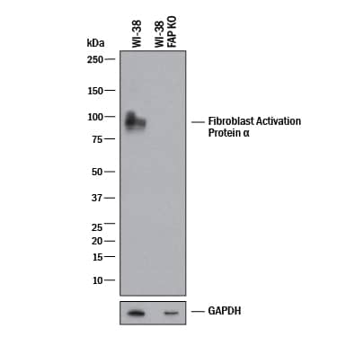
HAF016). A specific band was detected for Fibroblast Activation Protein alpha /FAP at approximately 97 kDa (as indicated) in the parental WI‑38 human lung fibroblast cell line, but is not detectable in knockout WI‑38 human lung fibroblast cell line. GAPDH (
AF5718) is shown as a loading control. This experiment was conducted under reducing conditions and using Western Blot Buffer Group 1." class="big_thumb" />
AF3715) at 5ug/mL with overnight incubation at 4 degrees Celsius followed by incubation with anti-sheep IgG VisUCyte HRP Polymer Antibody (Catalog #
VC006) and DAB chromogen (yellow-brown). Tissue was counterstained with hematoxylin (blue). Specific staining was localized to connective tissue. " class="big_lightbox">

AF3715) at 5ug/mL with overnight incubation at 4 degrees Celsius followed by incubation with anti-sheep IgG VisUCyte HRP Polymer Antibody (Catalog #
VC006) and DAB chromogen (yellow-brown). Tissue was counterstained with hematoxylin (blue). Specific staining was localized to connective tissue. " class="big_thumb" />
3715-SE) was serially diluted and captured by Mouse Anti-Human Fibroblast Activation Protein alpha /FAP Monoclonal Antibody (Catalog #
MAB3715) coated on a Clear Polystyrene Microplate (Catalog #
DY990). Sheep Anti-Human Fibroblast Activation Protein alpha /FAP Antigen Affinity-purified Polyclonal Antibody (Catalog # AF3715) was biotinylated and incubated with the protein captured on the plate. Detection of the standard curve was achieved by incubating Streptavidin-HRP (Catalog #
DY998)" class="big_lightbox">

3715-SE) was serially diluted and captured by Mouse Anti-Human Fibroblast Activation Protein alpha /FAP Monoclonal Antibody (Catalog #
MAB3715) coated on a Clear Polystyrene Microplate (Catalog #
DY990). Sheep Anti-Human Fibroblast Activation Protein alpha /FAP Antigen Affinity-purified Polyclonal Antibody (Catalog # AF3715) was biotinylated and incubated with the protein captured on the plate. Detection of the standard curve was achieved by incubating Streptavidin-HRP (Catalog #
DY998)" class="big_thumb" />
 HAF016). A specific band was detected for Fibroblast Activation Protein a/FAP at approximately 97 kDa (as indicated). This experiment was conducted under reducing conditions and using Immunoblot Buffer Group 1." alt="Western blot shows lysates of WI-38 human lung fibroblast cell line. PVDF membrane was probed with 0.5 µg/mL of Sheep Anti-Human Fibroblast Activation Protein a/FAP Antigen Affinity-purified Polyclonal Antibody (Catalog # AF3715) followed by HRP-conjugated Anti-Sheep IgG Secondary Antibody (Catalog # HAF016). A specific band was detected for Fibroblast Activation Protein a/FAP at approximately 97 kDa (as indicated). This experiment was conducted under reducing conditions and using Immunoblot Buffer Group 1." class="big_thumb" />
 MAB002, open histogram), followed by Phycoerythrin-conjugated Anti-Mouse IgG Secondary Antibody (Catalog # F0102B). View our protocol for Staining Membrane-associated Proteins." alt="WI-38 human lung fibroblast cell line was stained with Mouse Anti-Human Fibroblast Activation Protein a/FAP Monoclonal Antibody (Catalog # MAB3715, filled histogram) or isotype control antibody (Catalog # MAB002, open histogram), followed by Phycoerythrin-conjugated Anti-Mouse IgG Secondary Antibody (Catalog # F0102B). View our protocol for Staining Membrane-associated Proteins." class="big_thumb" />
 DR647RB) and counterstained with DAPI (blue; Lunaphore Catalog # DR100). Specific staining was localized to the membrane and cytoplasm of fibroblasts. Protocol available in COMET™ Panel Builder." alt="FAP was detected in immersion fixed paraffin-embedded sections of human Melanoma using Rabbit Anti-Human FAP, Monoclonal Antibody (Catalog# NBP3-14728) at 1:100 dilution at 37°Celsius for 4 minutes. Before incubation with the primary antibody, tissue underwent an all-in-one dewaxing and antigen retrieval preprocessing using PreTreatment Module (PT Module) and Dewax and HIER Buffer H (pH 9; Epredia Catalog # TA-999-DHBH). Tissue was stained using the Alexa Fluor™ Plus 647 Goat anti-Rabbit IgG Secondary Antibody at 1:200 at 37 ° Celsius for 2 minutes. (Yellow; Lunaphore Catalog # DR647RB) and counterstained with DAPI (blue; Lunaphore Catalog # DR100). Specific staining was localized to the membrane and cytoplasm of fibroblasts. Protocol available in COMET™ Panel Builder." class="big_thumb" />
 MAB005, open histogram), followed by Phycoerythrin-conjugated Anti-Rat IgG Secondary Antibody (Catalog # F0105B). View our protocol for Staining Membrane-associated Proteins." alt="C2C12 mouse myoblast cell line was stained with Rat Anti-Mouse Fibroblast Activation Protein alpha/FAP Monoclonal Antibody (Catalog # MAB9727, filled histogram) or isotype control antibody (Catalog # MAB005, open histogram), followed by Phycoerythrin-conjugated Anti-Rat IgG Secondary Antibody (Catalog # F0105B). View our protocol for Staining Membrane-associated Proteins." class="big_thumb" />


 CTS019) and counterstained with hematoxylin (blue). Specific staining was localized to connective tissue. View our protocol for
  CTS019) and counterstained with hematoxylin (blue). Specific staining was localized to cytoplasm. View our protocol for
  HAF016). This experiment was conducted under reducing conditions and using the 12-230 kDa separation system." class="big_thumb" />
 HAF016). A specific band was detected for Fibroblast Activation Protein alpha /FAP at approximately 97 kDa (as indicated) in the parental WI‑38 human lung fibroblast cell line, but is not detectable in knockout WI‑38 human lung fibroblast cell line. GAPDH (
 AF3715) at 5ug/mL with overnight incubation at 4 degrees Celsius followed by incubation with anti-sheep IgG VisUCyte HRP Polymer Antibody (Catalog #


 3715-SE) was serially diluted and captured by Mouse Anti-Human Fibroblast Activation Protein alpha /FAP Monoclonal Antibody (Catalog #
 ![N/A Fibroblast Activation Protein alpha/FAP [HRP]](http://images.novusbio.com/fullsize/elisa/DATA_Proprotein_Convertase_9_DPC900_ELISA_786.jpg "N/A Fibroblast Activation Protein alpha/FAP [HRP]")
![N/A Fibroblast Activation Protein alpha/FAP [HRP]](http://images.novusbio.com/fullsize/elisa/Proprotein_Convertase_9_DPC900_ELISA_157.jpg)
 MAB002, open histogram), followed by Fluorescein-conjugated Anti-Mouse IgG Secondary Antibody (Catalog #


 3715-SE) was serially diluted and captured by Mouse Anti-Human Fibroblast Activation Protein alpha /FAP Monoclonal Antibody (Catalog # MAB3715) coated on a Clear Polystyrene Microplate (Catalog #

 DR647RB) and counterstained with DAPI (blue; Lunaphore Catalog #
  DR647RB) and counterstained with DAPI (blue; Lunaphore Catalog #
 ![Immunoprecipitation: Fibroblast Activation Protein alpha/FAP Antibody [NBP3-14728] - Whole cell lysate (500 ug per IP reaction; 5% of IPloaded) from IMR-90 cells. Antibody: Rabbit anti-FAPrecombinant monoclonal antibody used for IP at 20 ul/mg lysate. For blottingimmunoprecipitated FAP, was used at 1:1000.Chemiluminescence with an exposure time of 30 seconds.](http://images.novusbio.com/fullsize/Fibroblast-Activation-Protein-alpha-FAP-Antibody-Immunoprecipitation-NBP3-14728-img0001.jpg)
![Western Blot-Fibroblast Activation Protein alpha/FAP Antibody [NBP3-14728] -Whole cell lysate (50 ug) from SNB-75, HeLa, IMR-90, HEK293T, and U2OS cells prepared using NETN lysis buffer. Antibody: Rabbit anti FAP recombinant monoclonal antibody [BLR150J] used at 1:1000. Secondary: HRP conjugated goat anti-rabbit IgG. Chemiluminescence with an exposure time of 30 seconds. Lower Panel: Rabbit anti-COPB2 antibody.](http://images.novusbio.com/fullsize/nbp3-14728_rabbit-fibroblast-activation-protein-alpha-fap-mab-western-blot-22320249348..jpg)
![Immunohistochemistry-Paraffin-Fibroblast Activation Protein alpha/FAP Antibody [NBP3-14728] - Rabbit anti-FibroblastActivation Protein alpha/FAP recombinant monoclonalantibody [BLR150J]. Secondary: HRP conjugated goat anti-rabbit IgG. Substrate:DAB.](http://images.novusbio.com/fullsize/nbp3-14728_rabbit-fibroblast-activation-protein-alpha-fap-mab-immunohistochemistry-paraffin-22320249549..jpg)
![ICC/IF- Fibroblast Activation Protein alpha/FAP Antibody [NBP3-14728] -Rabbit anti-FibroblastActivation Protein alpha/FAP recombinant monoclonalantibody [BLR150J]. Secondary: HRP conjugated goat anti-rabbit IgG. Substrate:DAB.](http://images.novusbio.com/fullsize/nbp3-14728_rabbit-fibroblast-activation-protein-alpha-fap-mab-immunocytochemistry-immunofluorescence-22320249755..jpg)
![Western Blot: Fibroblast Activation Protein alpha/FAP Antibody [NB110-85534] - Western blot using affinity purified anti-Fibroblast Activation protein alpha/Fibroblast Activation Protein alpha/FAP antibody shows detection of Fibroblast Activation Protein alpha/FAP protein in whole cell lysates from Fibroblast Activation Protein alpha/FAP expressing HEK cells (lane 4) but not control HEK cells (lane 3). Specific band staining is blocked when the primary antibody is pre-incubated with immunizing peptide (lanes 1 and 2 respectively). The band at ~90 kDa, indicated by the arrowhead, corresponds to the expected molecular weight of transfected Fibroblast Activation Protein alpha/FAP. Primary antibody was used at 1:1,000.](http://images.novusbio.com/fullsize/Fibroblast-Activation-Protein-alpha-FAP-Antibody-Western-Blot-NB110-85534-img0006.jpg "Western Blot: Fibroblast Activation Protein alpha/FAP Antibody [NB110-85534] - Western blot using affinity purified anti-Fibroblast Activation protein alpha/Fibroblast Activation Protein alpha/FAP antibody shows detection of Fibroblast Activation Protein alpha/FAP protein in whole cell lysates from Fibroblast Activation Protein alpha/FAP expressing HEK cells (lane 4) but not control HEK cells (lane 3). Specific band staining is blocked when the primary antibody is pre-incubated with immunizing peptide (lanes 1 and 2 respectively). The band at ~90 kDa, indicated by the arrowhead, corresponds to the expected molecular weight of transfected Fibroblast Activation Protein alpha/FAP. Primary antibody was used at 1:1,000.")
![ELISA: Fibroblast Activation Protein alpha/FAP Antibody [NB110-85534] - ELISA results of purified Fibroblast Activation protein alpha/FAP antibody tested against BSA-conjugated peptide of immunizing peptide. Each well was coated in duplicate with 0.1ug of conjugate. The starting dilution of antibody was 5ug/ml and the X-axis represents the Log10 of a 3-fold dilution. This titration is a 4-parameter curve fit where the IC50 is defined as the titer of the antibody. Assay performed using 3% fish gel, Goat anti-Rabbit IgG Antibody Peroxidase Conjugated (Min X Bv Ch Gt GP Ham Hs Hu Ms Rt & Sh Serum Proteins) and TMB ELISA Peroxidase Substrate.](http://images.novusbio.com/fullsize/Fibroblast-Activation-Protein-alpha-FAP-Antibody-ELISA-NB110-85534-img0007.jpg)
![Flow Cytometry: Fibroblast Activation Protein alpha/FAP Antibody [NB110-85534] - Specific co-culture system created to study human macrophage cancer cells' conduction of MMT process, and Fibroblast Activation Protein alpha/FAP Antibody [Alexa Fluor® 488] (Catalog # NB110-85534AF488) was used as a myo-CAFs marker. Image from a verified customer review.](http://images.novusbio.com/fullsize/nb110-85534_rabbit-polyclonal-fibroblast-activation-protein-alpha-fap-antibody-71020241239278.png)

![N/A Fibroblast Activation Protein alpha/FAP [Biotin]](http://images.novusbio.com/fullsize/elisa/DATA_Fibroblast_Activation_Protein_alpha_DY3715_ELISA_1975.jpg "N/A Fibroblast Activation Protein alpha/FAP [Biotin]")
 NL013) and counterstained with DAPI (blue). Specific staining was localized to plasma membrane and cytoplasm. View our protocol for


![Western Blot: Fibroblast Activation Protein alpha/FAP Antibody (1E5) [H00002191-M01] - Analysis of FAP expression in transfected 293T cell line by FAP monoclonal antibody (M01), clone 1E5.Lane 1: FAP transfected lysate(87.7 KDa).Lane 2: Non-transfected lysate.](http://images.novusbio.com/fullsize/Fibroblast-Activation-Protein-alpha-FAP-Antibody-1E5-Western-Blot-H00002191-M01-img0005.jpg "Western Blot: Fibroblast Activation Protein alpha/FAP Antibody (1E5) [H00002191-M01] - Analysis of FAP expression in transfected 293T cell line by FAP monoclonal antibody (M01), clone 1E5.Lane 1: FAP transfected lysate(87.7 KDa).Lane 2: Non-transfected lysate.")
![Western Blot: Fibroblast Activation Protein alpha/FAP Antibody (1E5) [H00002191-M01] - FAP monoclonal antibody (M01), clone 1E5 Analysis of FAP expression in HepG2.](http://images.novusbio.com/fullsize/Fibroblast-Activation-Protein-alpha-FAP-Antibody-1E5-Western-Blot-H00002191-M01-img0004.jpg)
![Immunoprecipitation: Fibroblast Activation Protein alpha/FAP Antibody (1E5) [H00002191-M01] - Analysis of FAP transfected lysate using anti-FAP monoclonal antibody and Protein A Magnetic Bead, and immunoblotted with FAP MaxPab rabbit polyclonal antibody.](http://images.novusbio.com/fullsize/Fibroblast-Activation-Protein-alpha-FAP-Antibody-1E5-Immunoprecipitation-H00002191-M01-img0002.jpg)
![ELISA: Fibroblast Activation Protein alpha/FAP Antibody (1E5) [H00002191-M01] - Fibroblast Activation Protein alpha/FAP Antibody (1E5) [H00002191-M01] - Detection limit for recombinant GST tagged FAP is approximately 0.1ng/ml as a capture antibody.](http://images.novusbio.com/fullsize/Fibroblast-Activation-Protein-alpha-FAP-Antibody-1E5-ELISA-H00002191-M01-img0006.jpg)

![Immunohistochemistry-Paraffin: Fibroblast Activation Protein alpha/FAP Antibody (JA56-11) [NBP2-66844] - Analysis of paraffin-embedded human colon cancer tissue with Rabbit anti-FAP antibody at 1/1,000 dilution.The section was pre-treated using heat mediated antigen retrieval with Tris-EDTA buffer (pH 9.0) for 20 minutes. The tissues were blocked in 1% BSA for 20 minutes at room temperature, washed with ddH2O and PBS, and then probed with the primary antibody at 1/1,000 dilution for 1 hour at room temperature. The detection was performed using an HRP conjugated compact polymer system. DAB was used as the chromogen. Tissues were counterstained with hematoxylin and mounted with DPX.](http://images.novusbio.com/fullsize/nbp2-66844_rabbit-monoclonal-fibroblast-activation-protein-alpha-fap-antibody-ja56-11-immunohistochemistry-paraffin-127202491856..jpg "<b>Biological Strategies Validation. </b>Immunohistochemistry-Paraffin: Fibroblast Activation Protein alpha/FAP Antibody (JA56-11) [NBP2-66844] - Analysis of paraffin-embedded human colon cancer tissue with Rabbit anti-FAP antibody at 1/1,000 dilution.The section was pre-treated using heat mediated antigen retrieval with Tris-EDTA buffer (pH 9.0) for 20 minutes. The tissues were blocked in 1% BSA for 20 minutes at room temperature, washed with ddH2O and PBS, and then probed with the primary antibody at 1/1,000 dilution for 1 hour at room temperature. The detection was performed using an HRP conjugated compact polymer system. DAB was used as the chromogen. Tissues were counterstained with hematoxylin and mounted with DPX.")
![<b>Biological Strategies Validation. </b>Western blot analysis of U-87 MG cell lysates with NBP2-66844 at 1/1,000 dilution. Lysates/proteins at 20 µg/Lane. Predicted band size: 88 kDa Observed band size: 100 kDa Exposure time: 5 minutes; 4-20% SDS-PAGE gel. Proteins were transferred to a PVDF membrane and blocked with 5% NFDM/TBST for 1 hour at room temperature. NBP2-66844 at 1/1,000 dilution was used in 5% NFDM/TBST at 4℃ overnight. Goat Anti-Rabbit IgG [HRP] Secondary Antibody at 1/50,000 dilution was used for 1 hour at room temperature.](http://images.novusbio.com/fullsize/nbp2-66844_-western-blot-639204586675186790.jpg)


