HAF017). A specific band was detected for E-Cadherin at approximately 110 kDa (as indicated). This experiment was conducted under reducing conditions and using
Immunoblot Buffer Group 1." class="big_lightbox">
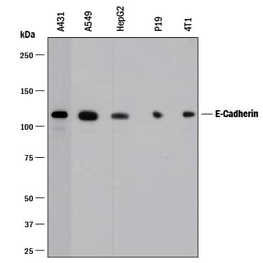
HAF017). A specific band was detected for E-Cadherin at approximately 110 kDa (as indicated). This experiment was conducted under reducing conditions and using
Immunoblot Buffer Group 1." alt="Western blot shows lysates of A431 human epithelial carcinoma cell line, A549 human lung carcinoma cell line, HepG2 human hepatocellular carcinoma cell line, P19 mouse embryonal carcinoma cell line, and 4T1 mouse breast cancer cell line. PVDF membrane was probed with 0.5 µg/mL of Goat Anti-Human/Mouse E-Cadherin Antigen Affinity-purified Polyclonal Antibody (Catalog # AF648) followed by HRP-conjugated Anti-Goat IgG Secondary Antibody (Catalog #
HAF017). A specific band was detected for E-Cadherin at approximately 110 kDa (as indicated). This experiment was conducted under reducing conditions and using
Immunoblot Buffer Group 1." class="big_thumb" />
042-206). This experiment was conducted under reducing conditions and using the 12-230kDa separation system." class="big_lightbox">

042-206). This experiment was conducted under reducing conditions and using the 12-230kDa separation system." class="big_thumb" />
MAB2018). Cells were incubated with primary antibodies for 3 hours at room temperature. Cells were stained for E-Cadherin using the NorthernLights™ 557-conjugated Anti-Goat IgG Secondary Antibody (green; Catalog #
NL001) and for SOX2 using the NorthernLights 493-conjugated Anti-Mouse Secondary Antibody (red; Catalog #
NL009). Cells were counterstained with DAPI (blue). View our protocol for
Fluorescent ICC Staining of Cells on Coverslips." class="big_lightbox">
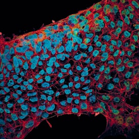
MAB2018). Cells were incubated with primary antibodies for 3 hours at room temperature. Cells were stained for E-Cadherin using the NorthernLights™ 557-conjugated Anti-Goat IgG Secondary Antibody (green; Catalog #
NL001) and for SOX2 using the NorthernLights 493-conjugated Anti-Mouse Secondary Antibody (red; Catalog #
NL009). Cells were counterstained with DAPI (blue). View our protocol for
Fluorescent ICC Staining of Cells on Coverslips." class="big_thumb" />
HAF017). This experiment was conducted under reducing conditions and using the 12-230 kDa separation system." class="big_lightbox">
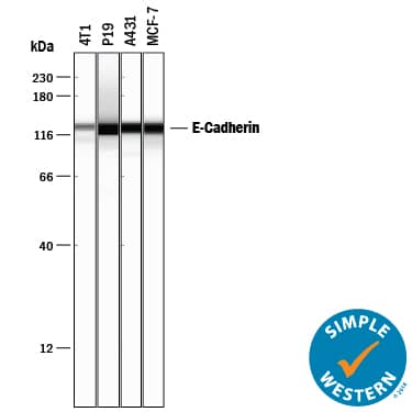
HAF017). This experiment was conducted under reducing conditions and using the 12-230 kDa separation system." class="big_thumb" />
VC004) and DAB chromogen (lower image, yellow-brown). Tissues were counterstained with hematoxylin (blue)." class="big_lightbox">
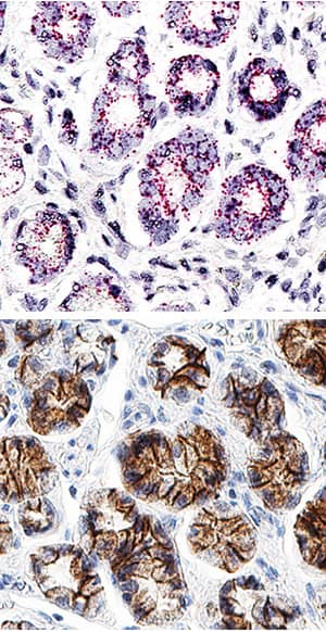
VC004) and DAB chromogen (lower image, yellow-brown). Tissues were counterstained with hematoxylin (blue)." class="big_thumb" />
MAB18382) coated on a Clear Polystyrene Microplate (Catalog #
DY990). Goat Anti-Human/Mouse E‑Cadherin Antigen Affinity-purified Polyclonal Antibody (Catalog # AF648) was biotinylated and incubated with the protein captured on the plate. Detection of the standard curve was achieved by incubating Streptavidin-HRP (Catalog #
DY998)" class="big_lightbox">

MAB18382) coated on a Clear Polystyrene Microplate (Catalog #
DY990). Goat Anti-Human/Mouse E‑Cadherin Antigen Affinity-purified Polyclonal Antibody (Catalog # AF648) was biotinylated and incubated with the protein captured on the plate. Detection of the standard curve was achieved by incubating Streptavidin-HRP (Catalog #
DY998)" class="big_thumb" />
 HAF017). A specific band was detected for E-Cadherin at approximately 110 kDa (as indicated). This experiment was conducted under reducing conditions and using Immunoblot Buffer Group 1." alt="Western blot shows lysates of A431 human epithelial carcinoma cell line, A549 human lung carcinoma cell line, HepG2 human hepatocellular carcinoma cell line, P19 mouse embryonal carcinoma cell line, and 4T1 mouse breast cancer cell line. PVDF membrane was probed with 0.5 µg/mL of Goat Anti-Human/Mouse E-Cadherin Antigen Affinity-purified Polyclonal Antibody (Catalog # AF748) followed by HRP-conjugated Anti-Goat IgG Secondary Antibody (Catalog # HAF017). A specific band was detected for E-Cadherin at approximately 110 kDa (as indicated). This experiment was conducted under reducing conditions and using Immunoblot Buffer Group 1." class="big_thumb" />
 HAF017). A specific band was detected for E-Cadherin at approximately 110 kDa (as indicated). This experiment was conducted under reducing conditions and using Immunoblot Buffer Group 1." alt="Western blot shows lysates of A431 human epithelial carcinoma cell line, A549 human lung carcinoma cell line, HepG2 human hepatocellular carcinoma cell line, P19 mouse embryonal carcinoma cell line, and 4T1 mouse breast cancer cell line. PVDF membrane was probed with 0.5 µg/mL of Goat Anti-Human/Mouse E-Cadherin Antigen Affinity-purified Polyclonal Antibody (Catalog # AF648) followed by HRP-conjugated Anti-Goat IgG Secondary Antibody (Catalog # HAF017). A specific band was detected for E-Cadherin at approximately 110 kDa (as indicated). This experiment was conducted under reducing conditions and using Immunoblot Buffer Group 1." class="big_thumb" />
 DR647MS) and counterstained with DAPI (blue; Lunaphore Catalog # DR100). Specific staining was localized to the membrane. Protocol available in COMET™ Panel Builder." alt="E-Cadherin was detected in immersion fixed paraffin-embedded sections of human breast using Mouse Anti-Human E-Cadherin Monoclonal Antibody (Novus Catalog # NBP2-19051) at 1:1000 at 37 ° Celsius for 4 minutes. Before incubation with the primary antibody, tissue underwent an all-in-one dewaxing and antigen retrieval preprocessing using PreTreatment Module (PT Module) and Dewax and HIER Buffer H (pH 9). Tissue was stained using the Alexa Fluor™ Plus 647 Goat anti-Mouse IgG Secondary Antibody at 1:200 at 37 ° Celsius for 2 minutes. (Yellow; Lunaphore Catalog # DR647MS) and counterstained with DAPI (blue; Lunaphore Catalog # DR100). Specific staining was localized to the membrane. Protocol available in COMET™ Panel Builder." class="big_thumb" />
 HAF018). A specific band was detected for E-Cadherin at approximately 110 kDa (as indicated). This experiment was conducted under reducing conditions and using Immunoblot Buffer Group 1." alt="Western blot shows lysates of A549 human lung carcinoma cell line and HepG2 human hepatocellular carcinoma cell line. PVDF membrane was probed with 0.5 µg/mL of Mouse Anti-Human E-Cadherin Monoclonal Antibody (Catalog # MAB1838) followed by HRP-conjugated Anti-Mouse IgG Secondary Antibody (Catalog # HAF018). A specific band was detected for E-Cadherin at approximately 110 kDa (as indicated). This experiment was conducted under reducing conditions and using Immunoblot Buffer Group 1." class="big_thumb" />
 DR647RT) and counterstained with DAPI (blue; Lunaphore Catalog # DR100). Specific staining was localized to the cell membrane. Protocol available in COMET™ Panel Builder." alt="E-Cadherin was detected in immersion fixed paraffin-embedded sections of mouse Thymus using Rat Anti-Mouse E-Cadherin Monoclonal Antibody (Catalog # MAB7481) at 1ug at 37° Celsius for 4 minutes. Before incubation with the primary antibody, tissue underwent an all-in-one dewaxing and antigen retrieval preprocessing using PreTreatment Module (PT Module) and Dewax and HIER Buffer H (pH 9; Epredia Catalog # TA-999-DHBH). Tissue was stained using the Alexa Fluor™ 647 Goat anti-Rat IgG Secondary Antibody at 1:200 at 37 ° Celsius for 2 minutes. (Yellow; Lunaphore Catalog # DR647RT) and counterstained with DAPI (blue; Lunaphore Catalog # DR100). Specific staining was localized to the cell membrane. Protocol available in COMET™ Panel Builder." class="big_thumb" />
 HAF018). A specific band was detected for E-Cadherin at approximately 120 kDa (as indicated). This experiment was conducted under reducing conditions and using Immunoblot Buffer Group 1." alt="Western blot shows lysates of MCF-7 human breast cancer cell line, BT-20 human breast cancer cell line, and T47D human breast cancer cell line. PVDF membrane was probed with 2 µg/mL of Mouse Anti-Human E-Cadherin Monoclonal Antibody (Catalog # MAB18381) followed by HRP-conjugated Anti-Mouse IgG Secondary Antibody (Catalog # HAF018). A specific band was detected for E-Cadherin at approximately 120 kDa (as indicated). This experiment was conducted under reducing conditions and using Immunoblot Buffer Group 1." class="big_thumb" />


 NL001) and counterstained with DAPI (blue). Specific staining was localized to cell surfaces. View our protocol for
  NL001) and counterstained with DAPI (blue). Specific staining was localized to plasma membranes in keratinocytes. View our protocol for
  NL001) and counterstained with DAPI (blue). Magnification shown at 100X (upper panel) and 40X (lower panel). Specific staining was localized to cell surfaces. View our protocol for
  NL003) and counterstained with DAPI (blue). Specific staining was localized to cell surfaces. View our protocol for
  CTS008) and counterstained with hematoxylin (blue). Specific staining was localized to dorsal horn. View our protocol for
  HAF109). This experiment was conducted under reducing conditions and using the 12-230 kDa separation system." class="big_thumb" />


 AF748) at 0.5ug/mL with overnight incubation at 4 degrees Celsius followed by incubation with anti-goat IgG VisUCyte HRP Polymer Antibody (Catalog #
 human intestinal organoid culture protocol. (A) The organoids were fixed and stained with a Mouse Anti-Human MUC2 Monoclonal Antibody (Novus Biologicals; Catalog # NBP2-44431; green) to visualize intestinal goblet cells and counterstained with a Goat Anti-Human/Mouse E-Cadherin Antigen Affinity-purified Polyclonal Antibody (Catalog # AF748; red) and DAPI (Catalog # 5748; blue). The image shown was taken at 10x magnification. (B) The organoids were fixed and stained with a Mouse Anti-Human Chromogranin A Monoclonal Antibody (Catalog # MAB90981; green) to visualize enteroendocrine cells and counterstained with a Goat Anti-Human/Mouse E-Cadherin Antigen Affinity-purified Polyclonal Antibody (Catalog # AF748; red) and DAPI (Catalog # 5748; blue). The image shown was taken at 20x magnification." class="big_thumb" />

 042-206). This experiment was conducted under reducing conditions and using the 12-230kDa separation system." class="big_thumb" />
 AB-108-C, open histogram), followed by Allophycocyanin-conjugated Anti-Goat IgG Secondary Antibody (Catalog #
  NL001) and counterstained with DAPI (blue). View our protocol for
  MAB2018). Cells were incubated with primary antibodies for 3 hours at room temperature. Cells were stained for E-Cadherin using the NorthernLights™ 557-conjugated Anti-Goat IgG Secondary Antibody (green; Catalog #
  VC004). Tissue was stained using DAB (brown) and counterstained with hematoxylin (blue). Specific staining was localized to cell membrane and cytoplasm in gastric glands. View our protocol for
  HAF017). This experiment was conducted under reducing conditions and using the 12-230 kDa separation system." class="big_thumb" />
 VC004) and DAB chromogen (lower image, yellow-brown). Tissues were counterstained with hematoxylin (blue)." class="big_thumb" />


 MAB18382) coated on a Clear Polystyrene Microplate (Catalog #
  DR647MS) and counterstained with DAPI (blue; Lunaphore Catalog #
  DR647MS) and counterstained with DAPI (blue; Lunaphore Catalog #
 ![Immunohistochemistry-Paraffin: E-Cadherin Antibody (7H12) [NBP2-19051] - Human kidney distal tubules stained with E-CAD antibody. Secondary antibody: Donkey anti-Mouse Alexa Fluor 555. Imaging with a 40x objective from Zeiss Observer microscope with Apotome2. Image from verified customer review.](http://images.novusbio.com/fullsize/E-Cadherin-Antibody-7H12-Immunohistochemistry-Paraffin-NBP2-19051-img0011.jpg)

![Western Blot: E-Cadherin Antibody (7H12) [NBP2-19051] - Analysis of E-Cadherin in LNCAP (1), A431 (2), DU145 (3), PC-3 (4), MCF-7 (5), PC-12 (6), NIH/3T3 (7), C6 (8) and COS7 (9) cell lysate.](http://images.novusbio.com/fullsize/E-Cadherin-Antibody-7H12-Western-Blot-NBP2-19051-img0002.jpg)
![Immunocytochemistry/Immunofluorescence: E-Cadherin Antibody (7H12) [NBP2-19051] - ICC staining of E-Cadherin in human colon cancer sphere. Image from verified customer review.](http://images.novusbio.com/fullsize/E-Cadherin-Antibody-7H12-Immunocytochemistry-Immunofluorescence-NBP2-19051-img0010.jpg)

![Immunocytochemistry/Immunofluorescence: E-Cadherin Antibody (7H12) [NBP2-19051] - Human colonic tissue was stained with E-Cadherin antibody. Image from verified customer review.](http://images.novusbio.com/fullsize/E-Cadherin-Antibody-7H12-Immunocytochemistry-Immunofluorescence-NBP2-19051-img0009.jpg)
![Immunohistochemistry-Paraffin: E-Cadherin Antibody (7H12) [NBP2-19051] - IHC staining of E-Cadherin antibody in mouse colon cancer tissue. Antibody dilution 1:500. Image from verified customer review.](http://images.novusbio.com/fullsize/E-Cadherin-Antibody-7H12-Immunohistochemistry-Paraffin-NBP2-19051-img0012.jpg)
![Flow Cytometry: E-Cadherin Antibody (7H12) [NBP2-19051] - Analysis of Hela cells using E-Cadherin mouse mAb (green) and negative control (purple).](http://images.novusbio.com/fullsize/E-Cadherin-Antibody-7H12-Flow-Cytometry-NBP2-19051-img0004.jpg)

![Immunohistochemistry-Paraffin: E-Cadherin Antibody (7H12) [NBP2-19051] - IHC staining of E-Cadherin in gastric cancer tissues (left) and lung cancer tissues (right) with DAB staining.](http://images.novusbio.com/fullsize/E-Cadherin-Antibody-7H12-Immunohistochemistry-Paraffin-NBP2-19051-img0003.jpg)

 CTS002) and counterstained with hematoxylin (blue). Specific labeling was localized to the plasma membrane of epithelial cells. View our protocol for


 HAF005). A specific band was detected for E-Cadherin at approximately 120 kDa (as indicated). This experiment was conducted under reducing conditions and using
  MAB006, open histogram), followed by Phycoerythrin-conjugated Anti-Rat IgG Secondary Antibody (
 VCTS021). Tissue was stained using the HRP-conjugated Anti-Rat IgG Secondary Antibody (
![Immunohistochemistry-Paraffin: E-Cadherin Antibody [NBP2-16258] - Mouse stomach. E-Cadherin stained by E-Cadherin antibody diluted at 1:500.Antigen Retrieval: Citrate buffer, pH 6.0, 15 min.](http://images.novusbio.com/fullsize/E-Cadherin-Antibody-Immunohistochemistry-Paraffin-NBP2-16258-img0020.jpg "Immunohistochemistry-Paraffin: E-Cadherin Antibody [NBP2-16258] - Mouse stomach. E-Cadherin stained by E-Cadherin antibody diluted at 1:500.Antigen Retrieval: Citrate buffer, pH 6.0, 15 min.")
![<b>Genetic Strategies Validation. </b>Western Blot: E-Cadherin Antibody [NBP2-16258] - Non-transfected (-) and transfected (+) MCF-7 whole cell extracts (30 ug) were separated by 5% SDS-PAGE, and the membrane was blotted with E-Cadherin antibody.](http://images.novusbio.com/fullsize/E-Cadherin-Antibody-Western-Blot-NBP2-16258-img0002.jpg)
![Immunohistochemistry-Paraffin: E-Cadherin Antibody [NBP2-16258] - Mouse cervix. E-Cadherin antibody diluted at 1:500. Antigen Retrieval: Citrate buffer, pH 6.0, 15 min.](http://images.novusbio.com/fullsize/E-Cadherin-Antibody-Immunohistochemistry-Paraffin-NBP2-16258-img0004.jpg)
![Immunohistochemistry-Paraffin: E-Cadherin Antibody [NBP2-16258] - Rat intestine. E-Cadherin antibody diluted at 1:500. Antigen Retrieval: Citrate buffer, pH 6.0, 15 min.](http://images.novusbio.com/fullsize/E-Cadherin-Antibody-Immunohistochemistry-Paraffin-NBP2-16258-img0005.jpg)
![Immunohistochemistry-Paraffin: E-Cadherin Antibody [NBP2-16258] - Breast cancer. E-cadherin antibody dilution: 1:500. Antigen Retrieval: Trilogy™ (EDTA based, pH 8.0) buffer, 15min.](http://images.novusbio.com/fullsize/E-Cadherin-Antibody-Immunohistochemistry-Paraffin-NBP2-16258-img0013.jpg)
![Immunohistochemistry-Paraffin: E-Cadherin Antibody [NBP2-16258] - Rat prostate. E-Cadherin antibody diluted at 1:500. Antigen Retrieval: Citrate buffer, pH 6.0, 15 min.](http://images.novusbio.com/fullsize/E-Cadherin-Antibody-Immunohistochemistry-Paraffin-NBP2-16258-img0017.jpg)
![Immunohistochemistry-Paraffin: E-Cadherin Antibody [NBP2-16258] - Mouse intestine. E-Cadherin stained by E-Cadherin antibody diluted at 1:500.Antigen Retrieval: Citrate buffer, pH 6.0, 15 min.](http://images.novusbio.com/fullsize/E-Cadherin-Antibody-Immunohistochemistry-Paraffin-NBP2-16258-img0019.jpg)
![Immunoprecipitation: E-Cadherin Antibody [NBP2-16258] - MCF-7 whole cell extract A. Control with 3 ug of preimmune Rabbit IgG B. Immunoprecipitation of E-cadherin protein by 3 ug E-cadherin antibody 5 % SDS-PAGE The immunoprecipitated E-cadherin protein was detected by E-cadherin antibody diluted at 1:500. [EasyBlot anti-rabbit IgG was used as a secondary reagent]](http://images.novusbio.com/fullsize/E-Cadherin-Antibody-Immunoprecipitation-NBP2-16258-img0008.jpg)
![Immunoprecipitation: E-Cadherin Antibody [NBP2-16258] - Immunoprecipitation of E-Cadherin protein from MCF-7 whole cell extracts using 5 ug of E-Cadherin antibody. Western blot analysis was performed using E-Cadherin antibody. EasyBlot anti-Rabbit IgG was used as a secondary reagent.](http://images.novusbio.com/fullsize/E-Cadherin-Antibody-Immunoprecipitation-NBP2-16258-img0011.jpg)
![Immunohistochemistry-Paraffin: E-Cadherin Antibody [NBP2-16258] - E-Cadherin antibody detects E-Cadherin protein at cell membrane and cytoplasm by immunohistochemical analysis.</br>Sample: Paraffin-embedded dog skin.</br>E-Cadherin stained by E-Cadherin antibody (NBP2-16258) diluted at 1:500.</br>Antigen Retrieval: Citrate buffer, pH 6.0, 15 min](http://images.novusbio.com/fullsize/nbp2-16258_rabbit-polyclonal-e-cadherin-antibody-910202420122059.jpg)
![Immunohistochemistry-Paraffin: E-Cadherin Antibody [NBP2-16258] - E-Cadherin antibody detects E-Cadherin protein at cell membrane in rat prostate by immunohistochemical analysis. <br>Sample: Paraffin-embedded rat prostate. <br>E-Cadherin antibody (NBP2-16258) diluted at 1:500.<br><br>Antigen Retrieval: Citrate buffer, pH 6.0, 15 min](http://images.novusbio.com/fullsize/nbp2-16258_rabbit-polyclonal-e-cadherin-antibody-91020242075979.jpg)
![Immunocytochemistry/ Immunofluorescence: E-Cadherin Antibody [NBP2-16258] - E-Cadherin antibody detects E-Cadherin protein at cell membrane by immunofluorescent analysis.</br>Sample: HCT116 cells were fixed in 4% paraformaldehyde at RT for 15 min.</br>Green: E-Cadherin stained by E-Cadherin antibody (NBP2-16258) diluted at 1:500.</br>Blue: Fluoroshield with DAPI .</br>Scale bar= 10 um.](http://images.novusbio.com/fullsize/nbp2-16258_rabbit-polyclonal-e-cadherin-antibody-9102024205666.jpg)
![Western Blot: E-Cadherin Antibody [NBP2-16258] - Whole cell extracts (30 ug) was separated by 7.5% SDS-PAGE, and the membrane was blotted with E-Cadherin antibody (NBP2-16258) diluted at 1:10000. The HRP-conjugated anti-rabbit IgG antibody was used to detect the primary antibody.](http://images.novusbio.com/fullsize/nbp2-16258_rabbit-polyclonal-e-cadherin-antibody-91020242021963.jpg)
![Immunohistochemistry-Paraffin: E-Cadherin Antibody [NBP2-16258] - E-Cadherin antibody detects E-Cadherin protein at cell membrane and cytoplasm by immunohistochemical analysis.</br>Sample: Paraffin-embedded rat duodenum.</br>E-Cadherin stained by E-Cadherin antibody (NBP2-16258) diluted at 1:1000.</br>Antigen Retrieval: Citrate buffer, pH 6.0, 15 min](http://images.novusbio.com/fullsize/nbp2-16258_rabbit-polyclonal-e-cadherin-antibody-910202420131274.jpg)
![Immunocytochemistry/ Immunofluorescence: E-Cadherin Antibody [NBP2-16258] - E-Cadherin antibody detects E-Cadherin protein at cell membrane by immunofluorescent analysis.</br>Sample: MDCK cells were fixed in 4% paraformaldehyde at RT for 15 min.</br>Green: E-Cadherin stained by E-Cadherin antibody (NBP2-16258) diluted at 1:500.](http://images.novusbio.com/fullsize/nbp2-16258_rabbit-polyclonal-e-cadherin-antibody-910202419593579.jpg)
![Western Blot: E-Cadherin Antibody [NBP2-16258] - Mouse tissue extract (50 ug) was separated by 5% SDS-PAGE, and the membrane was blotted with E-Cadherin antibody (NBP2-16258) diluted at 1:2000. The HRP-conjugated anti-rabbit IgG antibody was used to detect the primary antibody.](http://images.novusbio.com/fullsize/nbp2-16258_rabbit-polyclonal-e-cadherin-antibody-91020242010440.jpg)


![Western Blot: E-Cadherin Antibody (DECMA-1) [NB120-11512] - MDCK cell extract separated on SDS-PAGE and probed with Rat Anti-Uvomorulin/E-Cadherin Clone: DECMA-1. Detection by Rabbit Anti-Rat IgG-Peroxidase and a chemiluminescent substrate. Antibody dilution 1:250.](http://images.novusbio.com/fullsize/E-Cadherin-Antibody-DECMA-1-Western-Blot-NB120-11512-img0006.jpg "Western Blot: E-Cadherin Antibody (DECMA-1) [NB120-11512] - MDCK cell extract separated on SDS-PAGE and probed with Rat Anti-Uvomorulin/E-Cadherin Clone: DECMA-1. Detection by Rabbit Anti-Rat IgG-Peroxidase and a chemiluminescent substrate. Antibody dilution 1:250.")
![Immunocytochemistry/Immunofluorescence: E-Cadherin Antibody (DECMA-1) [NB120-11512] - Adult mouse small intestine stained with NB12-11512. Image from verified customer review.](http://images.novusbio.com/fullsize/E-Cadherin-Antibody-DECMA-1-Immunocytochemistry-Immunofluorescence-NB120-11512-img0009.jpg)
![Western Blot: E-Cadherin Antibody (DECMA-1) [NB120-11512] - Cat testis, cat ovary and HEK293 whole cell lysates. Image from verified customer review.](http://images.novusbio.com/fullsize/E-Cadherin-Antibody-DECMA-1-Western-Blot-NB120-11512-img0005.jpg)
![Immunocytochemistry/Immunofluorescence: E-Cadherin Antibody (DECMA-1) [NB120-11512] - MDCK cells were fixed and permeabilized with methanol followed by acetone. Fixed cells were stained with 1:3200 Rat Monoclonal Anti-Uvomorulin/E-Cadherin, Clone: DECMA-1. Detection using Goat Anti-Rat IgG, FITC conjugate.](http://images.novusbio.com/fullsize/E-Cadherin-Antibody-DECMA-1-Immunocytochemistry-Immunofluorescence-NB120-11512-img0007.jpg)
![Immunocytochemistry/Immunofluorescence: E-Cadherin Antibody (DECMA-1) [NB120-11512] - MDCK cells were fixed and permeabilized with cold methanol followed by aceton. Fixed cells were stained with Rat Anti-Uvomorulin/E-Cadherin Clone: DECMA-1 diluted to 1:3200. The antibody was developed using Rabbit Anti-Rat IgG, FITC-conjugate.](http://images.novusbio.com/fullsize/E-Cadherin-Antibody-DECMA-1-Immunocytochemistry-Immunofluorescence-NB120-11512-img0008.jpg)
![Immunohistochemistry-Paraffin: pan Cadherin Antibody [NB200-592] - Mouse cervical cancer tissue stained with N-Cadherin antibody (green). Nuclei were counterstained with DAPI (Blue). Antigen retrieval was done with pepsin treatment. Image from verified customer review.](http://images.novusbio.com/fullsize/N-Cadherin-Antibody-Immunohistochemistry-Paraffin-NB200-592-img0003.jpg "<b>Biological Strategies Validation. </b>Immunohistochemistry-Paraffin: pan Cadherin Antibody [NB200-592] - Mouse cervical cancer tissue stained with N-Cadherin antibody (green). Nuclei were counterstained with DAPI (Blue). Antigen retrieval was done with pepsin treatment. Image from verified customer review.")
![<b>Biological Strategies Validation. </b>Western Blot: pan Cadherin Antibody [NB200-592] - Whole extract of Chicken Cardiac Muscle were separated on SDS-PAGE and probed with Anti-Pan Cadherin antibody produced in Rabbit. The antibody was developed using 1:1000 Anti-Rabbit IgG (whole molecule)-Peroxidase antibody produced in Goat.Lanes:1. 1:200 antibody2. 1:400 antibody3. Negative Control](http://images.novusbio.com/fullsize/pan-Cadherin-Antibody-Western-Blot-NB200-592-img0005.jpg)
![Immunohistochemistry: pan Cadherin Antibody [NB200-592] - Immunoperoxidase staining of a formalin-fixed, paraffin-embedded section of human heart using Rabbit Anti-Pan Cadherin.](http://images.novusbio.com/fullsize/pan-Cadherin-Antibody-Immunohistochemistry-NB200-592-img0004.jpg)
![Western Blot: E-Cadherin Antibody (4A2) [NBP2-53229] - Western Blot Analysis of MCF-7 cell lysate using E-Cadherin Antibody (4A2).](http://images.novusbio.com/fullsize/E-Cadherin-Antibody-4A2-Western-Blot-NBP2-53229-img0005.jpg "Western Blot: E-Cadherin Antibody (4A2) [NBP2-53229] - Western Blot Analysis of MCF-7 cell lysate using E-Cadherin Antibody (4A2).")
![Immunocytochemistry/Immunofluorescence: E-Cadherin Antibody (4A2) [NBP2-53229] - Immunofluorescence analysis of MeOH-fixed MCF-7 cells. E-Cadherin Mouse Monoclonal Antibody (4A2) followed by goat anti-mouse IgG-CF488 (green). Nuclei counterstained with RedDot.](http://images.novusbio.com/fullsize/E-Cadherin-Antibody-4A2-Immunocytochemistry-Immunofluorescence-NBP2-53229-img0007.jpg)
![Immunohistochemistry: E-Cadherin Antibody (4A2) [NBP2-53229] - Staining of human pancreatic tumor. Image from a verified customer review.](http://images.novusbio.com/fullsize/E-Cadherin-Antibody-4A2-Immunohistochemistry-NBP2-53229-img0003.jpg)
![Flow Cytometry: E-Cadherin Antibody (4A2) [NBP2-53229] - Flow Cytometric Analysis of human trypsinized MCF-7 cells using E-Cadherin Antibody (4A2) followed by Goat anti-Mouse IgG-CF488 (Blue); Isotype control (Red).](http://images.novusbio.com/fullsize/E-Cadherin-Antibody-4A2-Flow-Cytometry-NBP2-53229-img0006.jpg)
![Immunohistochemistry-Paraffin: E-Cadherin Antibody (4A2) [NBP2-53229] - Formalin-fixed, paraffin-embedded human Skin stained with E-Cadherin Monoclonal Antibody (4A2).](http://images.novusbio.com/fullsize/E-Cadherin-Antibody-4A2-Immunohistochemistry-Paraffin-NBP2-53229-img0001.jpg)
![Western Blot: E-Cadherin Antibody (CDH1/1525) [NBP2-53354] - Western Blot Analysis (A) Recombinant Protein (B) human Stomach lysate Using E-Cadherin Monoclonal Antibody (CDH1/1525).](http://images.novusbio.com/fullsize/E-Cadherin-Antibody-CDH1-1525-Western-Blot-NBP2-53354-img0006.jpg "Western Blot: E-Cadherin Antibody (CDH1/1525) [NBP2-53354] - Western Blot Analysis (A) Recombinant Protein (B) human Stomach lysate Using E-Cadherin Monoclonal Antibody (CDH1/1525).")
![Immunocytochemistry/Immunofluorescence: E-Cadherin Antibody (CDH1/1525) [NBP2-53354] - Confocal Immunofluorescence of MCF-7 cells E-Cadherin Antibody (CDH1/1525). labeled with CF488 (Green); Red Dot is used to label the nuclei.](http://images.novusbio.com/fullsize/E-Cadherin-Antibody-CDH1-1525-Immunocytochemistry-Immunofluorescence-NBP2-53354-img0007.jpg)
![Immunohistochemistry-Paraffin: E-Cadherin Antibody (CDH1/1525) [NBP2-53354] - Formalin-fixed, paraffin-embedded human breast adenocarcinoma stained with E-Cadherin Mouse Monoclonal Antibody (CDH1/1525).](http://images.novusbio.com/fullsize/E-Cadherin-Antibody-CDH1-1525-Immunohistochemistry-Paraffin-NBP2-53354-img0009.jpg)
![Immunohistochemistry-Paraffin: E-Cadherin Antibody (CDH1/1525) [NBP2-53354] - Formalin-fixed, paraffin-embedded human Colon Carcinoma stained with E-Cadherin Monoclonal Antibody (CDH1/1525).](http://images.novusbio.com/fullsize/E-Cadherin-Antibody-CDH1-1525-Immunohistochemistry-Paraffin-NBP2-53354-img0001.jpg)
![Immunohistochemistry-Paraffin: E-Cadherin Antibody (CDH1/1525) [NBP2-53354] - Formalin-fixed, paraffin-embedded human Skin stained with E-Cadherin Monoclonal Antibody (CDH1/1525).](http://images.novusbio.com/fullsize/E-Cadherin-Antibody-CDH1-1525-Immunohistochemistry-Paraffin-NBP2-53354-img0002.jpg)
![Immunohistochemistry-Paraffin: E-Cadherin Antibody (CDH1/1525) [NBP2-53354] - Formalin-paraffin human Placenta stained with E-Cadherin MAb (CDH1/1525).](http://images.novusbio.com/fullsize/E-Cadherin-Antibody-CDH1-1525-Immunohistochemistry-Paraffin-NBP2-53354-img0004.jpg)
![Protein Array: E-Cadherin Antibody (CDH1/1525) [NBP2-53354] - Analysis of Protein Array containing more than 19,000 full-length human proteins using E-Cadherin Antibody (CDH1/1525).Z- and S- Score: The Z-score represents the strength of a signal that a monoclonal antibody (MAb) (in combination with a fluorescently-tagged anti-IgG secondary antibody) produces when binding to a particular protein on the HuProt(TM) array. Z-scores are described in units of standard deviations (SD's) above the mean value of all signals generated on that array. If targets on HuProt(TM) are arranged in descending order of the Z-score, the S-score is the difference (also in units of SD's) between the Z-score. S-score therefore represents the relative target specificity of a MAb to its intended target. A MAb is considered to specific to its intended target, if the MAb has an S-score of at least 2.5.](http://images.novusbio.com/fullsize/E-Cadherin-Antibody-CDH1-1525-Protein-Array-NBP2-53354-img0008.jpg)
![Immunohistochemistry-Paraffin: E-Cadherin Antibody (CDH1/1525) [NBP2-53354] - Human breast cancer tissue stained fro E-cadherin (white) and counterstained with DAPI (blue). Alexa Fluor 750 version of antibody used. Image from verified customer review.](http://images.novusbio.com/fullsize/nbp2-53354_mouse-monoclonal-e-cadherin-antibody-cdh1-1525-17720241044616.jpg)
 NL007) and counterstained with DAPI (blue). Specific staining was localized to cell membrane. View our protocol for
  VC001). Tissue was stained using DAB (brown) and counterstained with hematoxylin (blue). Specific staining was localized to plasma membrane. View our protocol for
  VC001). Tissue was stained using DAB (brown) and counterstained with hematoxylin (blue). Specific staining was localized to plasma membrane. View our protocol for


