MAB003, open histogram) followed by Allophycocyanin-conjugated Anti-Mouse IgG Secondary Antibody (Catalog #
F0101B). To facilitate intracellular staining, cells were fixed and permeabilized with FlowX FoxP3 Fixation & Permeabilization Buffer Kit (Catalog #
FC012). View our protocol for
Staining Intracellular Molecules." class="big_lightbox">
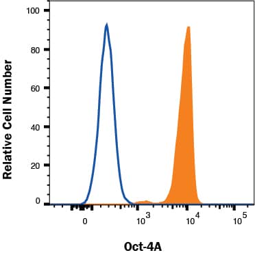
MAB003, open histogram) followed by Allophycocyanin-conjugated Anti-Mouse IgG Secondary Antibody (Catalog #
F0101B). To facilitate intracellular staining, cells were fixed and permeabilized with FlowX FoxP3 Fixation & Permeabilization Buffer Kit (Catalog #
FC012). View our protocol for
Staining Intracellular Molecules." alt="NTERA-2 cells were stained with Mouse Anti-Human Oct‑4A Monoclonal Antibody (Catalog # MAB17591, filled histogram) or isotype control antibody (Catalog #
MAB003, open histogram) followed by Allophycocyanin-conjugated Anti-Mouse IgG Secondary Antibody (Catalog #
F0101B). To facilitate intracellular staining, cells were fixed and permeabilized with FlowX FoxP3 Fixation & Permeabilization Buffer Kit (Catalog #
FC012). View our protocol for
Staining Intracellular Molecules." class="big_thumb" />
HAF007). A specific band was detected for Oct-4A at approximately 50 kDa (as indicated). This experiment was conducted under reducing conditions and using
Immunoblot Buffer Group 1." class="big_lightbox">
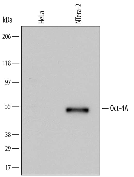
HAF007). A specific band was detected for Oct-4A at approximately 50 kDa (as indicated). This experiment was conducted under reducing conditions and using
Immunoblot Buffer Group 1." class="big_thumb" />
AF648). Cells were incubated with primary antibodies for 3 hours at room temperature. Cells were stained for Oct-4A using the Northern-Lights™ 493-conjugated Anti-Mouse IgG Secondary Antibody (green;
NL009), and stained for E-Cadherin using the Northern-Lights™ 557-conjugated Anti-Goat IgG Secondary Antibody (red;
NL001). Specific staining of Oct-4A was localized to nuclei. View our protocol for
Fluorescent ICC Staining of Cells on Coverslips." class="big_lightbox">
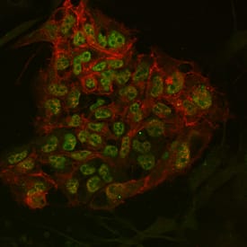
AF648). Cells were incubated with primary antibodies for 3 hours at room temperature. Cells were stained for Oct-4A using the Northern-Lights™ 493-conjugated Anti-Mouse IgG Secondary Antibody (green;
NL009), and stained for E-Cadherin using the Northern-Lights™ 557-conjugated Anti-Goat IgG Secondary Antibody (red;
NL001). Specific staining of Oct-4A was localized to nuclei. View our protocol for
Fluorescent ICC Staining of Cells on Coverslips." class="big_thumb" />
MAB003, open histogram), followed by Allophycocyanin-conjugated Anti-Mouse IgG F(ab')
2Secondary Antibody (
F0101B). To facilitate intracellular staining, cells were fixed with paraformaldehyde and permeabilized with saponin." class="big_lightbox">
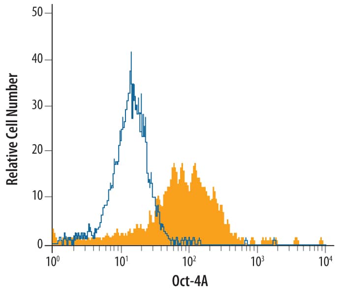
MAB003, open histogram), followed by Allophycocyanin-conjugated Anti-Mouse IgG F(ab')
2Secondary Antibody (
F0101B). To facilitate intracellular staining, cells were fixed with paraformaldehyde and permeabilized with saponin." class="big_thumb" />
MAB003, open histogram), followed by Allophycocyanin-conjugated Anti-Mouse IgG Secondary Antibody (Catalog #
F0101B). To facilitate intracellular staining, cells were fixed with Flow Cytometry Fixation Buffer (Catalog #
FC004) and permeabilized with Flow Cytometry Permeabilization/Wash Buffer I (Catalog #
FC005). View our protocol for
Staining Intracellular Molecules." class="big_lightbox">

MAB003, open histogram), followed by Allophycocyanin-conjugated Anti-Mouse IgG Secondary Antibody (Catalog #
F0101B). To facilitate intracellular staining, cells were fixed with Flow Cytometry Fixation Buffer (Catalog #
FC004) and permeabilized with Flow Cytometry Permeabilization/Wash Buffer I (Catalog #
FC005). View our protocol for
Staining Intracellular Molecules." class="big_thumb" />
 HAF017). A specific band was detected for Oct-3/4 at approximately 48 kDa (as indicated). This experiment was conducted under reducing conditions and using Immunoblot Buffer Group 1." alt="Western blot shows lysates of BG01V human embryonic stem cells and D3 mouse embryonic stem cell line. PVDF membrane was probed with 0.5 µg/mL of Goat Anti-Human/Mouse Oct-3/4 Antigen Affinity-purified Polyclonal Antibody (Catalog # AF1759) followed by HRP-conjugated Anti-Goat IgG Secondary Antibody (Catalog # HAF017). A specific band was detected for Oct-3/4 at approximately 48 kDa (as indicated). This experiment was conducted under reducing conditions and using Immunoblot Buffer Group 1." class="big_thumb" />
 MAB0061, open histogram) followed by Allophycocyanin-conjugated Anti-Rat IgG Secondary Antibody (Catalog # F0113). To facilitate intracellular staining, cells were fixed and permeabilized with FlowX FoxP3 Fixation & Permeabilization Buffer Kit (Catalog # FC012). View our protocol for Staining Intracellular Molecules." alt="NTERA-2 cells were stained with Rat Anti-Human/Mouse Oct‑3/4 Monoclonal Antibody (Catalog # MAB1759, filled histogram) or isotype control antibody (Catalog # MAB0061, open histogram) followed by Allophycocyanin-conjugated Anti-Rat IgG Secondary Antibody (Catalog # F0113). To facilitate intracellular staining, cells were fixed and permeabilized with FlowX FoxP3 Fixation & Permeabilization Buffer Kit (Catalog # FC012). View our protocol for Staining Intracellular Molecules." class="big_thumb" />
 MAB003, open histogram) followed by Allophycocyanin-conjugated Anti-Mouse IgG Secondary Antibody (Catalog # F0101B). To facilitate intracellular staining, cells were fixed and permeabilized with FlowX FoxP3 Fixation & Permeabilization Buffer Kit (Catalog # FC012). View our protocol for Staining Intracellular Molecules." alt="NTERA-2 cells were stained with Mouse Anti-Human Oct‑4A Monoclonal Antibody (Catalog # MAB17591, filled histogram) or isotype control antibody (Catalog # MAB003, open histogram) followed by Allophycocyanin-conjugated Anti-Mouse IgG Secondary Antibody (Catalog # F0101B). To facilitate intracellular staining, cells were fixed and permeabilized with FlowX FoxP3 Fixation & Permeabilization Buffer Kit (Catalog # FC012). View our protocol for Staining Intracellular Molecules." class="big_thumb" />


 MAB1448) and 10 µg/mL Goat Anti-Human/Mouse Oct-3/4 Antigen Affinity-purified Polyclonal Antibody (Catalog # AF1759). Cells were incubated with primary antibodies for 3 hours at room temperature. Cells were stained for ALPL using the NorthernLights™ 557-conjugated Anti-Mouse IgG Secondary Antibody (pseudo-stained green; Catalog #
  AB-108-C) for 15 minutes in an ultrasonic bath, followed by Biotinylated Anti-Goat IgG Secondary Antibody (Catalog #
  HAF109). This experiment was conducted under reducing conditions and using the 12-230 kDa separation system." class="big_thumb" />


 HAF005). A specific band was detected for Oct-3/4 at approximately 46 kDa (as indicated). This experiment was conducted under reducing conditions and using
  Fluorescent ICC Staining of Cells on Coverslips." class="big_thumb" />


![Western Blot: OCT4 Antibody [NB100-2379] - WB detection of OCT4 in mouse brain lysate using OCT4 antibody with ECL based detection. The antibody generated a specific band of OCT4 at the expected ~40kDa position.](http://images.novusbio.com/fullsize/OCT4-Antibody-Western-Blot-NB100-2379-img0011.jpg)
![Flow Cytometry: OCT4 Antibody [NB100-2379] - An intracellular stain was performed on Jurkat with NB100-2379 and a matched isotype control. Cells were fixed with 4% PFA and then permeablized with 0.1% saponin. Cells were incubated in an antibody dilution of 1 ug/mL for 30 minutes at room temperature, followed by Rabbit IgG (H+L) Cross-Adsorbed Secondary Antibody.](http://images.novusbio.com/fullsize/OCT4-Antibody-Flow-Cytometry-NB100-2379-img0012.jpg)
![Immunocytochemistry/Immunofluorescence: OCT4 Antibody [NB100-2379] - Ntera2 cells were fixed in 4% paraformaldehyde for 10 minutes and permeabilized in 0.5% Triton X-100 in PBS for 5 minutes. The cells were incubated with anti- NB100-2379 at 1 ug/ml overnight at 4C and detected with an anti-rabbit Dylight 488 (Green) at a 1:1000 dilution for 60 minutes. Nuclei were counterstained with DAPI (Blue). Cells were imaged using a 100X objective and digitally deconvolved.](http://images.novusbio.com/fullsize/OCT4-Antibody-Immunocytochemistry-Immunofluorescence-NB100-2379-img0018.jpg)
![<b>Biological Strategies Validation. </b>Western Blot: OCT4 Antibody [NB100-2379] - MDA-MB-231 subclones transfected with NTC or HIF-1 alpha shRNA vector were treated with vehicle (V) or paclitaxel (P) and Western blot assay was performed. WB image submitted by a verified customer review.](http://images.novusbio.com/fullsize/OCT4-Antibody-Western-Blot-NB100-2379-img0016.jpg)
![Immunohistochemistry-Paraffin: OCT4 Antibody [NB100-2379] - Analysis of a FFPE tissue section of mouse brain using 1:200 dilution of OCT4 antibody. The staining was developed using HRP labeled anti-rabbit secondary antibody and DAB reagent, and nuclei of cells were counter-stained with hematoxylin.](http://images.novusbio.com/fullsize/OCT4-Antibody-Immunohistochemistry-Paraffin-NB100-2379-img0017.jpg)
![Flow Cytometry: OCT4 Antibody - BSA Free [NB100-2379] - An intracellular stain was performed on U2-OS cells with OCT4 NB100-2379AF647 (blue) and a matched isotype control NBP2-24891 (orange). Cells were fixed with 4% PFA and then permeabilized with 0.1% saponin. Cells were incubated in an antibody dilution of 2.5 ug/mL for 30 minutes at room temperature. Both antibodies were conjugated to Alexa Fluor 647.](http://images.novusbio.com/fullsize/OCT4-Antibody---BSA-Free-Flow-Cytometry-NB100-2379-img0022.jpg)
![Western Blot: OCT4 Antibody [NB100-2379] - The expression of pluripotency factors Oct4 in human head and neck cancer cell lines.](http://images.novusbio.com/fullsize/OCT4-Antibody-Western-Blot-NB100-2379-img0013.jpg)
![Flow Cytometry: OCT4 Antibody [NB100-2379] - An intracellular stain was performed on Jurkat cells with OCT4 Antibody NB100-2379 (blue) and a matched isotype control NBP2-24891 (orange). Cells were fixed with 4% PFA and then permeabilized with 0.1% saponin. Cells were incubated in an antibody dilution of 1.0 ug/mL for 30 minutes at room temperature, followed by Rabbit IgG (H+L) Cross-Adsorbed Secondary Antibody, Dylight 550 (SA5-10033, Thermo Fisher).](http://images.novusbio.com/fullsize/OCT4-Antibody-Flow-Cytometry-NB100-2379-img0019.jpg)
![Flow Cytometry: OCT4 Antibody [NB100-2379] - An intracellular stain was performed on Neuro2a cells with OCT4 Antibody NB100-2379 (blue) and a matched isotype control NBP2-24891 (orange). Cells were fixed with 4% PFA and then permeabilized with 0.1% saponin. Cells were incubated in an antibody dilution of 1.0 ug/mL for 30 minutes at room temperature, followed by Rabbit IgG (H+L) Cross-Adsorbed Secondary Antibody, Dylight 550 (SA5-10033, Thermo Fisher).](http://images.novusbio.com/fullsize/OCT4-Antibody-Flow-Cytometry-NB100-2379-img0020.jpg)
![Flow Cytometry: OCT4 Antibody [NB100-2379] - An intracellular stain was performed on Ntera2 cells with OCT4 Antibody NB100-2379 (blue) and a matched isotype control NBP2-24891 (orange). Cells were fixed with 4% PFA and then permeabilized with 0.1% saponin. Cells were incubated in an antibody dilution of 1.0 ug/mL for 30 minutes at room temperature, followed by Rabbit IgG (H+L) Cross-Adsorbed Secondary Antibody, Dylight 550 (SA5-10033, Thermo Fisher).](http://images.novusbio.com/fullsize/OCT4-Antibody-Flow-Cytometry-NB100-2379-img0021.jpg)


![Western Blot: OCT4 Antibody - BSA Free [NB100-2379] - Protein expression of NANOG a, OCT4 b & SOX2 c in bovine uterine tissues obtained from control cows & from cows with adenomyosis. Data were normalized against glyceraldehyde-3-phosphate dehydrogenase (GAPDH). Bars represent the mean ± SEM. There were no statistical differences between uterine normal & adenomyotic tissues (P > 0.05), as determined by Student’s t-test. Representative blots for NANOG, OCT4, SOX2 & GAPDH are shown below the graphs d. MM – molecular weight marker, C – tissues obtained from control cows, ADENO – tissues obtained from cows with adenomyosis Image collected & cropped by CiteAb from the following publication (//www.rbej.com/content/13/1/110), licensed under a CC-BY license. Not internally tested by Novus Biologicals.](http://images.novusbio.com/fullsize/nb100-2379_rabbit-polyclonal-oct4-antibody-310202415394524.jpg)
![Western Blot: OCT4 Antibody - BSA Free [NB100-2379] - Protein expression of pluripotency markers in uterine cells isolated from control cows & from cows with adenomyosis. NANOG a OCT4 b & SOX2 c protein expression in uterine stromal cells. NANOG d OCT4 e & SOX2 f protein expression in uterine myometrial cells. Data were normalized against glyceraldehyde-3-phosphate dehydrogenase (GAPDH). Bars represent the mean ± SEM. Asterisks indicate statistical differences between uterine normal & adenomyotic tissue (*P < 0.05; **P < 0.01), as determined by Student’s t-test. Representative blots for NANOG, OCT4, SOX2 & GAPDH are shown below the graphs g. MM – molecular weight marker, C – cells obtained from control cows, ADENO – cells obtained from cows with adenomyosis Image collected & cropped by CiteAb from the following publication (//www.rbej.com/content/13/1/110), licensed under a CC-BY license. Not internally tested by Novus Biologicals.](http://images.novusbio.com/fullsize/nb100-2379_rabbit-polyclonal-oct4-antibody-310202415384144.jpg)

 HAF007). A specific band was detected for Oct-4A at approximately 50 kDa (as indicated). This experiment was conducted under reducing conditions and using
  AF648). Cells were incubated with primary antibodies for 3 hours at room temperature. Cells were stained for Oct-4A using the Northern-Lights™ 493-conjugated Anti-Mouse IgG Secondary Antibody (green;
  MAB003, open histogram), followed by Allophycocyanin-conjugated Anti-Mouse IgG F(ab')2Secondary Antibody (
 MAB003, open histogram), followed by Allophycocyanin-conjugated Anti-Mouse IgG Secondary Antibody (Catalog #
 ![Western Blot: OCT4 Antibody (486) [NBP2-15052] - Whole cell extract (30 ug) was separated by 10% SDS-PAGE, and the membrane was blotted with Oct4 antibody [GT486] diluted at 1:1000. The HRP-conjugated anti-mouse IgG antibody was used to detect the primary antibody.](http://images.novusbio.com/fullsize/OCT4-Antibody-486-Western-Blot-NBP2-15052-img0013.jpg "Western Blot: OCT4 Antibody (486) [NBP2-15052] - Whole cell extract (30 ug) was separated by 10% SDS-PAGE, and the membrane was blotted with Oct4 antibody [GT486] diluted at 1:1000. The HRP-conjugated anti-mouse IgG antibody was used to detect the primary antibody.")
![Immunohistochemistry-Paraffin: OCT4 Antibody (486) [NBP2-15052] - Paraffin-embedded human cervical carcinoma.Oct4 stained by Oct4 antibody [GT486] diluted at 1:200.Antigen Retrieval: Citrate buffer, pH 6.0, 15 min](http://images.novusbio.com/fullsize/OCT4-Antibody-486-Immunohistochemistry-Paraffin-NBP2-15052-img0016.jpg)
![Flow Cytometry: OCT4 Antibody (486) [NBP2-15052] - Human embryonic stem cells Black: Isotype control dilution: 1:50 Green: Oct4 antibody dilution: 1:50](http://images.novusbio.com/fullsize/OCT4-Antibody-486-Flow-Cytometry-NBP2-15052-img0009.jpg)
![Western Blot: OCT4 Antibody (486) [NBP2-15052] - Sample (10 ug of whole cell lysate) A: Non-transfected HeLa lysates B: HeLa transfected recombinant OCT3/4 protein 10% SDS PAGE; antibody diluted at 1:1000.](http://images.novusbio.com/fullsize/OCT4-Antibody-486-Western-Blot-NBP2-15052-img0003.jpg)
![Immunocytochemistry/Immunofluorescence: OCT4 Antibody (486) [NBP2-15052] - Human embryonic stem cell were fixed in 4% Paraformaldehyde for 15 min. Green: Oct4 protein stained by Oct4 antibody diluted at 1:200. Blue: Hoechst 33342 staining. Scale bar = 10 um.](http://images.novusbio.com/fullsize/OCT4-Antibody-486-Immunocytochemistry-Immunofluorescence-NBP2-15052-img0008.jpg)
![Immunohistochemistry-Paraffin: OCT4 Antibody (486) [NBP2-15052] - BT483 xenograft, using OCT3/4 antibody at 1:200 dilution. Antigen Retrieval: Trilogy™ (EDTA based, pH 8.0) buffer, 15min.](http://images.novusbio.com/fullsize/OCT4-Antibody-486-Immunohistochemistry-Paraffin-NBP2-15052-img0001.jpg)
![Immunohistochemistry-Paraffin: OCT4 Antibody (486) [NBP2-15052] - Imaging of Rabbit stem cels in Bone marrow of tibia. This image was submitted via customer Review.](http://images.novusbio.com/fullsize/OCT4-Antibody-486-Immunohistochemistry-Paraffin-NBP2-15052-img0012.jpg)
![Immunoprecipitation: OCT4 Antibody (486) [NBP2-15052] - Sample: cell lysate/extract of OCT4 gene transfected 293T cells A. Cell lysate/extract of transfected 293T cell, B. Control with 2 ug of preimmune mouse IgG, C. Immunoprecipitation of OCT4 by 2 ug of OCT4 antibody 12% SDS-PAGE gel.](http://images.novusbio.com/fullsize/OCT4-Antibody-486-Immunoprecipitation-NBP2-15052-img0010.jpg)
![Western Blot: OCT4 Antibody (486) [NBP2-15052] - Mouse embryonic stem cell extract (50 ug) was separated by 10% SDS-PAGE, and the membrane was blotted with Oct4 antibody [GT486] (NBP2-15052) diluted at 1:1000. The HRP-conjugated anti-mouse IgG antibody was used to detect the primary antibody.](http://images.novusbio.com/fullsize/nbp2-15052_mouse-monoclonal-oct4-antibody-486-910202420134071.jpg)
![Immunohistochemistry-Paraffin: OCT4 Antibody (486) [NBP2-15052] - Oct4 antibody [GT486] detects Oct4 protein at nucleus by immunohistochemical analysis.</br>Sample: Paraffin-embedded human breast carcinoma.</br>Oct4 stained by Oct4 antibody [GT486] (NBP2-15052) diluted at 1:500.</br>Antigen Retrieval: Citrate buffer, pH 6.0, 15 min](http://images.novusbio.com/fullsize/nbp2-15052_mouse-monoclonal-oct4-antibody-486-91020242011495.jpg)
![Immunocytochemistry/ Immunofluorescence: OCT4 Antibody (486) [NBP2-15052] - Oct4 antibody [GT486] detects Oct4 protein at nucleus by immunofluorescent analysis.</br>Sample: NT2D1 cells were fixed in 4% paraformaldehyde at RT for 15 min.</br>Green: Oct4 stained by Oct4 antibody [GT486] (NBP2-15052) diluted at 1:500.</br>Red: beta Tubulin, a cytoskeleton marker, stained by beta Tubulin antibody diluted at 1:1000.](http://images.novusbio.com/fullsize/nbp2-15052_mouse-monoclonal-oct4-antibody-486-9102024202468.jpg)
![<b>Genetic Strategies Validation. </b>Western Blot: OCT4 Antibody (486) [NBP2-15052] - Non-transfected (–) and transfected (+) NT2D1 whole cell extracts (30 ug) were separated by 10% SDS-PAGE, and the membrane was blotted with Oct4 antibody [GT486] diluted at 1:120000. The HRP-conjugated anti-mouse IgG antibody was used to detect the primary antibody.](http://images.novusbio.com/fullsize/nbp2-15052_mouse-monoclonal-oct4-antibody-486-1610202419405748.jpg)
![Oct4 antibody [GT486] detects Oct4 protein by western blot analysis.<br />A. 30 ug human ESC whole cell lysate/extract<br />10% SDS-PAGE<br /> (NBP2-15052) dilution: 1:1000 <br />The HRP-conjugated anti-mouse IgG antibody was used to detect the primary antibody.](http://images.novusbio.com/fullsize/nbp2-15052_mouse-monoclonal-oct4-antibody-486-western-blot-2611202518301214.jpg)
![Western Blot: OCT4 Antibody (OTI9B7) [NBP1-47923] - HEK293T cells were transfected with the pCMV6-ENTRY control (Left lane) or pCMV6-ENTRY OCT4 (Right lane) cDNA for 48 hrs and lysed. Equivalent amounts of cell lysates (5 ug per lane) were separated by SDS-PAGE and immunoblotted with anti-OCT4](http://images.novusbio.com/fullsize/OCT4-Antibody-OTI9B7-Western-Blot-NBP1-47923-img0007.jpg "Western Blot: OCT4 Antibody (OTI9B7) [NBP1-47923] - HEK293T cells were transfected with the pCMV6-ENTRY control (Left lane) or pCMV6-ENTRY OCT4 (Right lane) cDNA for 48 hrs and lysed. Equivalent amounts of cell lysates (5 ug per lane) were separated by SDS-PAGE and immunoblotted with anti-OCT4")
![Immunocytochemistry/Immunofluorescence: OCT4 Antibody (OTI9B7) [NBP1-47923] - Staining of COS7 cells transiently transfected by pCMV6-ENTRY 41186.](http://images.novusbio.com/fullsize/OCT4-Antibody-OTI9B7-Immunocytochemistry-Immunofluorescence-NBP1-47923-img0006.jpg)
![Immunohistochemistry-Paraffin: OCT4 Antibody (OTI9B7) [NBP1-47923] - Staining of paraffin-embedded Human Ovary tissue using anti-41186 mouse monoclonal antibody.](http://images.novusbio.com/fullsize/OCT4-Antibody-OTI9B7-Immunohistochemistry-Paraffin-NBP1-47923-img0004.jpg)
![Western Blot: OCT4 Antibody (OTI9B7) [NBP1-47923] - HEK293T cells were transfected with the pCMV6-ENTRY control (Left lane) or pCMV6-ENTRY OCT4 (Right lane) cDNA for 48 hrs and lysed. Equivalent amounts of cell lysates (5 ug per lane) were separated by SDS-PAGE and immunoblotted with anti-OCT4.](http://images.novusbio.com/fullsize/OCT4-Antibody-OTI9B7-Western-Blot-NBP1-47923-img0001.jpg)

![Immunohistochemistry-Paraffin: OCT4 Antibody (OTI9B7) [NBP1-47923] - Staining of paraffin-embedded Human endometrium tissue using anti-41186 mouse monoclonal antibody.](http://images.novusbio.com/fullsize/OCT4-Antibody-OTI9B7-Immunohistochemistry-Paraffin-NBP1-47923-img0002.jpg)
![Immunohistochemistry-Paraffin: OCT4 Antibody (OTI9B7) [NBP1-47923] - Staining of paraffin-embedded Human lung tissue using anti-41186 mouse monoclonal antibody.](http://images.novusbio.com/fullsize/OCT4-Antibody-OTI9B7-Immunohistochemistry-Paraffin-NBP1-47923-img0003.jpg)
![Immunohistochemistry: OCT4 Antibody (OTI9B7) [NBP1-47923] - Representative DAB IHC-stained sections of MDBMSCC demonstrating nuclear expression of EMA of cells within the tumor nests [(A), brown]. Expression of SOX2 was seen in cells within the tumor nests [(B), brown, thick arrows] & the stroma [(B), brown, arrowheads], & on the endothelium of the microvessels within the stroma [(B), brown, thin arrows]. Expression of SALL4 was limited to cells within the tumor nests [(C), brown, thick arrows] & the endothelium of the microvessels [(C), brown, thin arrows]. OCT4 was also expressed in cells within tumor nests [(D), brown, thick arrows] & the stroma [(D), brown, arrowheads], & the endothelium of the microvessels within the stroma [(D), brown, thin arrows]. Expression of pSTAT3 was detected on cells within the tumor nests [(E), brown, thick arrows] & the stroma [(E), brown, arrowheads], & the endothelium of the microvessels within the stroma [(E), brown, thin arrows]. NANOG was also seen in cells within the tumor nests [(F), brown, thick arrows] & the stroma [(F), brown, arrowheads], & the endothelium of the microvessels within the stroma [(F), brown, thin arrows]. CD44 expression was seen as membranous staining of the tumor nest cells [(G), brown]. Original magnification: 400×. Image collected & cropped by CiteAb from the following publication (//journal.frontiersin.org/Article/10.3389/fsurg.2016.00046/abstract), licensed under a CC-BY license. Not internally tested by Novus Biologicals.](http://images.novusbio.com/fullsize/nbp1-47923_mouse-oct4-mab-oti9b7-310202416165645.jpg)
![Representative IF IHC-stained sections of MDBMSCC demonstrating the expression of pSTAT3 [(A), red] and EMA [(A), green] by cells within the tumor nests. There was a CSC subpopulation remonstrating nuclear co-expression of STAT3 [(B), red] and CD34 [(B), green], appearing as orange, on the endothelium of the microvessels within the stroma; and another subpopulation staining only positively for pSTAT3 within the stroma [(B), red]. Nuclear expression of NANOG [(C), red] was demonstrated on the endothelium of the microvessels which expressed CD34 [(C), green] within stroma. The NANOG+ cells [(C), red] that do not express CD34 were seen within the tumor nests and the stroma. SOX2 [(D), red] was also expressed by cells within tumor nests and the stroma, and the endothelium of the microvessels expressing CD34 [(D), green]. Nuclear expression of both SOX2 [(E), red] and SALL4 [(E), green], appearing as orange, was seen on the cells within the tumor nests and the stroma. Expression of both SOX2 [(F), red] and OCT4 [(F), green], appearing as orange, was seen on cells within the tumor nests and the stroma, and the endothelium of the microvessels within the stroma. pSTAT3 [(G), red] and membranous staining CD44 [(G), green] were co-expressed by cells within the tumor nests. Scale bars: 20 μm. Image collected and cropped by CiteAb from the following open publication (//pubmed.ncbi.nlm.nih.gov/27532037), licensed under a CC-BY license. Not internally tested by Novus Biologicals.](http://images.novusbio.com/fullsize/nbp1-47923_mouse-oct4-mab-oti9b7-immunocytochemistry-immunofluorescence-13220261927378.jpg)
![Western Blot: OCT4 Antibody (7E7) [NBP1-51664] - Analysis using OCT4 mouse mAb against NTERA-2 cell lysate.](http://images.novusbio.com/fullsize/OCT4-Antibody-7E7-Western-Blot-NBP1-51664-img0007.jpg "Western Blot: OCT4 Antibody (7E7) [NBP1-51664] - Analysis using OCT4 mouse mAb against NTERA-2 cell lysate.")
![Immunocytochemistry/Immunofluorescence: OCT4 Antibody (7E7) [NBP1-51664] - Analysis of NTERA-2 cells using OCT4 mouse mAb (green). Actin filaments have been labeled with Alexa Fluor-555 phalloidin (red).](http://images.novusbio.com/fullsize/OCT4-Antibody-7E7-Immunocytochemistry-Immunofluorescence-NBP1-51664-img0008.jpg)
![Flow Cytometry: OCT4 Antibody (7E7) [NBP1-51664] - Flow cytometric analysis of Jurket cells using OCT4 mouse mAb (green) and negative control (purple).](http://images.novusbio.com/fullsize/OCT4-Antibody-7E7-Flow-Cytometry-NBP1-51664-img0005.jpg)
![Immunocytochemistry/Immunofluorescence: OCT4 Antibody (7E7) [NBP1-51664] - Analysis of NTERA-2 cells using OCT4 mouse mAb (green). Actin filaments have been labeled with Alexa Fluor-555 phalloidin (red).](http://images.novusbio.com/fullsize/OCT4-Antibody-7E7-Immunocytochemistry-Immunofluorescence-NBP1-51664-img0006.jpg)
![Immunohistochemistry-Paraffin: OCT4 Antibody [NBP2-15053] - Oct4 antibody detects Oct4 protein at nucleus by immunohistochemical analysis.Sample: Paraffin-embedded human breast carcinoma. Oct4 stained by Oct4 antibody diluted at 1:1000. Antigen Retrieval: Citrate buffer, pH 6.0, 15 min.](http://images.novusbio.com/fullsize/OCT4-Antibody-Immunohistochemistry-Paraffin-Negative-NBP2-15053-img0015.jpg "Immunohistochemistry-Paraffin: OCT4 Antibody [NBP2-15053] - Oct4 antibody detects Oct4 protein at nucleus by immunohistochemical analysis.Sample: Paraffin-embedded human breast carcinoma. Oct4 stained by Oct4 antibody diluted at 1:1000. Antigen Retrieval: Citrate buffer, pH 6.0, 15 min.")
![Flow Cytometry: OCT4 Antibody [NBP2-15053] - Human embryonic stem cells, staining with OCT3/4 or rabbit IgG (black).](http://images.novusbio.com/fullsize/OCT4-Antibody-Flow-Cytometry-NBP2-15053-img0001.jpg)
![Western Blot: OCT4 Antibody [NBP2-15053] - Sample: cell lysate/extract of OCT4 gene transfected 293T cells A. Cell lysate/extract of transfected 293T cell, B. Control with 2 ug of preimmune rabbit IgG, C. Immunoprecipitation of OCT4 by 2 ug of OCT4 antibody 12% SDS-PAGE gel.](http://images.novusbio.com/fullsize/OCT4-Antibody-Western-Blot-NBP2-15053-img0007.jpg)
![Western Blot: OCT4 Antibody [NBP2-15053] - Sample (20 ug of whole cell lysate) A: mouse ESC 12% SDS PAGE; antibody diluted at 1:10000.](http://images.novusbio.com/fullsize/OCT4-Antibody-Western-Blot-NBP2-15053-img0008.jpg)
![Immunocytochemistry/Immunofluorescence: OCT4 Antibody [NBP2-15053] - Analysis of hESCs on CF1 feeders, using antibody at 1:200 dilution.](http://images.novusbio.com/fullsize/OCT4-Antibody-Immunocytochemistry-Immunofluorescence-NBP2-15053-img0003.jpg)
![<b>Genetic Strategies Validation. </b>Western Blot: OCT4 Antibody [NBP2-15053] - Non-transfected (–) and transfected (+) NT2D1 whole cell extracts (30 ug) were separated by 10% SDS-PAGE, and the membrane was blotted with Oct4 antibody diluted at 1:200000. The HRP-conjugated anti-rabbit IgG antibody was used to detect the primary antibody.](http://images.novusbio.com/fullsize/nbp2-15053_rabbit-polyclonal-oct4-antibody-1610202419414555.jpg)


![Western Blot: OCT4 Antibody (735) [NBP2-15051] - A. 30 ug human ESC whole cell lysate/extract 10 % SDS-PAGE dilution: 1:1000](http://images.novusbio.com/fullsize/OCT4-Antibody-735-Western-Blot-NBP2-15051-img0005.jpg "Western Blot: OCT4 Antibody (735) [NBP2-15051] - A. 30 ug human ESC whole cell lysate/extract 10 % SDS-PAGE dilution: 1:1000")
![Flow Cytometry: OCT4 Antibody (735) [NBP2-15051] - Sample: Human embryonic stem cells Black: Isotype control dilution: 1:50 Green: Oct4 antibody [735] dilution: 1:50](http://images.novusbio.com/fullsize/OCT4-Antibody-735-Flow-Cytometry-NBP2-15051-img0004.jpg)
![Western Blot: OCT4 Antibody (GT735) [NBP2-15051] - Sample: cell lysate/extract of OCT4 gene transfected 293T cells A. Cell lysate/extract of transfected 293T cell, B. Control with 2 ug of preimmune mouse IgG, C. Immunoprecipitation of OCT4 by 2 ug of OCT4 antibody 12% SDS-PAGE gel.](http://images.novusbio.com/fullsize/OCT4-Antibody-GT735-Western-Blot-NBP2-15051-img0001.jpg)
![Western Blot: OCT4 Antibody (GT735) [NBP2-15051] - Sample (10 ug of whole cell lysate) A: Non-transfected HeLa lysates B: OCT 3/4 transfected HeLa lysates 10% SDS PAGE; antibody diluted at 1:5000.](http://images.novusbio.com/fullsize/OCT4-Antibody-GT735-Western-Blot-NBP2-15051-img0002.jpg)
![Western Blot: OCT4 Antibody (GT735) [NBP2-15051] - Sample (30 ug of whole cell lysate) A: NT2D1 10% SDS PAGE; antibody diluted at 1:1000.](http://images.novusbio.com/fullsize/OCT4-Antibody-GT735-Western-Blot-NBP2-15051-img0003.jpg)
![<b>Genetic Strategies Validation. </b>Western Blot: OCT4 Antibody (735) [NBP2-15051] - Non-transfected (–) and transfected (+) NT2D1 whole cell extracts (30 ug) were separated by 10% SDS-PAGE, and the membrane was blotted with Oct4 antibody [GT735] diluted at 1:2000. The HRP-conjugated anti-mouse IgG antibody was used to detect the primary antibody.](http://images.novusbio.com/fullsize/nbp2-15051_mouse-monoclonal-oct4-antibody-735-1610202419405787.jpg)

![Immunocytochemistry/Immunofluorescence: OCT4 Antibody [NBP3-05882] - Staining of OCT4 in HESS9 cells. Cells were fixed with 4% PFA, permeabilzed with 0.3% Triton X-100 in PBS,blocked with 10% serum, and incubated with rabbit anti-human OCT4 polyclonal antibody (1:1000) at 4? overnight. Then cells were stained with the Alexa Fluor®488-conjugated Goat Anti-rabbit IgG secondary antibody (green)Positive staining was localized to nucleus.](http://images.novusbio.com/fullsize/OCT4-Antibody-Immunocytochemistry-Immunofluorescence-NBP3-05882-img0001.jpg "Immunocytochemistry/Immunofluorescence: OCT4 Antibody [NBP3-05882] - Staining of OCT4 in HESS9 cells. Cells were fixed with 4% PFA, permeabilzed with 0.3% Triton X-100 in PBS,blocked with 10% serum, and incubated with rabbit anti-human OCT4 polyclonal antibody (1:1000) at 4? overnight. Then cells were stained with the Alexa Fluor®488-conjugated Goat Anti-rabbit IgG secondary antibody (green)Positive staining was localized to nucleus.")
![Western Blot: Oct-4B Antibody (OTI7H1) [NBP2-45376] - Analysis of HEK293T cells were transfected with the pCMV6-ENTRY control (Left lane) or pCMV6-ENTRY POU5F1P1 .](http://images.novusbio.com/fullsize/Oct-4B-Antibody-OTI7H1-Western-Blot-NBP2-45376-img0003.jpg "Western Blot: Oct-4B Antibody (OTI7H1) [NBP2-45376] - Analysis of HEK293T cells were transfected with the pCMV6-ENTRY control (Left lane) or pCMV6-ENTRY POU5F1P1 .")
![Western Blot: Oct-4B Antibody (OTI7H1) [NBP2-45376] - Analysis of extracts (35ug) from 9 different cell lines. (HepG2: human; HeLa: human; SVT2: mouse; A549: human; COS7: monkey; Jurkat: human; MDCK: canine; PC12: rat; MCF7: human).](http://images.novusbio.com/fullsize/Oct-4B-Antibody-OTI7H1-Western-Blot-NBP2-45376-img0004.jpg)
![Immunofluorescence: Oct-4B Antibody (OTI7H1) [NBP2-45376] - Analysis of COS7 cells transiently transfected by pCMV6-ENTRY POU5F1P1.](http://images.novusbio.com/fullsize/Oct-4B-Antibody-OTI7H1-Immunofluorescence-NBP2-45376-img0001.jpg)
![Immunofluorescence: Oct-4B Antibody (OTI7H1) [NBP2-45376] - Analysis of CRL1973 cells.](http://images.novusbio.com/fullsize/Oct-4B-Antibody-OTI7H1-Immunofluorescence-NBP2-45376-img0002.jpg)
![Western Blot: OCT4 Antibody [NBP2-59164] - Nuclear extracts (15 ug) from human embryonic stem cells were analysed by Western blot using the antibody against OCT4 diluted 1:1,000 in TBS- Tween containing 5% skimmed milk. The position of the protein of interest is indicated on the right; the marker (kDa) is shown on the left.](http://images.novusbio.com/fullsize/OCT4-Antibody-Western-Blot-NBP2-59164-img0001.jpg "Western Blot: OCT4 Antibody [NBP2-59164] - Nuclear extracts (15 ug) from human embryonic stem cells were analysed by Western blot using the antibody against OCT4 diluted 1:1,000 in TBS- Tween containing 5% skimmed milk. The position of the protein of interest is indicated on the right; the marker (kDa) is shown on the left.")
![Chromatin Immunoprecipitation: OCT4 Antibody [NBP2-59164] - ChIP assays were performed using E14Tg2a mouse embryonic stem cells, the antibody against OCT4 and optimized PCR primer sets for qPCR. ChIP was performed using sheared chromatin from 4 million cells. A titration of the antibody consisting of 1, 2. 5 and 10 ug per ChIP experiment was analysed. IgG (2 ug/IP) was used as negative IP control. Quantitative PCR was performed with primers for known OCT4 targets UTF1, YES1 and ZSCAN10, used as positive control targets, and for the promoter of the GAPDH gene, used as a negative control. Figure shows the recovery, expressed as a % of input (the relative amount of immunoprecipitated DNA compared to input DNA after qPCR analysis).](http://images.novusbio.com/fullsize/OCT4-Antibody-Chromatin-Immunoprecipitation-NBP2-59164-img0003.jpg)
![ELISA: OCT4 Antibody [NBP2-59164] - To determine the titer of the antibody, an ELISA was performed using a serial dilution of the antibody directed against OCT4. The plates were coated with the peptides used for immunization of the rabbit. By plotting the absorbance against the antibody dilution, the titer of the antibody was estimated to be 1:100,000.](http://images.novusbio.com/fullsize/OCT4-Antibody-ELISA-NBP2-59164-img0002.jpg)


