HAF017). A specific band was detected for EpCAM/TROP-1 at approximately 40 kDa (as indicated). This experiment was conducted under reducing conditions and using
Immunoblot Buffer Group 1." class="big_lightbox">
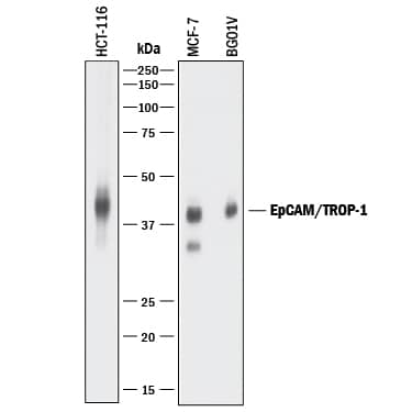
HAF017). A specific band was detected for EpCAM/TROP-1 at approximately 40 kDa (as indicated). This experiment was conducted under reducing conditions and using
Immunoblot Buffer Group 1." alt="Western blot shows lysates of HCT-116 human colorectal carcinoma cell line, MCF-7 human breast cancer cell line, and BG01V human embryonic stem cells. PVDF membrane was probed with 0.2 µg/mL of Goat Anti-Human EpCAM/TROP-1 Antigen Affinity-purified Polyclonal Antibody (Catalog # AF960) followed by HRP-conjugated Anti-Goat IgG Secondary Antibody (
HAF017). A specific band was detected for EpCAM/TROP-1 at approximately 40 kDa (as indicated). This experiment was conducted under reducing conditions and using
Immunoblot Buffer Group 1." class="big_thumb" />
AB-108-C, open histogram), followed by Phycoerythrin-conjugated Anti-Goat IgG Secondary Antibody (
F0107)." class="big_lightbox">
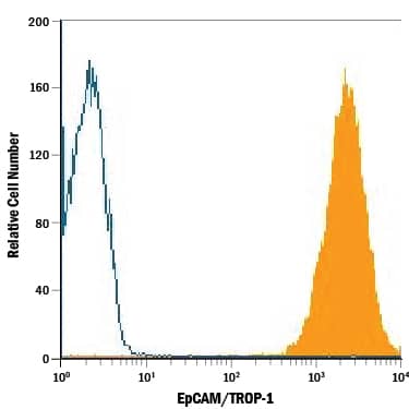
AB-108-C, open histogram), followed by Phycoerythrin-conjugated Anti-Goat IgG Secondary Antibody (
F0107)." class="big_thumb" />
VC004). Before incubation with the primary antibody, tissue was subjected to heat-induced epitope retrieval using Antigen Retrieval Reagent-Basic (Catalog #
CTS013). Tissue was stained using DAB (brown) and counterstained with hematoxylin (blue). Specific staining was localized to cytoplasm and plasma membrane. View our protocol for
IHC Staining with VisUCyte HRP Polymer Detection Reagents." class="big_lightbox">
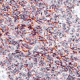
VC004). Before incubation with the primary antibody, tissue was subjected to heat-induced epitope retrieval using Antigen Retrieval Reagent-Basic (Catalog #
CTS013). Tissue was stained using DAB (brown) and counterstained with hematoxylin (blue). Specific staining was localized to cytoplasm and plasma membrane. View our protocol for
IHC Staining with VisUCyte HRP Polymer Detection Reagents." class="big_thumb" />
VC004). Before incubation with the primary antibody, tissue was subjected to heat-induced epitope retrieval using Antigen Retrieval Reagent-Basic (
CTS013). Tissue was stained using DAB (brown) and counterstained with hematoxylin (blue). Specific staining was localized to cytoplasm and plasma membrane. View our protocol for
IHC Staining with VisUCyte HRP Polymer Detection Reagents." class="big_lightbox">
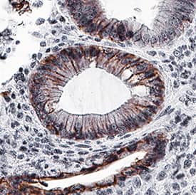
VC004). Before incubation with the primary antibody, tissue was subjected to heat-induced epitope retrieval using Antigen Retrieval Reagent-Basic (
CTS013). Tissue was stained using DAB (brown) and counterstained with hematoxylin (blue). Specific staining was localized to cytoplasm and plasma membrane. View our protocol for
IHC Staining with VisUCyte HRP Polymer Detection Reagents." class="big_thumb" />
960-EP) was serially diluted and captured by Mouse Anti-Human
EpCAM/TROP‑1 Monoclonal Antibody (Catalog #
MAB9601) coated on a Clear Polystyrene Microplate (Catalog #
DY990). Goat Anti-Human
EpCAM/TROP‑1 Antigen Affinity-purified Polyclonal Antibody (Catalog # AF960) was biotinylated and incubated with the protein captured on the plate. Detection of the standard curve was achieved by incubating Streptavidin-HRP (Catalog #
DY998)" class="big_lightbox">

960-EP) was serially diluted and captured by Mouse Anti-Human
EpCAM/TROP‑1 Monoclonal Antibody (Catalog #
MAB9601) coated on a Clear Polystyrene Microplate (Catalog #
DY990). Goat Anti-Human
EpCAM/TROP‑1 Antigen Affinity-purified Polyclonal Antibody (Catalog # AF960) was biotinylated and incubated with the protein captured on the plate. Detection of the standard curve was achieved by incubating Streptavidin-HRP (Catalog #
DY998)" class="big_thumb" />
 HAF017). A specific band was detected for EpCAM/TROP-1 at approximately 40 kDa (as indicated). This experiment was conducted under reducing conditions and using Immunoblot Buffer Group 1." alt="Western blot shows lysates of HCT-116 human colorectal carcinoma cell line, MCF-7 human breast cancer cell line, and BG01V human embryonic stem cells. PVDF membrane was probed with 0.2 µg/mL of Goat Anti-Human EpCAM/TROP-1 Antigen Affinity-purified Polyclonal Antibody (Catalog # AF960) followed by HRP-conjugated Anti-Goat IgG Secondary Antibody (HAF017). A specific band was detected for EpCAM/TROP-1 at approximately 40 kDa (as indicated). This experiment was conducted under reducing conditions and using Immunoblot Buffer Group 1." class="big_thumb" />
 DR555MS) and counterstained with DAPI (blue; Lunaphore Catalog # DR100). Specific staining was localized to the membrane. Protocol available in COMET™ Panel Builder." alt="EpCAM/TROP-1 was detected in immersion fixed paraffin-embedded sections of human Colon using Mouse Anti-Human EpCAM/TROP-1, Monoclonal Antibody (Catalog # MAB960) at 1ug/mL at 37° Celsius for 4 minutes. Before incubation with the primary antibody, tissue underwent an all-in-one dewaxing and antigen retrieval preprocessing using PreTreatment Module (PT Module) and Dewax and HIER Buffer H (pH 9; Epredia Catalog # TA-999-DHBH). Tissue was stained using the Alexa Fluor™ 555 Goat anti-Mouse IgG Secondary Antibody at 1:100 at 37° Celsius for 2 minutes. (Yellow; Lunaphore Catalog # DR555MS) and counterstained with DAPI (blue; Lunaphore Catalog # DR100). Specific staining was localized to the membrane. Protocol available in COMET™ Panel Builder." class="big_thumb" />


 AB-108-C, open histogram), followed by Phycoerythrin-conjugated Anti-Goat IgG Secondary Antibody (
 NL001) and counterstained with DAPI (blue). Specific staining was localized to cytoplasm and cell membrane. View our protocol for
  VC004). Before incubation with the primary antibody, tissue was subjected to heat-induced epitope retrieval using Antigen Retrieval Reagent-Basic (Catalog #
  VC004). Before incubation with the primary antibody, tissue was subjected to heat-induced epitope retrieval using Antigen Retrieval Reagent-Basic (
 NL001) and counterstained with DAPI (blue). Specific staining was localized to cell surface . View our protocol for
  960-EP) was serially diluted and captured by Mouse Anti-Human
 ![Immunohistochemistry-Paraffin: EpCAM/TROP1 Antibody (AUA1) [NB600-1182] - Analysis of a FFPE human breast carcinoma tissue section using 1:100 dilution of EpCAM/TROP1 antibody (clone AUA1) on a Bond Rx autostainer (Leica Biosystems). The assay involved 20 minutes of heat induced antigen retrieval (HIER) with 10mM sodium citrate buffer (pH 6.0) and endogenous peroxidase quenching using peroxide block. The sections were incubated with primary antibody for 30 minutes. Bond Polymer Refine Detection (Leica Biosystems) and DAB were used for signal detection which followed counterstaining with hematoxylin. Whole slide scanning and capturing of representative images (20X) were performed using Aperio AT2 (Leica Biosystems). This EpCAM antibody generated an expected immunostaining of EpCAM (CD326) protein in the membranes of the cancer cells and the inter-cellular spaces. The tumor stroma and the stromal cells did not show EpCAM immunopositivity.](http://images.novusbio.com/fullsize/EpCAM-TROP1-Antibody-AUA1-Immunohistochemistry-Paraffin-NB600-1182-img0009.jpg "Immunohistochemistry-Paraffin: EpCAM/TROP1 Antibody (AUA1) [NB600-1182] - Analysis of a FFPE human breast carcinoma tissue section using 1:100 dilution of EpCAM/TROP1 antibody (clone AUA1) on a Bond Rx autostainer (Leica Biosystems). The assay involved 20 minutes of heat induced antigen retrieval (HIER) with 10mM sodium citrate buffer (pH 6.0) and endogenous peroxidase quenching using peroxide block. The sections were incubated with primary antibody for 30 minutes. Bond Polymer Refine Detection (Leica Biosystems) and DAB were used for signal detection which followed counterstaining with hematoxylin. Whole slide scanning and capturing of representative images (20X) were performed using Aperio AT2 (Leica Biosystems). This EpCAM antibody generated an expected immunostaining of EpCAM (CD326) protein in the membranes of the cancer cells and the inter-cellular spaces. The tumor stroma and the stromal cells did not show EpCAM immunopositivity.")
![Western Blot: EpCAM/TROP1 Antibody (AUA1) [NB600-1182] - Detection of EpCAM/CD326 in two Human mammary tumor cell lines, MDA-MB-231 (mesenchyme-like) and MCF-7 (epithelium-like). Dilution: 1:500 in PBS with 5% BSA. Secondary Ab: anti-Mouse IgG 1:5,000. This image was submitted via customer Review.](http://images.novusbio.com/fullsize/EpCAM-TROP1-Antibody-AUA1-Western-Blot-NB600-1182-img0015.jpg)
![Immunocytochemistry/Immunofluorescence: EpCAM/TROP1 Antibody (AUA1) [NB600-1182] - A431 cells were fixed for 10 minutes using 10% formalin and then permeabilized for 5 minutes using 1X PBS + 0.05% Triton X-100. The cells were incubated with anti-EpCAM/TROP1 (AUA1) conjugated to Alexa Fluor 488 [NB600-1182AF488] at 10ug/ml for 1 hour at room temperature. Nuclei were counterstained with DAPI (blue). Cells were imaged using a 40X objective.](http://images.novusbio.com/fullsize/EpCAM-TROP1-Antibody-AUA1-Immunocytochemistry-Immunofluorescence-NB600-1182-img0016.jpg)
![Immunohistochemistry-Paraffin: EpCAM/TROP1 Antibody (AUA1) [NB600-1182] - Analysis of a FFPE human breast carcinoma tissue with 1:500 dilution of EpCAM/TROP1 antibody (clone AUA1) on a Bond Rx autostainer (Leica Biosystems). The assay involved 20 minutes of heat induced antigen retrieval (HIER) with 10mM sodium citrate buffer (pH 6.0) and endogenous peroxidase quenching using peroxide block. The sections were incubated with primary antibody for 30 minutes. Bond Polymer Refine Detection (Leica Biosystems) and DAB were used for signal detection which followed counterstaining with hematoxylin. Whole slide scanning and capturing of representative images (20X) were performed using Aperio AT2 (Leica Biosystems). The staining was primarily localized to the membranes and inter-cellular spaces of the cancer cells. Staining intensity for 1:500 dilution was lower than what was seen in sections tested with 1:100 dilution. Staining was performed by Histowiz.](http://images.novusbio.com/fullsize/EpCAM-TROP1-Antibody-AUA1-Immunohistochemistry-Paraffin-NB600-1182-img0014.jpg)
![Flow Cytometry: EpCAM/TROP1 Antibody (AUA1) [NB600-1182] - Using the PE direct conjugate A cell surface stain was performed on HT-29 cells with EpCAM (AUA1) antibody NB600-1182PE (blue) and a matched isotype control NBP2-27287PE (orange). Cells were incubated in an antibody dilution of 5 ug/mL for 20 minutes at room temperature. Both antibodies were conjugated to phycoerythrin.](http://images.novusbio.com/fullsize/EpCAM-TROP1-Antibody-AUA1-Flow-Cytometry-NB600-1182-img0008.jpg)
![Western Blot: EpCAM/TROP1 Antibody (AUA1) [NB600-1182] - Analysis of A431 lysate (A) and Hek293 lysate (B) using EpCAM/CD326 antibody (AUA1) at 2 ug/ml. Non-reduced lysate was used in each lane.](http://images.novusbio.com/fullsize/EpCAM-TROP1-Antibody-AUA1-Western-Blot-NB600-1182-img0001.jpg)
![Flow Cytometry: EpCAM/TROP1 Antibody (AUA1) [NB600-1182] - Intracellular flow cytometric staining of 1 x 10^6 CHO (A) and HeLa (B) cells using EpCAM/CD326 antibody (blue) and matched Isotype control (orange). An antibody concentration of 1 ug/1x10^6 cells was used.](http://images.novusbio.com/fullsize/EpCAM-TROP1-Antibody-AUA1-Flow-Cytometry-NB600-1182-img0007.jpg)
 DR555MS) and counterstained with DAPI (blue; Lunaphore Catalog #
  DR555MS) and counterstained with DAPI (blue; Lunaphore Catalog #
  NL009) and counterstained with DAPI (blue). View our protocol for
  NL007) and counterstained with DAPI (blue). View our protocol for
  CTS002) and counterstained with hematoxylin (blue). Specific staining was localized to cancer cells. View our protocol for
  NL007) and counterstained with DAPI (blue). Specific staining was localized to cell surface and cytoplasm. View our protocol for
 ![Immunocytochemistry/Immunofluorescence: EpCAM/TROP1 Antibody (VU-1D9) [NBP2-33051] - Immunofluorescence Analysis of MCF-7 cells EpCAM/TROP1 Antibody (VU-1D9) labeled with CF488 (green); NucSpot is used to label the nuclei (red).](http://images.novusbio.com/fullsize/EpCAM-TROP1-Antibody-VU-1D9-Immunocytochemistry-Immunofluorescence-NBP2-33051-img0010.jpg "Immunocytochemistry/Immunofluorescence: EpCAM/TROP1 Antibody (VU-1D9) [NBP2-33051] - Immunofluorescence Analysis of MCF-7 cells EpCAM/TROP1 Antibody (VU-1D9) labeled with CF488 (green); NucSpot is used to label the nuclei (red).")
![Immunohistochemistry-Paraffin: EpCAM/TROP1 Antibody (VU-1D9) [NBP2-33051] - Formalin-fixed, paraffin-embedded human Ovarian Carcinoma stained with Ep-CAM monoclonal antibody (VU-1D9).](http://images.novusbio.com/fullsize/EpCAM-TROP1-Antibody-VU-1D9-Immunohistochemistry-Paraffin-NBP2-33051-img0003.jpg)
![Flow Cytometry: EpCAM/TROP1 Antibody (VU-1D9) [NBP2-33051] - Flow Cytometric analysis of human EpCAM/TROP1 on HT29 cells. Black: cells alone; Green: Isotype Control; Red: PE-labeled EpCAM/TROP1 antibody (VU-1D9).](http://images.novusbio.com/fullsize/EpCAM-TROP1-Antibody-VU-1D9-Flow-Cytometry-NBP2-33051-img0015.jpg)
![Flow Cytometry: EpCAM/TROP1 Antibody (VU-1D9) [NBP2-33051] - Analysis using Azide and BSA Free version of NBP2-33051. Surface staining of HT29 cells using Ep-CAM MAb (VU-1D9) (red) and isotype control antibody (green).](http://images.novusbio.com/fullsize/EpCAM-TROP1-Antibody-VU-1D9-Flow-Cytometry-NBP2-33051-img0005.jpg)
![Flow Cytometry: EpCAM/TROP1 Antibody (VU-1D9) [NBP2-33051] - Surface staining of Human Colon HT29 cells using EpCAM/TROP1 Antibody (VU-1D9) (red) and isotype control antibody (green).](http://images.novusbio.com/fullsize/EpCAM-TROP1-Antibody-VU-1D9-Flow-Cytometry-NBP2-33051-img0008.jpg)
![Flow Cytometry: EpCAM/TROP1 Antibody (VU-1D9) [NBP2-33051] - Flow Cytometric Analysis of PFA-fixed MCF-7 cells using EpCAM/TROP1 Mouse Monoclonal Antibody (VU-1D9) followed by goat anti-Mouse IgG-CF488 (Blue); Isotype Control (Red).](http://images.novusbio.com/fullsize/EpCAM-TROP1-Antibody-VU-1D9-Flow-Cytometry-NBP2-33051-img0011.jpg)
![Flow Cytometry: EpCAM/TROP1 Antibody (VU-1D9) [NBP2-33051] - Flow cytometry of MCF-7 cells unstained (gray) or stained with CF555-labeled EpCAM/TROP1 monoclonal antibody (VU-1D9) (orange)](http://images.novusbio.com/fullsize/EpCAM-TROP1-Antibody-VU-1D9-Flow-Cytometry-NBP2-33051-img0012.jpg)
![Flow Cytometry: EpCAM/TROP1 Antibody (VU-1D9) [NBP2-33051] - Flow cytometry of bead-bound exosomes derived from MCF-7 cells. Unstained exosomes (gray) or stained with CF555-labeled EpCAM/TROP1 monoclonal antibody (VU-1D9) (orange)](http://images.novusbio.com/fullsize/EpCAM-TROP1-Antibody-VU-1D9-Flow-Cytometry-NBP2-33051-img0013.jpg)
![Western Blot: EpCAM/TROP1 Antibody (MOC-31) [NBP2-44640] - Western Blot analysis of human lung tissue lysate using EpCAM/TROP1 Antibody (MOC-31).](http://images.novusbio.com/fullsize/EpCAM-TROP1-Antibody-MOC-31-Western-Blot-NBP2-44640-img0005.jpg "Western Blot: EpCAM/TROP1 Antibody (MOC-31) [NBP2-44640] - Western Blot analysis of human lung tissue lysate using EpCAM/TROP1 Antibody (MOC-31).")
![Immunohistochemistry-Paraffin: EpCAM/TROP1 Antibody (MOC-31) [NBP2-44640] - Human Ovarian Carcinoma stained with Ep-CAM Monoclonal Antibody (MOC-31).](http://images.novusbio.com/fullsize/EpCAM-TROP1-Antibody-MOC-31-Immunohistochemistry-Paraffin-NBP2-44640-img0003.jpg)
![Immunohistochemistry-Paraffin: EpCAM/TROP1 Antibody (MOC-31) [NBP2-44640] - Human Colon Carcinoma stained with Ep-CAM Monoclonal Antibody (MOC-31).](http://images.novusbio.com/fullsize/EpCAM-TROP1-Antibody-MOC-31-Immunohistochemistry-Paraffin-NBP2-44640-img0001.jpg)
![Immunohistochemistry-Paraffin: EpCAM/TROP1 Antibody (MOC-31) [NBP2-44640] - Human Endometrial Carcinoma stained with Ep-CAM Monoclonal Antibody (MOC-31).](http://images.novusbio.com/fullsize/EpCAM-TROP1-Antibody-MOC-31-Immunohistochemistry-Paraffin-NBP2-44640-img0002.jpg)
![Immunohistochemistry-Paraffin: EpCAM/TROP1 Antibody (EGP40/1110) - (ECD) [NBP2-44635] - Formalin-fixed, paraffin-embedded feline (cat) small intestine stained with EpCAM/TROP1 Antibody (EGP40/1110) - (ECD).](http://images.novusbio.com/fullsize/EpCAM-TROP1-Antibody-EGP40-1110-ECD-Immunohistochemistry-Paraffin-NBP2-44635-img0009.jpg "Immunohistochemistry-Paraffin: EpCAM/TROP1 Antibody (EGP40/1110) - (ECD) [NBP2-44635] - Formalin-fixed, paraffin-embedded feline (cat) small intestine stained with EpCAM/TROP1 Antibody (EGP40/1110) - (ECD).")
![Immunohistochemistry-Paraffin: EpCAM/TROP1 Antibody (EGP40/1110) - (ECD) [NBP2-44635] - Human Ovarian Carcinoma stained with Ep-CAM Monoclonal Antibody (EGP40/1110).](http://images.novusbio.com/fullsize/EpCAM-TROP1-Antibody-EGP40-1110-ECD-Immunohistochemistry-Paraffin-NBP2-44635-img0001.jpg)
![Immunohistochemistry-Paraffin: EpCAM/TROP1 Antibody (EGP40/1110) - (ECD) [NBP2-44635] - Rat Colon stained with Ep-CAM Monoclonal Antibody (EGP40/1110).](http://images.novusbio.com/fullsize/EpCAM-TROP1-Antibody-EGP40-1110-ECD-Immunohistochemistry-Paraffin-NBP2-44635-img0002.jpg)
![Immunohistochemistry-Paraffin: EpCAM/TROP1 Antibody (EGP40/1110) - (ECD) [NBP2-44635] - Rat Lung stained with Ep-CAM Monoclonal Antibody (EGP40/1110).](http://images.novusbio.com/fullsize/EpCAM-TROP1-Antibody-EGP40-1110-ECD-Immunohistochemistry-Paraffin-NBP2-44635-img0003.jpg)
![Immunohistochemistry-Paraffin: EpCAM/TROP1 Antibody (EGP40/1110) - (ECD) [NBP2-44635] - Formalin-fixed, paraffin-embedded Mouse Colon stained with EpCAM Mouse Monoclonal Antibody (EGP40/1110).](http://images.novusbio.com/fullsize/EpCAM-TROP1-Antibody-EGP40-1110-ECD-Immunohistochemistry-Paraffin-NBP2-44635-img0006.jpg)
![Immunohistochemistry-Paraffin: EpCAM/TROP1 Antibody (EGP40/1110) - (ECD) [NBP2-44635] - Rat Oviduct stained with Ep-CAM Monoclonal Antibody (EGP40/1110).](http://images.novusbio.com/fullsize/EpCAM-TROP1-Antibody-EGP40-1110-ECD-Immunohistochemistry-Paraffin-NBP2-44635-img0004.jpg)
![Immunohistochemistry-Paraffin: EpCAM/TROP1 Antibody (EGP40/1110) - (ECD) [NBP2-44635] - Formalin-fixed, paraffin-embedded canine (dog) colon stained with EpCAM/TROP1 Antibody (EGP40/1110) - (ECD).](http://images.novusbio.com/fullsize/EpCAM-TROP1-Antibody-EGP40-1110-ECD-Immunohistochemistry-Paraffin-NBP2-44635-img0008.jpg)
![Protein Array: EpCAM/TROP1 Antibody (EGP40/1110) - (ECD) [NBP2-44635] - Analysis of Protein Array containing >19,000 full-length human proteins using EpCAM/TROP1 Antibody (EGP40/1110)Z- and S- Score: The Z-score represents the strength of a signal that a monoclonal antibody (MAb) (in combination with a fluorescently-tagged anti-IgG secondary antibody) produces when binding to a particular protein on the HuProt(TM) array. Z-scores are described in units of standard deviations (SD's) above the mean value of all signals generated on that array. If targets on HuProt(TM) are arranged in descending order of the Z-score, the S-score is the difference (also in units of SD's) between the Z-score. S-score therefore represents the relative target specificity of a MAb to its intended target. A MAb is considered to specific to its intended target, if the MAb has an S-score of at least 2.5.](http://images.novusbio.com/fullsize/EpCAM-TROP1-Antibody-EGP40-1110-ECD-Protein-Array-NBP2-44635-img0007.jpg)
![Immunohistochemistry: Mouse Monoclonal EpCAM/TROP1 Antibody (EGP40/1110) - (ECD) [NBP2-44635] - EpCAM/TROP1 signal in lung alveoli. Analysis in postfixed macaque lung tissue at a 1:100 concentration. Image from a verified customer review.](http://images.novusbio.com/fullsize/antibody/nbp2-44635_mouse-monoclonal-epcam-trop1-antibody-egp40-1110-ecd-immunohistochemistry-712202411034..png)
![Western Blot: EpCAM/TROP1 Antibody [NBP2-27107] - Analysis of EpCAM using EpCAM antibody. Human HCT116 cell lysate in the 1) absence and 2) presence of immunizing peptide and 3) mouse NIH 3T3 lysate probed with 3 ug/mL of EpCAM antibody. Goat anti-rabbit Ig HRP secondary antibody and PicoTect ECL substrate solution were used for this test.](http://images.novusbio.com/fullsize/EpCAM-TROP1-Antibody-Western-Blot-NBP2-27107-img0006.jpg "Western Blot: EpCAM/TROP1 Antibody [NBP2-27107] - Analysis of EpCAM using EpCAM antibody. Human HCT116 cell lysate in the 1) absence and 2) presence of immunizing peptide and 3) mouse NIH 3T3 lysate probed with 3 ug/mL of EpCAM antibody. Goat anti-rabbit Ig HRP secondary antibody and PicoTect ECL substrate solution were used for this test.")
![Immunohistochemistry-Paraffin: EpCAM/TROP1 Antibody [NBP2-27107] - FFPE human colon stained with EpCAM antibody at 1:200, peroxidase-conjugate and DAB chromogen.](http://images.novusbio.com/fullsize/EpCAM-TROP1-Antibody-Immunohistochemistry-Paraffin-NBP2-27107-img0005.jpg)
![Immunocytochemistry/Immunofluorescence: EpCAM/TROP1 Antibody (EGP40/1555R) - (ECD) [NBP2-53370] - Confocal immunofluorescence analysis of MeOH-fixed HeLa cells. EpCAM/TROP1 antibody (EGP40/1555R) - (ECD) followed by goat anti-mouse IgG-CF488 (green). Nuclei counterstained with RedDot](http://images.novusbio.com/fullsize/EpCAM-TROP1-Antibody-EGP40-1555R-ECD-Immunocytochemistry-Immunofluorescence-NBP2-53370-img0003.jpg "Immunocytochemistry/Immunofluorescence: EpCAM/TROP1 Antibody (EGP40/1555R) - (ECD) [NBP2-53370] - Confocal immunofluorescence analysis of MeOH-fixed HeLa cells. EpCAM/TROP1 antibody (EGP40/1555R) - (ECD) followed by goat anti-mouse IgG-CF488 (green). Nuclei counterstained with RedDot")
![Immunohistochemistry-Paraffin: EpCAM/TROP1 Antibody (EGP40/1555R) - (ECD) [NBP2-53370] - Formalin-fixed, paraffin-embedded human colon carcinoma stained with EpCAM/TROP1 Antibody (EGP40/1555R) - (ECD).](http://images.novusbio.com/fullsize/EpCAM-TROP1-Antibody-EGP40-1555R-ECD-Immunohistochemistry-Paraffin-NBP2-53370-img0005.jpg)
![Flow Cytometry: EpCAM/TROP1 Antibody (EGP40/1555R) - (ECD) [NBP2-53370] - Flow Cytometric Analysis of trypsinized-MCF-7 cells. EpCAM/TROP1 Antibody (EGP40/1555R) followed by goat anti-Mouse IgG-CF488 (Blue); Isotype Control (Red).](http://images.novusbio.com/fullsize/EpCAM-TROP1-Antibody-EGP40-1555R-ECD-Flow-Cytometry-NBP2-53370-img0002.jpg)
![Immunohistochemistry-Paraffin: EpCAM/TROP1 Antibody (EGP40/1555R) - (ECD) [NBP2-53370] - Formalin-fixed, paraffin-embedded human Thyroid Carcinoma stained with Ep-CAM Recombinant Rabbit Monoclonal Antibody (EGP40/1555R).](http://images.novusbio.com/fullsize/EpCAM-TROP1-Antibody-EGP40-1555R-ECD-Immunohistochemistry-Paraffin-NBP2-53370-img0001.jpg)
![Immunohistochemistry-Paraffin: EpCAM/TROP1 Antibody (EGP40/1555R) - (ECD) [NBP2-53370] - Formalin-fixed, paraffin-embedded human prostate carcinoma stained with EpCAM/TROP1 Antibody (EGP40/1555R) - (ECD).](http://images.novusbio.com/fullsize/EpCAM-TROP1-Antibody-EGP40-1555R-ECD-Immunohistochemistry-Paraffin-NBP2-53370-img0004.jpg)
![Western Blot: EpCAM/TROP1 Antibody (EGP40/1372) - (ECD) [NBP2-53264] - Western Blot of human lung lysate using NBP2-53264.](http://images.novusbio.com/fullsize/EpCAM-TROP1-Antibody-EGP40-1372-ECD-Western-Blot-NBP2-53264-img0004.jpg "Western Blot: EpCAM/TROP1 Antibody (EGP40/1372) - (ECD) [NBP2-53264] - Western Blot of human lung lysate using NBP2-53264.")
![Immunocytochemistry/Immunofluorescence: EpCAM/TROP1 Antibody (EGP40/1372) - (ECD) [NBP2-53264] - Immunofluorescent staining of MCF-7 cells. NBP2-53264 labeled with CF488 (green). Nuclei are stained with Reddot (red)](http://images.novusbio.com/fullsize/EpCAM-TROP1-Antibody-EGP40-1372-ECD-Immunocytochemistry-Immunofluorescence-NBP2-53264-img0007.jpg)
![Immunohistochemistry-Paraffin: EpCAM/TROP1 Antibody (EGP40/1372) - (ECD) [NBP2-53264] - Formalin-fixed, paraffin-embedded feline (cat) small intestine stained with EpCAM/TROP1 Antibody (EGP40/1372) - (ECD).](http://images.novusbio.com/fullsize/EpCAM-TROP1-Antibody-EGP40-1372-ECD-Immunohistochemistry-Paraffin-NBP2-53264-img0013.jpg)
![Flow Cytometry: EpCAM/TROP1 Antibody (EGP40/1372) - (ECD) [NBP2-53264] - Flow Cytometric Analysis of PFA-fixed MCF-7 cells using EpCAM/TROP1 Antibody (EGP40/1372).followed by goat anti-Mouse IgG-CF488 (Blue); Isotype Control (Red).](http://images.novusbio.com/fullsize/EpCAM-TROP1-Antibody-EGP40-1372-ECD-Flow-Cytometry-NBP2-53264-img0010.jpg)
![Western Blot: EpCAM/TROP1 Antibody (EGP40/1372) - (ECD) [NBP2-53264] - Western Blot of 293, A431 and HCT116 Cell Lysate using EpCAM, Monoclonal Antibody (EGP40/1372).](http://images.novusbio.com/fullsize/EpCAM-TROP1-Antibody-EGP40-1372-ECD-Western-Blot-NBP2-53264-img0002.jpg)
![Immunocytochemistry/Immunofluorescence: EpCAM/TROP1 Antibody (EGP40/1372) - (ECD) [NBP2-53264] - Confocal Immunofluorescence of human Colorectal Carcinoma. NBP2-53264 labeled with CF488 (green); Nuclei are labeled with Reddot (red).](http://images.novusbio.com/fullsize/EpCAM-TROP1-Antibody-EGP40-1372-ECD-Immunocytochemistry-Immunofluorescence-NBP2-53264-img0006.jpg)
![Immunohistochemistry-Paraffin: EpCAM/TROP1 Antibody (EGP40/1372) - (ECD) [NBP2-53264] - Formalin-fixed, paraffin-embedded Human Colorectal Carcinoma stained with EpCAM Monoclonal Antibody (EGP40/1372).](http://images.novusbio.com/fullsize/EpCAM-TROP1-Antibody-EGP40-1372-ECD-Immunohistochemistry-Paraffin-NBP2-53264-img0001.jpg)
![Immunohistochemistry-Paraffin: EpCAM/TROP1 Antibody (EGP40/1372) - (ECD) [NBP2-53264] - Formalin-fixed, paraffin-embedded human Hepatocellular Carcinoma stained with NBP2-53264.](http://images.novusbio.com/fullsize/EpCAM-TROP1-Antibody-EGP40-1372-ECD-Immunohistochemistry-Paraffin-NBP2-53264-img0003.jpg)
![Immunohistochemistry-Paraffin: EpCAM/TROP1 Antibody (EGP40/1372) - (ECD) [NBP2-53264] - Formalin-fixed, paraffin-embedded canine (dog) bladder stained with EpCAM/TROP1 Antibody (EGP40/1372) - (ECD).](http://images.novusbio.com/fullsize/EpCAM-TROP1-Antibody-EGP40-1372-ECD-Immunohistochemistry-Paraffin-NBP2-53264-img0012.jpg)
![Protein Array: EpCAM/TROP1 Antibody (EGP40/1372) - (ECD) [NBP2-53264] - Analysis of Protein Array containing >19,000 full-length human proteins using EpCAM/TROP1 Antibody (EGP40/1372) Z- and S- Score: The Z-score represents the strength of a signal that a monoclonal antibody (MAb) (in combination with a fluorescently-tagged anti-IgG secondary antibody) produces when binding to a particular protein on the HuProt(TM) array. Z-scores are described in units of standard deviations (SD's) above the mean value of all signals generated on that array. If targets on HuProt(TM) are arranged in descending order of the Z-score, the S-score is the difference (also in units of SD's) between the Z-score. S-score therefore represents the relative target specificity of a MAb to its intended target. A MAb is considered to specific to its intended target, if the MAb has an S-score of at least 2.5.](http://images.novusbio.com/fullsize/EpCAM-TROP1-Antibody-EGP40-1372-ECD-Protein-Array-NBP2-53264-img0011.jpg)
![Immunohistochemistry-Paraffin: EpCAM/TROP1 Antibody (CL6313) [NBP2-76486] - Analysis in human small intestine and tonsil tissues. Corresponding EPCAM RNA-seq data are presented for the same tissues.](http://images.novusbio.com/fullsize/EpCAM-TROP1-Antibody-CL6313-Immunohistochemistry-Paraffin-NBP2-76486-img0002.jpg "<b>Orthogonal Strategies Validation. </b>Immunohistochemistry-Paraffin: EpCAM/TROP1 Antibody (CL6313) [NBP2-76486] - Analysis in human small intestine and tonsil tissues. Corresponding EPCAM RNA-seq data are presented for the same tissues.")
![<b>Orthogonal Strategies Validation. </b>Western Blot: EpCAM/TROP1 Antibody (CL6313) [NBP2-76486] - Analysis in human cell line CAPAN-2.](http://images.novusbio.com/fullsize/EpCAM-TROP1-Antibody-CL6313-Western-Blot-NBP2-76486-img0001.jpg)
![Immunohistochemistry-Paraffin: EpCAM/TROP1 Antibody (CL6313) [NBP2-76486] - Staining of human tonsil shows no positivity in lymphoid cells as expected.](http://images.novusbio.com/fullsize/EpCAM-TROP1-Antibody-CL6313-Immunohistochemistry-Paraffin-NBP2-76486-img0004.jpg)
![Immunohistochemistry-Paraffin: EpCAM/TROP1 Antibody (CL6313) [NBP2-76486] - Staining of human small intestine shows strong membranous positivity in glandular cells.](http://images.novusbio.com/fullsize/EpCAM-TROP1-Antibody-CL6313-Immunohistochemistry-Paraffin-NBP2-76486-img0003.jpg)
![Western Blot: EpCAM/TROP1 Antibody (BLR077G) [NBP3-14685] - Whole cell lysate (50 ug) from BW5147.3, CTLL-2, EL4, and mIMCD-3 cells prepared using NETN lysis buffer. Antibody: Rabbit anti-EpCAM recombinant monoclonal antibody [BLR077G] used at 1:1000. Secondary: HRP-conjugated goat anti-rabbit IgG. Chemiluminescence with an exposure time of 30 seconds.](http://images.novusbio.com/fullsize/EpCAM-TROP1-Antibody-BLR077G-Western-Blot-NBP3-14685-img0003.jpg "Western Blot: EpCAM/TROP1 Antibody (BLR077G) [NBP3-14685] - Whole cell lysate (50 ug) from BW5147.3, CTLL-2, EL4, and mIMCD-3 cells prepared using NETN lysis buffer. Antibody: Rabbit anti-EpCAM recombinant monoclonal antibody [BLR077G] used at 1:1000. Secondary: HRP-conjugated goat anti-rabbit IgG. Chemiluminescence with an exposure time of 30 seconds.")
![Immunocytochemistry/Immunofluorescence: EpCAM/TROP1 Antibody (BLR077G) [NBP3-14685] - Section of OVCAR-3 cells. Antibody: Rabbit anti-EpCAM recombinant monoclonal antibody [BLR077G] used at 1:100. Secondary: HRP-conjugated goat anti-rabbit IgG Substrate: DAB](http://images.novusbio.com/fullsize/EpCAM-TROP1-Antibody-BLR077G-Immunocytochemistry-Immunofluorescence-NBP3-14685-img0008.jpg)
![Immunohistochemistry-Paraffin: EpCAM/TROP1 Antibody (BLR077G) [NBP3-14685] - Section of human breast carcinoma. Antibody: Rabbit anti-EpCAM recombinant monoclonal antibody [BLR077G] used at 1:100. Secondary: HRP-conjugated goat anti-rabbit IgG. Substrate: DAB.](http://images.novusbio.com/fullsize/EpCAM-TROP1-Antibody-BLR077G-Immunohistochemistry-Paraffin-NBP3-14685-img0006.jpg)
![Western Blot: EpCAM/TROP1 Antibody (BLR077G) [NBP3-14685] - Whole cell lysate (50 ug) from HeLa, MCF-7, K-562, LNCaP, RKO, Hep-G2, Jurkat, HT-29, and SW620 cells prepared using NETN lysis buffer. Antibody: Rabbit anti-EpCAM recombinant monoclonal antibody [BLR077G]used at 1:1000. Secondary: HRP-conjugated goat anti-rabbit IgG. Chemiluminescence with anexposure time of 10 seconds. Lower Panel: Rabbit antiRPL23.](http://images.novusbio.com/fullsize/EpCAM-TROP1-Antibody-BLR077G-Western-Blot-NBP3-14685-img0002.jpg)
![Immunocytochemistry/Immunofluorescence: EpCAM/TROP1 Antibody (BLR077G) [NBP3-14685] - Section of HT-29 cells. Antibody: Rabbit anti-EpCAM recombinant monoclonal antibody [BLR077G] used at 1:100. Secondary: HRP-conjugated goat anti-rabbit IgG. Substrate: DA](http://images.novusbio.com/fullsize/EpCAM-TROP1-Antibody-BLR077G-Immunocytochemistry-Immunofluorescence-NBP3-14685-img0007.jpg)
![Immunohistochemistry-Paraffin: EpCAM/TROP1 Antibody (BLR077G) [NBP3-14685] - Rabbit anti-EpCAM recombinant monoclonal antibody [BLR077G]. Secondary: HRP-conjugated goat anti-rabbit IgG. Substrate: Opal™. Counterstain: DAPI.](http://images.novusbio.com/fullsize/EpCAM-TROP1-Antibody-BLR077G-Immunohistochemistry-Paraffin-NBP3-14685-img0004.jpg)
![Immunohistochemistry-Paraffin: EpCAM/TROP1 Antibody (BLR077G) [NBP3-14685] - Human ovarian carcinoma. Antibody: Rabbit anti-EpCAM recombinant monoclonalantibody [BLR077G] used at 1:100. Secondary: HRP-conjugated goat anti-rabbit IgG(A120-501P). Substrate: DA](http://images.novusbio.com/fullsize/EpCAM-TROP1-Antibody-BLR077G-Immunohistochemistry-Paraffin-NBP3-14685-img0005.jpg)
![Immunoprecipitation: EpCAM/TROP1 Antibody [NBP3-14685] - Whole cell lysate (1.0 mg per IP reaction; 20% of IP loaded) from MCF-7 cellsprepared using NETN lysis buffer. Antibody: Rabbit antiEpCAM recombinant monoclonal antibody used at 12 ul per reaction. For blottingimmunoprecipitated EpCAM was used at1:1000. Chemiluminescence with an exposure time of 30seconds.](http://images.novusbio.com/fullsize/EpCAM-TROP1-Antibody-Immunoprecipitation-NBP3-14685-img0001.jpg)
![Immunohistochemistry-Paraffin: EpCAM/TROP1 Antibody (EGP40/826) [NBP2-44643] - Human Breast Carcinoma stained with Ep-CAM Monoclonal Antibody (EGP40/826).](http://images.novusbio.com/fullsize/EpCAM-TROP1-Antibody-EGP40-826-Immunohistochemistry-Paraffin-NBP2-44643-img0003.jpg "Immunohistochemistry-Paraffin: EpCAM/TROP1 Antibody (EGP40/826) [NBP2-44643] - Human Breast Carcinoma stained with Ep-CAM Monoclonal Antibody (EGP40/826).")
![Immunofluorescence: EpCAM/TROP1 Antibody (EGP40/826) [NBP2-44643] - Confocal Immunofluorescent analysis of SK-OV-3 cells using AF488-labeled EpCAM Monoclonal Antibody (EGP40/826) (Green). F-actin filaments were labeled with DyLight 554 Phalloidin (red). DAPI was used to stain the cell nuclei (blue).](http://images.novusbio.com/fullsize/EpCAM-TROP1-Antibody-EGP40-826-Immunofluorescence-NBP2-44643-img0001.jpg)
![Immunofluorescence: EpCAM/TROP1 Antibody (EGP40/826) [NBP2-44643] - Confocal Immunofluorescent analysis of SK-OV-3 cells using AF488-labeled Isotype Control Monoclonal Antibody (IgG1) (Green). DAPI was used to stain the cell nuclei (blue). (Negative Control)](http://images.novusbio.com/fullsize/EpCAM-TROP1-Antibody-EGP40-826-Immunofluorescence-NBP2-44643-img0002.jpg)
![SDS-Page: EpCAM/TROP1 Antibody (EGP40/826) [NBP2-44643]](http://images.novusbio.com/fullsize/EpCAM-TROP1-Antibody-EGP40-826-SDS-Page-NBP2-44643-img0004.jpg)


