NL001) and counterstained with DAPI (blue). Specific staining was localized to cytoplasm. View our protocol for
Fluorescent ICC Staining of Cells on Coverslips." class="big_lightbox">
NL001) and counterstained with DAPI (blue). Specific staining was localized to cytoplasm. View our protocol for
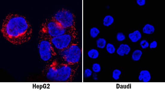
Fluorescent ICC Staining of Cells on Coverslips." alt="Osteopontin/OPN was detected in immersion fixed HepG2 human hepatocellular carcinoma cell line (positive staining; left panel) and Daudi human Burkitt's lymphoma cell line (negative staining; right panel) using Goat Anti-Human Osteopontin/OPN Antigen Affinity-purified Polyclonal Antibody (Catalog # AF1433) at 1.7 µg/mL for 3 hours at room temperature. Cells were stained using the NorthernLights™ 557-conjugated Anti-Goat IgG Secondary Antibody (red; Catalog #
NL001) and counterstained with DAPI (blue). Specific staining was localized to cytoplasm. View our protocol for
Fluorescent ICC Staining of Cells on Coverslips." class="big_thumb" />
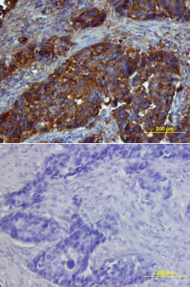
CTS008) and counterstained with hematoxylin (blue). Lower panel shows a lack of labeling if primary antibodies are omitted and tissue is stained only with secondary antibody followed by incubation with detection reagents. View our protocol for
Chromogenic IHC Staining of Paraffin-embedded Tissue Sections." class="big_lightbox">
CTS008) and counterstained with hematoxylin (blue). Lower panel shows a lack of labeling if primary antibodies are omitted and tissue is stained only with secondary antibody followed by incubation with detection reagents. View our protocol for
Chromogenic IHC Staining of Paraffin-embedded Tissue Sections." class="big_thumb" />
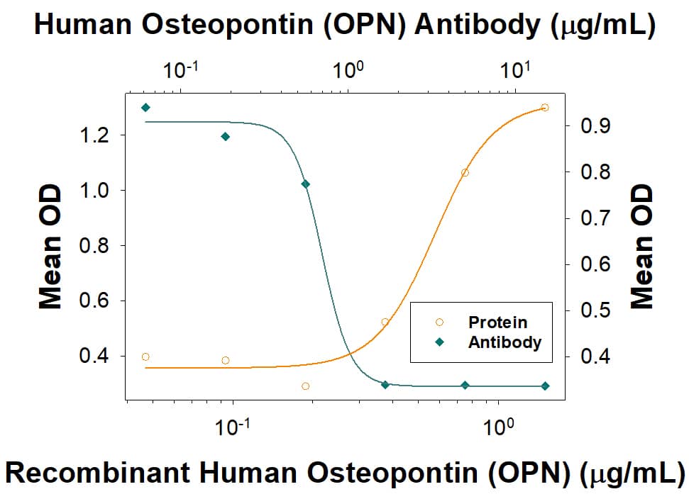
1433-OP), immobilized onto a microplate, supports the adhesion of the HEK293 human embryonic kidney cell line in a dose-dependent manner (orange line). Adhesion elicited by Recombinant Human Osteopontin/OPN (1 µg/mL) is neutralized (green line) by increasing concentrations of Goat Anti-Human Osteopontin/OPN Antigen Affinity-purified Polyclonal Antibody (Catalog # AF1433). The ND
50 is typically 0.300 - 3.60 µg/mL." class="big_lightbox">
1433-OP), immobilized onto a microplate, supports the adhesion of the HEK293 human embryonic kidney cell line in a dose-dependent manner (orange line). Adhesion elicited by Recombinant Human Osteopontin/OPN (1 µg/mL) is neutralized (green line) by increasing concentrations of Goat Anti-Human Osteopontin/OPN Antigen Affinity-purified Polyclonal Antibody (Catalog # AF1433). The ND
50 is typically 0.300 - 3.60 µg/mL." class="big_thumb" />
MAB14332R) coated on a Clear Polystyrene Microplate (Catalog #
DY990). Goat Anti-Human Osteopontin/OPN Antigen Affinity-purified Polyclonal Antibody (Catalog # AF1433) was biotinylated and incubated with the protein captured on the plate. Detection of the standard curve was achieved by incubating Streptavidin-HRP (Catalog #
DY998) followed by Substrate Solution (Catalog #
DY999) and stopping the enzymatic reaction with Stop Solution (Catalog #
DY994)." class="big_lightbox">
MAB14332R) coated on a Clear Polystyrene Microplate (Catalog #
DY990). Goat Anti-Human Osteopontin/OPN Antigen Affinity-purified Polyclonal Antibody (Catalog # AF1433) was biotinylated and incubated with the protein captured on the plate. Detection of the standard curve was achieved by incubating Streptavidin-HRP (Catalog #
DY998) followed by Substrate Solution (Catalog #
DY999) and stopping the enzymatic reaction with Stop Solution (Catalog #
DY994)." class="big_thumb" />
AF1433) at 1ug/mL with overnight incubation at 4 degrees Celsius followed by incubation with anti-goat IgG VisUCyte HRP Polymer Antibody (Catalog #
VC004) and DAB chromogen (yellow-brown). Tissue was counterstained with hematoxylin (blue). Specific staining was localized to cytoplasm in tubules. " class="big_lightbox">

AF1433) at 1ug/mL with overnight incubation at 4 degrees Celsius followed by incubation with anti-goat IgG VisUCyte HRP Polymer Antibody (Catalog #
VC004) and DAB chromogen (yellow-brown). Tissue was counterstained with hematoxylin (blue). Specific staining was localized to cytoplasm in tubules. " class="big_thumb" />
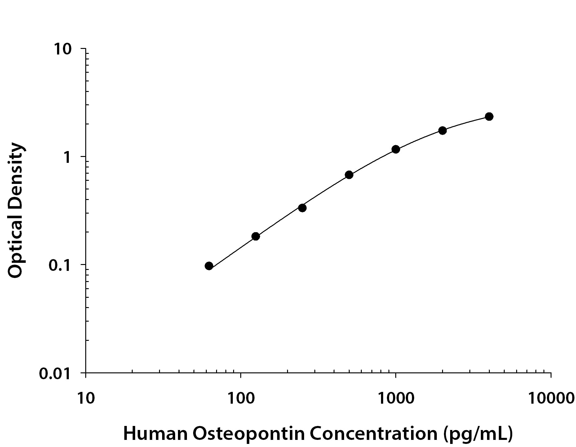
1433-OP) was serially diluted and captured by Mouse Anti-Human Osteopontin/OPN Monoclonal Antibody (Catalog #
MAB14332) coated on a Clear Polystyrene Microplate (Catalog #
DY990). Goat Anti-Human Osteopontin/OPN Antigen Affinity-purified Polyclonal Antibody (Catalog # AF1433) was biotinylated and incubated with the protein captured on the plate. Detection of the standard curve was achieved by incubating Streptavidin-HRP (Catalog #
DY998)" class="big_lightbox">

1433-OP) was serially diluted and captured by Mouse Anti-Human Osteopontin/OPN Monoclonal Antibody (Catalog #
MAB14332) coated on a Clear Polystyrene Microplate (Catalog #
DY990). Goat Anti-Human Osteopontin/OPN Antigen Affinity-purified Polyclonal Antibody (Catalog # AF1433) was biotinylated and incubated with the protein captured on the plate. Detection of the standard curve was achieved by incubating Streptavidin-HRP (Catalog #
DY998)" class="big_thumb" />
 NL001) and counterstained with DAPI (blue). Specific staining was localized to cytoplasm and secreted molecule. View our protocol for Fluorescent ICC Staining of Cells on Coverslips." alt="Osteopontin/OPN was detected in immersion fixed C2C12 mouse myoblast cell line (left panel, positive stain) and mouse splenocytes (right panel, negative stain) using Goat Anti-Mouse Osteopontin/OPN Antigen Affinity-purified Polyclonal Antibody (Catalog # AF808) at 5 µg/mL for 3 hours at room temperature. Cells were stained using the NorthernLights™ 557-conjugated Anti-Goat IgG Secondary Antibody (red; NL001) and counterstained with DAPI (blue). Specific staining was localized to cytoplasm and secreted molecule. View our protocol for Fluorescent ICC Staining of Cells on Coverslips." class="big_thumb" />
 NL001) and counterstained with DAPI (blue). Specific staining was localized to cytoplasm. View our protocol for Fluorescent ICC Staining of Cells on Coverslips." alt="Osteopontin/OPN was detected in immersion fixed HepG2 human hepatocellular carcinoma cell line (positive staining; left panel) and Daudi human Burkitt's lymphoma cell line (negative staining; right panel) using Goat Anti-Human Osteopontin/OPN Antigen Affinity-purified Polyclonal Antibody (Catalog # AF1433) at 1.7 µg/mL for 3 hours at room temperature. Cells were stained using the NorthernLights™ 557-conjugated Anti-Goat IgG Secondary Antibody (red; Catalog # NL001) and counterstained with DAPI (blue). Specific staining was localized to cytoplasm. View our protocol for Fluorescent ICC Staining of Cells on Coverslips." class="big_thumb" />
 CTS002) and counterstained with hematoxylin (blue). Specific staining was localized to cytoplasm in epithelial cells. View our protocol for Chromogenic IHC Staining of Paraffin-embedded Tissue Sections." alt="Osteopontin/OPN was detected in immersion fixed paraffin-embedded sections of human breast cancer tissue using Mouse Anti-Human Osteopontin/OPN Monoclonal Antibody (Catalog # MAB1433) at 25 µg/mL overnight at 4 °C. Tissue was stained using the Anti-Mouse HRP-DAB Cell & Tissue Staining Kit (brown; Catalog # CTS002) and counterstained with hematoxylin (blue). Specific staining was localized to cytoplasm in epithelial cells. View our protocol for Chromogenic IHC Staining of Paraffin-embedded Tissue Sections." class="big_thumb" />
 HAF007). A specific band was detected for Osteopontin/OPN at approximately 60 to 65 kDa (as indicated). This experiment was conducted under reducing conditions and using Immunoblot Buffer Group 1." alt="Western blot shows lysates of human adipose (diabetes) tissue. PVDF membrane was probed with 2 µg/mL of Mouse Anti-Human Osteopontin/OPN Monoclonal Antibody (Catalog # MAB14331) followed by HRP-conjugated Anti-Mouse IgG Secondary Antibody (Catalog # HAF007). A specific band was detected for Osteopontin/OPN at approximately 60 to 65 kDa (as indicated). This experiment was conducted under reducing conditions and using Immunoblot Buffer Group 1." class="big_thumb" />


 CTS008) and counterstained with hematoxylin (blue). View our protocol for
  441-OP), immobilized onto a microplate, supports the adhesion of the HEK293 human embryonic kidney cell line in a dose-dependent manner (orange line). Adhesion elicited by Recombinant Mouse Osteopontin/OPN (2 µg/mL) is neutralized (green line) by increasing concentrations of Mouse Osteopontin/OPN Antigen Affinity-purified Polyclonal Antibody (Catalog # AF808). The ND50 is typically 1-3 µg/mL." class="big_thumb" />


![<b>Biological Strategies Validation. </b>Osteopontin (OPN) drives enhancement in macrophage (Mφ) M2 polarization and angiogenic capacity. (A) Representative images of protein expression profiles obtained by comprehensive protein array in each Mφ subset. Red arrowheads indicate OPN. (B) The mRNA expression level of Spp1 relative to glyceraldehyde-3-phosphate dehydrogenase (Gapdh) was analyzed by real-time reverse transcription polymerase chain reaction in each Mφ subset and was normalized to Mφ (–), n = 6 [***p < 0.001 vs. untreated, #p < 0.05 vs. interleukin (IL)-10 alone]. (C) The protein expression level of OPN relative to GAPDH was measured by western blotting and was normalized to Mφ (–), n = 10. Lower panels are typical images of each protein (***p < 0.001 vs. untreated, #p < 0.05 vs. IL-10 alone). (D) Representative confocal laser scanning immunofluorescence overlay images of OPN (red) and DAPI (blue) in each Mφ subset. Scale bar represents 20 µm. Images in the right row are magnified regions from white or yellow rectangles in the panels of corresponding groups. Scale bar represents 10 µm. (E) Relative mean fluorescence intensity (MFI) of CD163 was measured by FACS analysis in each Mφ subset. An anti-OPN antibody (Ab) and its isotype-matched control Ab were used at 3 µg/mL, n = 4 (***p < 0.001 vs. untreated, ##p < 0.01, #p < 0.05 vs. IL-10 alone, †††p < 0.001 vs. IL-10 + IL-18). (F) The total areas and lengths of tube-like structures were determined by the Matrigel tube formation assay where b.End5 was cocultured with each Mφ subset, n = 12 (***p < 0.001, **p < 0.01, *p < 0.05 vs. untreated, #p < 0.05 vs. IL-10 alone, †††p < 0.001 vs. IL-10 + IL-18). All data are expressed as means ± SEM and were analyzed by a one-way ANOVA followed by Tukey’s test. Image collected and cropped by CiteAb from the following publication (//pubmed.ncbi.nlm.nih.gov/29559970), licensed under a CC-BY license. Not internally tested by R&D Systems.](http://images.novusbio.com/fullsize/af808_mouse-osteopontin-opn-affinity-purified-polyclonal-ab-41202410331992.jpg)
![<b>Biological Strategies Validation. </b>Thrombin contributes to macrophage (Mφ) M2 polarization and angiogenic capacity through proteolytic modification for osteopontin (OPN). (A) The mRNA expression level of Prothrombin relative to glyceraldehyde-3-phosphate dehydrogenase (Gapdh) was analyzed by reverse transcription polymerase chain reaction in each Mφ subset and were normalized to Mφ (–), n = 7 [***p < 0.001, **p < 0.01 vs. untreated, ##p < 0.01 vs. interleukin (IL)-10 alone]. (B,C) The protein expression levels of (B) thrombin or (C) OPN N-Half relative to GAPDH were measured by western blotting in each Mφ subset and were normalized to Mφ (–). Lower panels are typical images of each protein. (B)n = 8 (***p < 0.001, *p < 0.05 vs. untreated, #p < 0.05 vs. IL-10 alone). (C)n = 16 (***p < 0.001, *p < 0.05 vs. untreated, ##p < 0.01, #p < 0.05 vs. IL-10 alone, †††p < 0.001 vs. IL-10 + IL-18). (D) Representative confocal laser scanning immunofluorescence images of OPN (red), thrombin (green), and their merge with DAPI (blue) in each Mφ subset. Scale bar represents 20 µm. Higher magnification images are from the white rectangle region in merged panel of Mφ (IL-10 + IL-18). Scale bar represents 10 µm. (E) Relative mean fluorescence intensity (MFI) of CD163 was measured by FACS analysis in each Mφ subset. Hirudin, a specific thrombin inhibitor, was used at 1 µg/mL, n = 3 (***p < 0.001 vs. untreated, ###p < 0.001 vs. IL-10 alone, †††p < 0.001 vs. IL-10 + IL-18). (F) The total areas and lengths of tube-like structures were determined by the Matrigel tube formation assay where b.End5 were cocultured with each Mφ subset. Hirudin was used at 1 µg/mL, n = 6 (***p < 0.001, **p < 0.01 vs. untreated, ##p < 0.01, #p < 0.05 vs. IL-10 alone, †††p < 0.001 vs. IL-10 + IL-18). All data are expressed as means ± SEM and were analyzed by a one-way ANOVA followed by Tukey’s test. Image collected and cropped by CiteAb from the following publication (//pubmed.ncbi.nlm.nih.gov/29559970), licensed under a CC-BY license. Not int](http://images.novusbio.com/fullsize/af808_mouse-osteopontin-opn-affinity-purified-polyclonal-ab-41202410331959.jpg)
![<b>Biological Strategies Validation. </b>Osteopontin (OPN) drives enhancement in macrophage (Mφ) M2 polarization and angiogenic capacity. (A) Representative images of protein expression profiles obtained by comprehensive protein array in each Mφ subset. Red arrowheads indicate OPN. (B) The mRNA expression level of Spp1 relative to glyceraldehyde-3-phosphate dehydrogenase (Gapdh) was analyzed by real-time reverse transcription polymerase chain reaction in each Mφ subset and was normalized to Mφ (–), n = 6 [***p < 0.001 vs. untreated, #p < 0.05 vs. interleukin (IL)-10 alone]. (C) The protein expression level of OPN relative to GAPDH was measured by western blotting and was normalized to Mφ (–), n = 10. Lower panels are typical images of each protein (***p < 0.001 vs. untreated, #p < 0.05 vs. IL-10 alone). (D) Representative confocal laser scanning immunofluorescence overlay images of OPN (red) and DAPI (blue) in each Mφ subset. Scale bar represents 20 µm. Images in the right row are magnified regions from white or yellow rectangles in the panels of corresponding groups. Scale bar represents 10 µm. (E) Relative mean fluorescence intensity (MFI) of CD163 was measured by FACS analysis in each Mφ subset. An anti-OPN antibody (Ab) and its isotype-matched control Ab were used at 3 µg/mL, n = 4 (***p < 0.001 vs. untreated, ##p < 0.01, #p < 0.05 vs. IL-10 alone, †††p < 0.001 vs. IL-10 + IL-18). (F) The total areas and lengths of tube-like structures were determined by the Matrigel tube formation assay where b.End5 was cocultured with each Mφ subset, n = 12 (***p < 0.001, **p < 0.01, *p < 0.05 vs. untreated, #p < 0.05 vs. IL-10 alone, †††p < 0.001 vs. IL-10 + IL-18). All data are expressed as means ± SEM and were analyzed by a one-way ANOVA followed by Tukey’s test. Image collected and cropped by CiteAb from the following publication (//pubmed.ncbi.nlm.nih.gov/29559970), licensed under a CC-BY license. Not internally tested by R&D Systems.](http://images.novusbio.com/fullsize/af808_mouse-osteopontin-opn-affinity-purified-polyclonal-ab-41202410331947.jpg)

![Aging results in diminished OPN production in response to 2-20 RSV infection.Young and aged BALB/c mice (n = 4/group) were intranasally infected with a dose of 106 pfu/mouse of A2 or 105 pfu/mouse of 2-20) and total lung RNA was collected on time points indicated. (A-B) RNA transcripts of OPN were analyzed with qRT-PCR and represented as a relative ratio of target gene expression to endogenous mouse HPRT and examined with rA2-L19F, 2-20, or A2. (B) 5 µm lung sections obtained at 4, 5 and 8 dpi with either rA2-L19F, 2-20, or A2 infected young and aged mice. Lung sections were immunostained for anti-mouse OPN and nickel-DAB reagent before counterstaining with hematoxylin and eosin. Representative images shown are at 100x magnification with inset [400x] displaying nickel-DAB (dark brown/black staining) positive cells and contrast the hematoxylin (light blue) nuclear stain. Representative images are shown with scale bar indicating 100 µm. (C) Enumeration of OPN-positive cells was performed with ImmunoRatio ImageJ analysis on 200X magnified lung sections from 8 dpi and values are shown as a percentage of total hematoxylin-stained cells in an individual box plot with mean interval bars. Within a single frame, at least 5 frames per mouse (n = 4/group) were collected and individual dot plots are shown of either aged or young mice with interval bars and significance, determined with ANOVA and Fisher's test (p<0.05). (D) qRT-PCR was performed on total lung RNA from young and aged 2–20 RSV infected mice for mRNA expression of OPN receptor CD44. Statistical significance was determine with ANOVA 2-way analysis with p<0.05. All experiments were performed in triplicate. Image collected and cropped by CiteAb from the following open publication (//pubmed.ncbi.nlm.nih.gov/24558422), licensed under a CC-BY license. Not internally tested by R&D Systems.](http://images.novusbio.com/fullsize/af808_mouse-osteopontin-opn-affinity-purified-polyclonal-ab-812025557278.jpg)


 441-OP) was serially diluted and captured by Goat Anti-Mouse Osteopontin/OPN Antigen Affinity-purified Polyclonal Antibody (Catalog # AF808) coated on a Clear Polystyrene Microplate (Catalog #
  441-OP) was serially diluted and captured by Goat Anti-Mouse Osteopontin/OPN Antigen Affinity-purified Polyclonal Antibody (Catalog #
 ![N/A Osteopontin/OPN [HRP]](http://images.novusbio.com/fullsize/elisa/DATA_Osteopontin_DOST00_ELISA_783.jpg "N/A Osteopontin/OPN [HRP]")
![N/A Osteopontin/OPN [HRP]](http://images.novusbio.com/fullsize/elisa/Osteopontin_DOST00_ELISA_155.jpg)
![Western Blot: Osteopontin/OPN Antibody (1B20) - BSA Free [NB110-89062] - Analysis of Osteopontin expression in U2OS whole cell lysate.](http://images.novusbio.com/fullsize/Osteopontin-OPN-Antibody-1B20-BSA-Free-Western-Blot-NB110-89062-img0007.jpg "Western Blot: Osteopontin/OPN Antibody (1B20) - BSA Free [NB110-89062] - Analysis of Osteopontin expression in U2OS whole cell lysate.")
![Immunocytochemistry/Immunofluorescence: Osteopontin/OPN Antibody (1B20) - BSA Free [NB110-89062] - Osteopontin antibody was tested at 1:50 in U2OS cells with FITC (green). Nuclei and actin were counterstained with Dapi (blue) and Phalloidin (red).](http://images.novusbio.com/fullsize/Osteopontin-OPN-Antibody-1B20-BSA-Free-Immunocytochemistry-Immunofluorescence-NB110-89062-img0008.jpg)


![Immunohistochemistry: Osteopontin/OPN Antibody (1B20) - BSA Free [NB110-89062] - Analysis of Osteopontin on human lung adenocarcinoma.](http://images.novusbio.com/fullsize/Osteopontin-OPN-Antibody-1B20-BSA-Free-Immunohistochemistry-NB110-89062-img0009.jpg)

![Simple Western: Osteopontin/OPN Antibody (1B20) - BSA Free [NB110-89062] - Simple Western lane view shows a specific band for Osteopontin in 0.5 mg/ml of Human Breast Cancer lysate. This experiment was performed under reducing conditions using the 12-230 kDa separation system. * Non-specific interaction with the 230 kDa Simple Western standard may be seen with this antibody](http://images.novusbio.com/fullsize/Osteopontin-OPN-Antibody-1B20-BSA-Free-Simple-Western-NB110-89062-img0010.jpg)
![Immunohistochemistry: Osteopontin/OPN Antibody (1B20) - BSA Free [NB110-89062] - Photomicrographs of immunostaining for osteocalcin & osteopontin in (A) Group I, (B) Group II (C) Group III at 6 weeks.The pictures are arranged by staining technique (columns) & the investigated treatment (rows). Areas that stained positive for osteocalcin & osteopontin are indicated by red arrowheads. NB, new bone; OS, osteoid; FM, fatty marrow. Image collected & cropped by CiteAb from the following publication (//peerj.com/articles/3513), licensed under a CC-BY license. Not internally tested by Novus Biologicals.](http://images.novusbio.com/fullsize/nb110-89062_mouse-monoclonal-osteopontin-opn-antibody-1b20-3102024153560.jpg)
![Immunohistochemistry: Osteopontin/OPN Antibody (1B20) - BSA Free [NB110-89062] - Photomicrographs of immunostaining for osteocalcin & osteopontin in (A) Group I, (B) Group II (C) Group III at 6 weeks.The pictures are arranged by staining technique (columns) & the investigated treatment (rows). Areas that stained positive for osteocalcin & osteopontin are indicated by red arrowheads. NB, new bone; OS, osteoid; FM, fatty marrow. Image collected & cropped by CiteAb from the following publication (//peerj.com/articles/3513), licensed under a CC-BY license. Not internally tested by Novus Biologicals.](http://images.novusbio.com/fullsize/nb110-89062_mouse-monoclonal-osteopontin-opn-antibody-1b20-31020241534321.jpg)
![Western Blot: Osteopontin/OPN Antibody (1B20) - BSA Free [NB110-89062] - Photomicrographs of immunostaining for osteocalcin & osteopontin in (A) Group I, (B) Group II (C) Group III at 6 weeks.The pictures are arranged by staining technique (columns) & the investigated treatment (rows). Areas that stained positive for osteocalcin & osteopontin are indicated by red arrowheads. NB, new bone; OS, osteoid; FM, fatty marrow. Image collected & cropped by CiteAb from the following publication (//peerj.com/articles/3513), licensed under a CC-BY license. Not internally tested by Novus Biologicals.](http://images.novusbio.com/fullsize/nb110-89062_mouse-monoclonal-osteopontin-opn-antibody-1b20-310202415394515.jpg)

 CTS008) and counterstained with hematoxylin (blue). Specific labeling was localized to the surface of epithelial cells in the intralobular duct. View our protocol for
  CTS008) and counterstained with hematoxylin (blue). Lower panel shows a lack of labeling if primary antibodies are omitted and tissue is stained only with secondary antibody followed by incubation with detection reagents. View our protocol for
  1433-OP), immobilized onto a microplate, supports the adhesion of the HEK293 human embryonic kidney cell line in a dose-dependent manner (orange line). Adhesion elicited by Recombinant Human Osteopontin/OPN (1 µg/mL) is neutralized (green line) by increasing concentrations of Goat Anti-Human Osteopontin/OPN Antigen Affinity-purified Polyclonal Antibody (Catalog # AF1433). The ND50 is typically 0.300 - 3.60 µg/mL." class="big_thumb" />
 MAB14332R) coated on a Clear Polystyrene Microplate (Catalog #
  AF1433) at 1ug/mL with overnight incubation at 4 degrees Celsius followed by incubation with anti-goat IgG VisUCyte HRP Polymer Antibody (Catalog #


 1433-OP) was serially diluted and captured by Mouse Anti-Human Osteopontin/OPN Monoclonal Antibody (Catalog #
 ![N/A Osteopontin/OPN [HRP]](http://images.novusbio.com/fullsize/elisa/DATA_Osteopontin_MOST00_ELISA_1013.jpg "N/A Osteopontin/OPN [HRP]")
![N/A Osteopontin/OPN [HRP]](http://images.novusbio.com/fullsize/elisa/Osteopontin_MOST00_ELISA_305.jpg)
![N/A Osteopontin/OPN [HRP]](http://images.novusbio.com/fullsize/elisa/Osteopontin_MOST00_ELISA_304.jpg)


![N/A Osteopontin/OPN [Biotin]](http://images.novusbio.com/fullsize/elisa/DATA_Osteopontin_DY1433_ELISA_1646.jpg "N/A Osteopontin/OPN [Biotin]")
![N/A Osteopontin/OPN [Biotin]](http://images.novusbio.com/fullsize/elisa/DATA_Osteopontin_DY441_ELISA_2030.jpg "N/A Osteopontin/OPN [Biotin]")

![Western Blot: Osteopontin/OPN Antibody [NB600-1043] - Western Blot of Osteopontin/OPN/OPN antibody. Lane 1: Molecular Weight Marker. Lane 2: Human Osteopontin/OPN [250ng]. Lane 3: MMP-cleaved Osteopontin/OPN. Primary Antibody: Anti-Osteopontin/OPN at 1:1000 ovenight at 2-8C. Secondary Antibody: Goat Anti-Rabbit IgG HRP at 1:10,000. Observed MW: full length 66kDa, MMP-cleaved 32kDa.](http://images.novusbio.com/fullsize/Osteopontin-OPN-Antibody-Western-Blot-NB600-1043-img0009.jpg "Western Blot: Osteopontin/OPN Antibody [NB600-1043] - Western Blot of Osteopontin/OPN/OPN antibody. Lane 1: Molecular Weight Marker. Lane 2: Human Osteopontin/OPN [250ng]. Lane 3: MMP-cleaved Osteopontin/OPN. Primary Antibody: Anti-Osteopontin/OPN at 1:1000 ovenight at 2-8C. Secondary Antibody: Goat Anti-Rabbit IgG HRP at 1:10,000. Observed MW: full length 66kDa, MMP-cleaved 32kDa.")
![Immunohistochemistry: Osteopontin/OPN Antibody [NB600-1043] - Osteopontin/OPN/OPN antibody was used at a 1:100-1:300 dilution to detect osteopontin by immunohistochemistry. Osteopontin/OPN is a normal component of elastic fibers in the breast (shown heavily stained in this section of human breast tumor cells). There is also weak staining of the extracellular matrix. Osteopontin/OPN is not expressed in breast tumor cells, and there is no staining of the breast cells in this section. No antigen retrieval is required.](http://images.novusbio.com/fullsize/Osteopontin-OPN-Antibody-Immunohistochemistry-NB600-1043-img0010.jpg)
![Osteopontin/OPN Antibody [NB600-1043]](http://images.novusbio.com/fullsize/Osteopontin-OPN-Antibody-N-A-NB600-1043-img0008.jpg)


 MAB003, open histogram) followed by Allophycocyanin-conjugated Anti-Mouse IgG Secondary Antibody (Catalog #


