HAF008). A specific band was detected for VE‑Cadherin at approximately 125 kDa (as indicated). This experiment was conducted under reducing conditions and using
Western Blot Buffer Group 1." class="big_lightbox">

HAF008). A specific band was detected for VE‑Cadherin at approximately 125 kDa (as indicated). This experiment was conducted under reducing conditions and using
Western Blot Buffer Group 1." alt="Western Blot shows lysates of HUVEC human umbilical vein endothelial cells. PVDF membrane was probed with 1 µg/ml of Rabbit Anti-Human VE‑Cadherin Monoclonal Antibody (Catalog # MAB11726) followed by HRP-conjugated Anti-Rabbit IgG Secondary Antibody (Catalog #
HAF008). A specific band was detected for VE‑Cadherin at approximately 125 kDa (as indicated). This experiment was conducted under reducing conditions and using
Western Blot Buffer Group 1." class="big_thumb" />
VC003) or the HRP-conjugated Anti-Rabbit IgG Secondary Antibody (Catalog #
HAF008). Before incubation with the primary antibody, tissue was subjected to heat-induced epitope retrieval using VisUCyte Antigen Retrieval Reagent-Basic (Catalog #
VCTS021). Tissue was stained using DAB (brown) and counterstained with hematoxylin (blue). Specific staining was localized to the membrane. View our protocol for
IHC Staining with VisUCyte HRP Polymer Detection Reagents." class="big_lightbox">
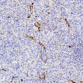
VC003) or the HRP-conjugated Anti-Rabbit IgG Secondary Antibody (Catalog #
HAF008). Before incubation with the primary antibody, tissue was subjected to heat-induced epitope retrieval using VisUCyte Antigen Retrieval Reagent-Basic (Catalog #
VCTS021). Tissue was stained using DAB (brown) and counterstained with hematoxylin (blue). Specific staining was localized to the membrane. View our protocol for
IHC Staining with VisUCyte HRP Polymer Detection Reagents." class="big_thumb" />
VC003) or the HRP-conjugated Anti-Rabbit IgG Secondary Antibody (Catalog #
HAF008). Before incubation with the primary antibody, tissue was subjected to heat-induced epitope retrieval using VisUCyte Antigen Retrieval Reagent-Basic (Catalog #
VCTS021). Tissue was stained using DAB (brown) and counterstained with hematoxylin (blue). Specific staining was localized to the membrane. View our protocol for
IHC Staining with VisUCyte HRP Polymer Detection Reagents." class="big_lightbox">

VC003) or the HRP-conjugated Anti-Rabbit IgG Secondary Antibody (Catalog #
HAF008). Before incubation with the primary antibody, tissue was subjected to heat-induced epitope retrieval using VisUCyte Antigen Retrieval Reagent-Basic (Catalog #
VCTS021). Tissue was stained using DAB (brown) and counterstained with hematoxylin (blue). Specific staining was localized to the membrane. View our protocol for
IHC Staining with VisUCyte HRP Polymer Detection Reagents." class="big_thumb" />
VC003) or the HRP-conjugated Anti-Rabbit IgG Secondary Antibody (Catalog #
HAF008). Before incubation with the primary antibody, tissue was subjected to heat-induced epitope retrieval using VisUCyte Antigen Retrieval Reagent-Basic (Catalog #
VCTS021). Tissue was stained using DAB (brown) and counterstained with hematoxylin (blue). Specific staining was localized to the membrane. View our protocol for
IHC Staining with VisUCyte HRP Polymer Detection Reagents." class="big_lightbox">

VC003) or the HRP-conjugated Anti-Rabbit IgG Secondary Antibody (Catalog #
HAF008). Before incubation with the primary antibody, tissue was subjected to heat-induced epitope retrieval using VisUCyte Antigen Retrieval Reagent-Basic (Catalog #
VCTS021). Tissue was stained using DAB (brown) and counterstained with hematoxylin (blue). Specific staining was localized to the membrane. View our protocol for
IHC Staining with VisUCyte HRP Polymer Detection Reagents." class="big_thumb" />
 HAF017). A specific band was detected for VE‑Cadherin at approximately 120 kDa (as indicated). This experiment was conducted under reducing conditions and using Immunoblot Buffer Group 1." alt="Western blot shows lysates of mouse lung tissue and bEnd.3 mouse endothelioma cell line. PVDF membrane was probed with 0.2 µg/mL of Goat Anti-Mouse VE‑Cadherin Antigen Affinity-purified Polyclonal Antibody (Catalog # AF1002) followed by HRP-conjugated Anti-Goat IgG Secondary Antibody (Catalog # HAF017). A specific band was detected for VE‑Cadherin at approximately 120 kDa (as indicated). This experiment was conducted under reducing conditions and using Immunoblot Buffer Group 1." class="big_thumb" />
 HAF017). A specific band was detected for VE-Cadherin at approximately 125 kDa (as indicated). This experiment was conducted under reducing conditions and using Immunoblot Buffer Group 1." alt="Western blot shows lysate of HUVEC human umbilical vein endothelial cells. PVDF membrane was probed with 0.25 µg/mL of Goat Anti-Human VE-Cadherin Antigen Affinity-purified Polyclonal Antibody (Catalog # AF938) followed by HRP-conjugated Anti-Goat IgG Secondary Antibody (HAF017). A specific band was detected for VE-Cadherin at approximately 125 kDa (as indicated). This experiment was conducted under reducing conditions and using Immunoblot Buffer Group 1." class="big_thumb" />
 HAF018). A specific band was detected for VE-Cadherin at approximately 125 kDa (as indicated). This experiment was conducted under reducing conditions and using Immunoblot Buffer Group 1." alt="Western blot shows lysate of HUVEC human umbilical vein endothelial cells. PVDF membrane was probed with 1 µg/mL of Mouse Anti-Human VE-Cadherin Monoclonal Antibody (Catalog # MAB9381) followed by HRP-conjugated Anti-Mouse IgG Secondary Antibody (HAF018). A specific band was detected for VE-Cadherin at approximately 125 kDa (as indicated). This experiment was conducted under reducing conditions and using Immunoblot Buffer Group 1." class="big_thumb" />
 HAF018). A specific band was detected for VE‑Cadherin at approximately 125 kDa (as indicated). This experiment was conducted under reducing conditions and using Western Blot Buffer Group 1." alt="Western Blot shows lysates of HUVEC human umbilical vein endothelial cells. PVDF membrane was probed with 2 µg/ml of Mouse Anti-Human VE‑Cadherin Monoclonal Antibody (Catalog # MAB11663) followed by HRP-conjugated Anti-Mouse IgG Secondary Antibody (Catalog # HAF018). A specific band was detected for VE‑Cadherin at approximately 125 kDa (as indicated). This experiment was conducted under reducing conditions and using Western Blot Buffer Group 1." class="big_thumb" />
 MAB0061, open histogram), followed by Phycoerythrin-conjugated Anti-Rat IgG Secondary Antibody (Catalog # F0105B). View our protocol for Staining Membrane-associated Proteins." alt="bEnd.3 mouse endothelioma cell line was stained with Rat Anti-Mouse VE-Cadherin Monoclonal Antibody (Catalog # MAB1002, filled histogram) or isotype control antibody (Catalog # MAB0061, open histogram), followed by Phycoerythrin-conjugated Anti-Rat IgG Secondary Antibody (Catalog # F0105B). View our protocol for Staining Membrane-associated Proteins." class="big_thumb" />
 HAF008). A specific band was detected for VE‑Cadherin at approximately 125 kDa (as indicated). This experiment was conducted under reducing conditions and using Western Blot Buffer Group 1." alt="Western Blot shows lysates of HUVEC human umbilical vein endothelial cells. PVDF membrane was probed with 1 µg/ml of Rabbit Anti-Human VE‑Cadherin Monoclonal Antibody (Catalog # MAB11726) followed by HRP-conjugated Anti-Rabbit IgG Secondary Antibody (Catalog # HAF008). A specific band was detected for VE‑Cadherin at approximately 125 kDa (as indicated). This experiment was conducted under reducing conditions and using Western Blot Buffer Group 1." class="big_thumb" />


 VC004). Tissue was stained using DAB (brown) and counterstained with hematoxylin (blue). Specific staining was localized to cell membranes. View our protocol for


 AF1002) at 3ug/mL with 1 hour incubation at room temperature followed by incubation with anti-goat IgG VisUCyte HRP Polymer Antibody (Catalog #


 NL001) and counterstained with DAPI (blue). Specific staining was localized to the plasma membrane. View our protocol for
  AB-108-C, open histogram) followed by Allophycocyanin-conjugated Anti-Goat IgG Secondary Antibody (


 NL007) and counterstained with DAPI (blue). Specific staining was localized to plasma membrane. View our protocol for
  MAB004, open histogram) followed by Allophycocyanin-conjugated Anti-Mouse IgG Secondary Antibody (
![Western Blot: VE-Cadherin Antibody (BV14) [NBP1-43347] - Non-reduced (left) and reduced (right) bEnd.3 cell line lysates were loaded at 1x10^5 cells/lane, probed with 2 ug/mL of Anti-Mouse CD144 (VE-Cadherin) Purified and revealed with Anti-Rat IgG HRP.](http://images.novusbio.com/fullsize/VE-Cadherin-Antibody-BV14-Western-Blot-NBP1-43347-img0003.jpg "Western Blot: VE-Cadherin Antibody (BV14) [NBP1-43347] - Non-reduced (left) and reduced (right) bEnd.3 cell line lysates were loaded at 1x10^5 cells/lane, probed with 2 ug/mL of Anti-Mouse CD144 (VE-Cadherin) Purified and revealed with Anti-Rat IgG HRP.")


![Immunocytochemistry/Immunofluorescence: VE Cadherin Antibody (BV9) [NB600-1409] - Endothelial cells were fixed with 4% PAF (15 min, room temp), and then permeabilized with 0.5% TritionX-100 (3 min, room temperature). Cells were incubated with a final concentration of 10mg/ml BV9. Secondary detection was performed with anti-mouse Alexa-Fluor 647 and counterstained using DAPI.](http://images.novusbio.com/fullsize/VE-Cadherin-Antibody-BV9-Immunocytochemistry-Immunofluorescence-NB600-1409-img0008.jpg "Immunocytochemistry/Immunofluorescence: VE Cadherin Antibody (BV9) [NB600-1409] - Endothelial cells were fixed with 4% PAF (15 min, room temp), and then permeabilized with 0.5% TritionX-100 (3 min, room temperature). Cells were incubated with a final concentration of 10mg/ml BV9. Secondary detection was performed with anti-mouse Alexa-Fluor 647 and counterstained using DAPI.")
![Flow Cytometry: VE Cadherin Antibody (BV9) [NB600-1409] - HUVEC cells were stained with antibody BV9 in PBS/0.1% saponin for 1h at 4 degrees C.(Black- no stain, Red- isotype control, Blue-HM2032-BV9)](http://images.novusbio.com/fullsize/VE-Cadherin-Antibody-BV9-Flow-Cytometry-NB600-1409-img0007.jpg)

 VC001) or the HRP-conjugated Anti-Mouse IgG Secondary Antibody (Catalog #
  VC001) or the HRP-conjugated Anti-Mouse IgG Secondary Antibody (Catalog #
  NL007) and counterstained with DAPI (blue). Specific staining was localized to the cell membrane of HUVEC cells. View our protocol for
  VC003) or the HRP-conjugated Anti-Rabbit IgG Secondary Antibody (Catalog #
  VC003) or the HRP-conjugated Anti-Rabbit IgG Secondary Antibody (Catalog #
  VC003) or the HRP-conjugated Anti-Rabbit IgG Secondary Antibody (Catalog #
  NL004) and counterstained with DAPI (blue). Specific staining was localized to the cell surface of HUVEC cells. View our protocol for
 ![N/A VE-Cadherin [HRP]](http://images.novusbio.com/fullsize/elisa/DATA_VECadherin_DCADV0_ELISA_606.jpg "N/A VE-Cadherin [HRP]")
![ELISA: Mouse VE-Cadherin ELISA Kit (Colorimetric) [NBP3-20089] - Samples were spiked with high concentrations of Mouse VE-Cadherin and diluted with Reference Standard & Sample Diluent to produce samples with values within the range of the assay.](http://images.novusbio.com/fullsize/nbp3-20089_mouse-ve-cadherin-elisa-kit-colorimetric-132202416102247.jpg "ELISA: Mouse VE-Cadherin ELISA Kit (Colorimetric) [NBP3-20089] - Samples were spiked with high concentrations of Mouse VE-Cadherin and diluted with Reference Standard & Sample Diluent to produce samples with values within the range of the assay.")
![ELISA: Mouse VE-Cadherin ELISA Kit (Colorimetric) [NBP3-20089] - Standard Curve Reference](http://images.novusbio.com/fullsize/nbp3-20089_mouse-ve-cadherin-elisa-kit-colorimetric-432024855052.jpg)
![Flow Cytometry: VE-Cadherin Antibody (BV13) [NBP1-43348] - Analysis using the Biotin conjugate of NBP1-43348. Staining of bEnd.3 cell line with 0.5 micrograms of Rat IgG1 k Isotype Control Biotin (blue histogram) or 0.125 micrograms of Anti-Mouse CD144 (VE-Cadherin) Biotin purple histogram) followed by Streptavidin PE. Total viable cells were used for analysis.](http://images.novusbio.com/fullsize/VE-Cadherin-Antibody-BV13-Flow-Cytometry-NBP1-43348-img0003.jpg "Flow Cytometry: VE-Cadherin Antibody (BV13) [NBP1-43348] - Analysis using the Biotin conjugate of NBP1-43348. Staining of bEnd.3 cell line with 0.5 micrograms of Rat IgG1 k Isotype Control Biotin (blue histogram) or 0.125 micrograms of Anti-Mouse CD144 (VE-Cadherin) Biotin purple histogram) followed by Streptavidin PE. Total viable cells were used for analysis.")
![Flow Cytometry: VE-Cadherin Antibody (BV13) [NBP1-43348] - Staining of bEnd.3 cell line with 0.5 ug of Rat IgG1 K Isotype Control Purified (open histogram) or 0.5 ug of AntiMouse CD144 (VE-Cadherin) Purified (filled histogram) followed by F(ab')2 Anti-Rat IgG PE. Total viable cells were used for analysis.](http://images.novusbio.com/fullsize/VE-Cadherin-Antibody-BV13-Flow-Cytometry-NBP1-43348-img0001.jpg)


