NL007) and counterstained with DAPI (blue). Specific staining was localized to nuclei. View our protocol for
Fluorescent ICC Staining of Cells on Coverslips." class="big_lightbox">
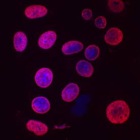
NL007) and counterstained with DAPI (blue). Specific staining was localized to nuclei. View our protocol for
Fluorescent ICC Staining of Cells on Coverslips." alt="Bromodeoxyuridine/BrdU was detected in immersion fixed MCF-7 human breast cancer cell line stimulated with BrdU using Mouse Anti-Bromodeoxyuridine/ BrdU Antigen Affinity-purified Monoclonal Antibody (Catalog # MAB7225) at 10 µg/mL for 3 hours at room temperature. Cells were stained using the NorthernLights™ 557-conjugated Anti-Mouse IgG Secondary Antibody (red; Catalog #
NL007) and counterstained with DAPI (blue). Specific staining was localized to nuclei. View our protocol for
Fluorescent ICC Staining of Cells on Coverslips." class="big_thumb" />
VC001). Before incubation with the primary antibody, tissue was subjected to heat-induced epitope retrieval using Antigen Retrieval Reagent-Basic (
CTS013). Tissue was stained using DAB (brown) and counterstained with hematoxylin (blue). Specific staining was localized to cell nuclei. Staining was performed using our protocol for IHC Staining with VisUCyte HRP Polymer Detection Reagents." class="big_lightbox">
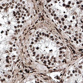
VC001). Before incubation with the primary antibody, tissue was subjected to heat-induced epitope retrieval using Antigen Retrieval Reagent-Basic (
CTS013). Tissue was stained using DAB (brown) and counterstained with hematoxylin (blue). Specific staining was localized to cell nuclei. Staining was performed using our protocol for IHC Staining with VisUCyte HRP Polymer Detection Reagents." class="big_thumb" />
VC001). Before incubation with the primary antibody, tissue was subjected to heat-induced epitope retrieval using Antigen Retrieval Reagent-Basic (
CTS013). Tissue was stained using DAB (brown) and counterstained with hematoxylin (blue). Specific staining was localized to cell nuclei. Staining was performed using our protocol for IHC Staining with VisUCyte HRP Polymer Detection Reagents." class="big_lightbox">
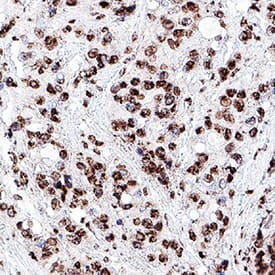
VC001). Before incubation with the primary antibody, tissue was subjected to heat-induced epitope retrieval using Antigen Retrieval Reagent-Basic (
CTS013). Tissue was stained using DAB (brown) and counterstained with hematoxylin (blue). Specific staining was localized to cell nuclei. Staining was performed using our protocol for IHC Staining with VisUCyte HRP Polymer Detection Reagents." class="big_thumb" />
MAB003, open histo-gram), followed by Allophycocyanin-conjugated Anti-Mouse IgG Secondary Antibody (Catalog #
F0101B). To facilitate intracellular staining, cells were fixed with cold, 70% ethanol for 5 minutes, DNA was denatured with 1.5M HCl for 30 minutes, and then cells were permeabilized with Flow Cytometry Permeabilization/Wash Buffer I (Catalog #
FC005)." class="big_lightbox">
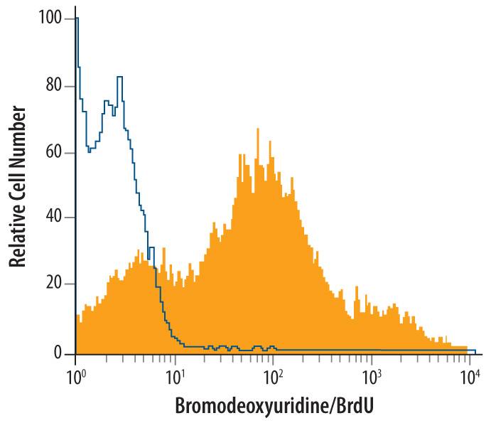
MAB003, open histo-gram), followed by Allophycocyanin-conjugated Anti-Mouse IgG Secondary Antibody (Catalog #
F0101B). To facilitate intracellular staining, cells were fixed with cold, 70% ethanol for 5 minutes, DNA was denatured with 1.5M HCl for 30 minutes, and then cells were permeabilized with Flow Cytometry Permeabilization/Wash Buffer I (Catalog #
FC005)." class="big_thumb" />
 NL007) and counterstained with DAPI (blue). Specific staining was localized to nuclei. View our protocol for Fluorescent ICC Staining of Cells on Coverslips." alt="Bromodeoxyuridine/BrdU was detected in immersion fixed MCF-7 human breast cancer cell line stimulated with BrdU using Mouse Anti-Bromodeoxyuridine/ BrdU Antigen Affinity-purified Monoclonal Antibody (Catalog # MAB7225) at 10 µg/mL for 3 hours at room temperature. Cells were stained using the NorthernLights™ 557-conjugated Anti-Mouse IgG Secondary Antibody (red; Catalog # NL007) and counterstained with DAPI (blue). Specific staining was localized to nuclei. View our protocol for Fluorescent ICC Staining of Cells on Coverslips." class="big_thumb" />


![Immunocytochemistry/Immunofluorescence: Bromodeoxyuridine/BrdU Antibody [NB500-235] - IF analysis of BrdU in Proteinase K pretreated mouse lacrimal gland. Image courtesy of anonymous customer review.](http://images.novusbio.com/fullsize/Bromodeoxyuridine-BrdU-Antibody-Immunocytochemistry-Immunofluorescence-NB500-235-img0006.jpg "<b>Biological Strategies Validation. </b>Immunocytochemistry/Immunofluorescence: Bromodeoxyuridine/BrdU Antibody [NB500-235] - IF analysis of BrdU in Proteinase K pretreated mouse lacrimal gland. Image courtesy of anonymous customer review.")
![<b>Biological Strategies Validation. </b>Immunoprecipitation: Bromodeoxyuridine/BrdU Antibody [NB500-235] - Lysate was immunoprecipitated with anti-brdu antibody (NB500-235), 5-methyl cytosine (5-Me), or a no antibody control (No A). An ELISA was then performed to measure total activity.](http://images.novusbio.com/fullsize/Bromodeoxyuridine-BrdU-Antibody-Immunoprecipitation-NB500-235-img0005.jpg)


 VC001). Before incubation with the primary antibody, tissue was subjected to heat-induced epitope retrieval using Antigen Retrieval Reagent-Basic (
 VC001). Before incubation with the primary antibody, tissue was subjected to heat-induced epitope retrieval using Antigen Retrieval Reagent-Basic (
 MAB003, open histo-gram), followed by Allophycocyanin-conjugated Anti-Mouse IgG Secondary Antibody (Catalog #


![Immunohistochemistry-Paraffin: Bromodeoxyuridine/BrdU Antibody [NBP2-14890] - Mouse intestinal tissue pulsed with BrdU. BrdU stained by BrdU antibody diluted at 1:100. Antigen Retrieval: Citrate buffer, pH 6.0, 15 min](http://images.novusbio.com/fullsize/Bromodeoxyuridine-BrdU-Antibody-Immunohistochemistry-Paraffin-NBP2-14890-img0009.jpg "Immunohistochemistry-Paraffin: Bromodeoxyuridine/BrdU Antibody [NBP2-14890] - Mouse intestinal tissue pulsed with BrdU. BrdU stained by BrdU antibody diluted at 1:100. Antigen Retrieval: Citrate buffer, pH 6.0, 15 min")
![<b>Biological Strategies Validation. </b>Flow Cytometry: Bromodeoxyuridine/BrdU Antibody [NBP2-14890] - BrdU antibody detects BrdU protein from HeLa treated with 10 uM BrdU for 30 minutes and fixed in 4% paraformaldehyde at 4C for 15 minutes by flow cytometry analysis.<br></br>Black: Unlabelled sample was used as a control.<br>Red: BrdU antibody<br>Acquisition of 20,000 events were collected using a Dylight 488-conjugated secondary antibody for FACS analysis.](http://images.novusbio.com/fullsize/Bromodeoxyuridine-BrdU-Antibody-Flow-Cytometry-NBP2-14890-img0004.jpg)
![Immunohistochemistry-Paraffin: Bromodeoxyuridine/BrdU Antibody (MoBu-1) [NB500-439] - Mouse Small Intestine stained with BrdU Monoclonal Antibody (MoBu-1).](http://images.novusbio.com/fullsize/Bromodeoxyuridine-BrdU-Antibody-MoBu-1-Immunohistochemistry-Paraffin-NB500-439-img0003.jpg "Immunohistochemistry-Paraffin: Bromodeoxyuridine/BrdU Antibody (MoBu-1) [NB500-439] - Mouse Small Intestine stained with BrdU Monoclonal Antibody (MoBu-1).")
![Immunohistochemistry: Bromodeoxyuridine/BrdU Antibody (MoBu-1) [NB500-439] - Staining of bromodeoxyuridine/BrdU-labeled cells (chick embryo; paraffin-embedded sections)](http://images.novusbio.com/fullsize/Bromodeoxyuridine-BrdU-Antibody-MoBu-1-Immunohistochemistry-NB500-439-img0002.jpg)
![Western Blot: Bromodeoxyuridine/BrdU Antibody (29G6.E8) [NBP2-44055] - Lane 1: loading control. Lane 2: FdU. Lane 3: BrdU. Lane 4: IdU. Lane 5 CldU. Load: 20 Ug per lane.](http://images.novusbio.com/fullsize/Bromodeoxyuridine-BrdU-Antibody-29G6-E8-Western-Blot-NBP2-44055-img0001.jpg "Western Blot: Bromodeoxyuridine/BrdU Antibody (29G6.E8) [NBP2-44055] - Lane 1: loading control. Lane 2: FdU. Lane 3: BrdU. Lane 4: IdU. Lane 5 CldU. Load: 20 Ug per lane.")
![Immunocytochemistry/Immunofluorescence: Bromodeoxyuridine/BrdU Antibody (29G6.E8) [NBP2-44055] - Tissue: OCT-embedded E10.5 mouse embryo. Localization: 20X, section through the developing hindbrain. Fixation: 4% PFA. Antigen retrieval: not required. Staining: Double labeled (green/blue) cells represent cells that were actively dividing.](http://images.novusbio.com/fullsize/Bromodeoxyuridine-BrdU-Antibody-29G6-E8-Immunocytochemistry-Immunofluorescence-NBP2-44055-img0008.jpg)
![Immunocytochemistry/Immunofluorescence: Bromodeoxyuridine/BrdU Antibody (29G6.E8) [NBP2-44055] - Tissue: OCT-embedded E10.5 mouse embryo. Localization: 20X, section through the developing limb bud. Fixation: 4% PFA. Antigen retrieval: not required.](http://images.novusbio.com/fullsize/Bromodeoxyuridine-BrdU-Antibody-29G6-E8-Immunocytochemistry-Immunofluorescence-NBP2-44055-img0003.jpg)
![Immunocytochemistry/Immunofluorescence: Bromodeoxyuridine/BrdU Antibody (29G6.E8) [NBP2-44055] - Tissue: OCT-embedded E10.5 mouse embryo. Localization: 40X, section through the developing limb bud. Fixation: 4% PFA. Antigen retrieval: not required. Primary antibody: BrdU antibody at 1:500 in 0.4% PBS+Triton with 1% normal sheep serum overnight at 4C. Staining: Double labeled (green/blue) cells represent cells that were actively dividing.](http://images.novusbio.com/fullsize/Bromodeoxyuridine-BrdU-Antibody-29G6-E8-Immunocytochemistry-Immunofluorescence-NBP2-44055-img0007.jpg)
![Bromodeoxyuridine/BrdU Antibody (29G6.E8) [NBP2-44055]](http://images.novusbio.com/fullsize/Bromodeoxyuridine-BrdU-Antibody-29G6-E8-N-A-NBP2-44055-img0009.jpg)
![Immunohistochemistry-Paraffin: Bromodeoxyuridine/BrdU Antibody (BU20a) [NBP2-32922] - Formalin-fixed, paraffin-embedded Mouse Small Intestine stained with BrdU Monoclonal Antibody (BU20a).](http://images.novusbio.com/fullsize/Bromodeoxyuridine-BrdU-Antibody-BU20a-Immunohistochemistry-Paraffin-NBP2-32922-img0001.jpg "Immunohistochemistry-Paraffin: Bromodeoxyuridine/BrdU Antibody (BU20a) [NBP2-32922] - Formalin-fixed, paraffin-embedded Mouse Small Intestine stained with BrdU Monoclonal Antibody (BU20a).")
![Immunohistochemistry-Paraffin: Bromodeoxyuridine/BrdU Antibody (SPM166) [NBP2-32919] - Formalin-fixed, paraffin-embedded mouse intestine tissue stained with BrdU MAb (SPM166).](http://images.novusbio.com/fullsize/Bromodeoxyuridine-BrdU-Antibody-SPM166-Immunohistochemistry-Paraffin-NBP2-32919-img0001.jpg "Immunohistochemistry-Paraffin: Bromodeoxyuridine/BrdU Antibody (SPM166) [NBP2-32919] - Formalin-fixed, paraffin-embedded mouse intestine tissue stained with BrdU MAb (SPM166).")
![Immunohistochemistry-Paraffin: Bromodeoxyuridine/BrdU Antibody (BRD494) [NBP2-32920] - Formalin-fixed, paraffin-embedded Mouse Small Intestine stained with BrdU Monoclonal Antibody (BRD494).](http://images.novusbio.com/fullsize/Bromodeoxyuridine-BrdU-Antibody-BRD494-Immunohistochemistry-Paraffin-NBP2-32920-img0001.jpg "Immunohistochemistry-Paraffin: Bromodeoxyuridine/BrdU Antibody (BRD494) [NBP2-32920] - Formalin-fixed, paraffin-embedded Mouse Small Intestine stained with BrdU Monoclonal Antibody (BRD494).")
![Immunohistochemistry: Bromodeoxyuridine/BrdU Antibody (A9C7-R) [NBP3-32092] - Immunofluorescence analysis of paraffin-embedded Bromodeoxyuridine/BrdU treated mouse embryo tissue labeling BrdU with Mouse anti-Bromodeoxyuridine/BrdU antibody (NBP3-32092) at 1/500 dilution.<br/><br/> The section was pre-treated using heat mediated antigen retrieval with sodium citrate buffer (pH 6.0) for 2 minutes. The tissues were blocked in 10% negative goat serum for 1 hour at room temperature, washed with PBS, and then probed with the primary antibody (NBP3-32092, green) at 1/500 dilution overnight at 4 ℃, washed with PBS. Goat Anti-Mouse IgG H&L (iFluor™ 488) was used as the secondary antibody at 1/1,000 dilution. Nuclei were counterstained with DAPI (blue).](http://images.novusbio.com/fullsize/nbp3-32092_mouse-bromodeoxyuridine-brdu-mab-a9c7-r-298202413585576.jpg "Immunohistochemistry: Bromodeoxyuridine/BrdU Antibody (A9C7-R) [NBP3-32092] - Immunofluorescence analysis of paraffin-embedded Bromodeoxyuridine/BrdU treated mouse embryo tissue labeling BrdU with Mouse anti-Bromodeoxyuridine/BrdU antibody (NBP3-32092) at 1/500 dilution.<br/><br/> The section was pre-treated using heat mediated antigen retrieval with sodium citrate buffer (pH 6.0) for 2 minutes. The tissues were blocked in 10% negative goat serum for 1 hour at room temperature, washed with PBS, and then probed with the primary antibody (NBP3-32092, green) at 1/500 dilution overnight at 4 ℃, washed with PBS. Goat Anti-Mouse IgG H&L (iFluor™ 488) was used as the secondary antibody at 1/1,000 dilution. Nuclei were counterstained with DAPI (blue).")
![Immunohistochemistry: Bromodeoxyuridine/BrdU Antibody (A9C7-R) [NBP3-32092] - Immunohistochemical analysis of paraffin-embedded Bromodeoxyuridine/BrdU treated mouse embryo tissue with Mouse anti-Bromodeoxyuridine/BrdU antibody (NBP3-32092) at 1/5,000 dilution.<br/><br/> The section was pre-treated using heat mediated antigen retrieval with sodium citrate buffer (pH 6.0) for 2 minutes. The tissues were blocked in 1% BSA for 20 minutes at room temperature, washed with ddH2O and PBS, and then probed with the primary antibody (NBP3-32092) at 1/5,000 dilution for 1 hour at room temperature. The detection was performed using an HRP conjugated compact polymer system. DAB was used as the chromogen. Tissues were counterstained with hematoxylin and mounted with DPX.](http://images.novusbio.com/fullsize/nbp3-32092_mouse-bromodeoxyuridine-brdu-mab-a9c7-r-298202414161891.jpg)
![Immunocytochemistry/ Immunofluorescence: Bromodeoxyuridine/BrdU Antibody (A9C7-R) [NBP3-32092] - Immunocytochemistry analysis of Bromodeoxyuridine/BrdU treated NIH/3T3 cells labeling Bromodeoxyuridine/BrdU with Mouse anti-Bromodeoxyuridine/BrdU antibody (NBP3-32092) at 1/250 dilution.<br/><br/> Cells were fixed in 4% paraformaldehyde for 20 minutes at room temperature, permeabilized with 0.1% Triton X-100 in PBS for 5 minutes at room temperature, then blocked with 1% BSA in 10% negative goat serum for 1 hour at room temperature. Cells were then incubated with Mouse anti-Bromodeoxyuridine/BrdU antibody (NBP3-32092) at 1/250 dilution in 1% BSA in PBST overnight at 4 ℃. Goat Anti-Mouse IgG H&L (iFluor™ 488) was used as the secondary antibody at 1/1,000 dilution. PBS instead of the primary antibody was used as the secondary antibody only control. Nuclear DNA was labelled in blue with DAPI.<br/><br/> beta Tubulin (red) was stained at 1/100 dilution overnight at +4℃. Goat Anti-Rabbit IgG H&L (iFluor™ 594) were used as the secondary antibody at 1/1,000 dilution.](http://images.novusbio.com/fullsize/nbp3-32092_mouse-bromodeoxyuridine-brdu-mab-a9c7-r-2920241114823.jpg)
![Flow Cytometry: Bromodeoxyuridine/BrdU Antibody (A9C7-R) [NBP3-32092] - Dot plot showing Bromodeoxyuridine/BrdU treated NIH/3T3 cells stained with NBP3-32092. Cells were incubated with 10 µM BrdU for 30 minutes prior to being harvested, washed twice in 1x PBS and fixed in 70% ethanol at 4℃ for 30 minutes. Once fixed, pellets were acid denatured with 2M HCl for 30 minutes at room temperature and then neutralised with borate buffer (0.1M, pH8.5) for 15 minutes.<br/><br/> Samples were washed and incubated in 10% normal goat serum to block non-specific protein-protein interactions followed by the antibody (1µg/ml) for 30 min at room temperature. The secondary antibody used was iFluor™ 488 conjugate-Goat anti-Mouse IgG at 1/1,000 dilution for 30 minutes at room temperature.<br/><br/> PI was added to cells 15 min prior to data acquisition.](http://images.novusbio.com/fullsize/nbp3-32092_mouse-bromodeoxyuridine-brdu-mab-a9c7-r-29202416372151.jpg)
![Immunohistochemistry: Bromodeoxyuridine/BrdU Antibody (PSH0-18) [NBP3-32093] - Immunofluorescence analysis of paraffin-embedded Bromodeoxyuridine/BrdU treated mouse embryo tissue labeling BrdU with Rabbit anti-Bromodeoxyuridine/BrdU antibody (NBP3-32093) at 1/2,000 dilution.<br/><br/> The section was pre-treated using heat mediated antigen retrieval with sodium citrate buffer (pH 6.0) for 20 minutes. The tissues were blocked in 10% negative goat serum for 1 hour at room temperature, washed with PBS, and then probed with the primary antibody (NBP3-32093, green) at 1/2,000 dilution overnight at 4 ℃, washed with PBS.<br/><br/> Goat Anti-Rabbit IgG H&L (iFluor™ 488) was used as the secondary antibody at 1/1,000 dilution. Nuclei were counterstained with DAPI (blue).](http://images.novusbio.com/fullsize/nbp3-32093_rabbit-bromodeoxyuridine-brdu-mab-psh0-18-29820241435287.jpg "Immunohistochemistry: Bromodeoxyuridine/BrdU Antibody (PSH0-18) [NBP3-32093] - Immunofluorescence analysis of paraffin-embedded Bromodeoxyuridine/BrdU treated mouse embryo tissue labeling BrdU with Rabbit anti-Bromodeoxyuridine/BrdU antibody (NBP3-32093) at 1/2,000 dilution.<br/><br/> The section was pre-treated using heat mediated antigen retrieval with sodium citrate buffer (pH 6.0) for 20 minutes. The tissues were blocked in 10% negative goat serum for 1 hour at room temperature, washed with PBS, and then probed with the primary antibody (NBP3-32093, green) at 1/2,000 dilution overnight at 4 ℃, washed with PBS.<br/><br/> Goat Anti-Rabbit IgG H&L (iFluor™ 488) was used as the secondary antibody at 1/1,000 dilution. Nuclei were counterstained with DAPI (blue).")
![Immunohistochemistry: Bromodeoxyuridine/BrdU Antibody (PSH0-18) [NBP3-32093] - Immunohistochemical analysis of paraffin-embedded Brdu treated / Untreated / Edu treated mouse embryo brain tissue with Rabbit anti-Bromodeoxyuridine/BrdU antibody (NBP3-32093) at 1/10,000 dilution.<br/><br/> The section was pre-treated using heat mediated antigen retrieval with sodium citrate buffer (pH 6.0) for 2 minutes. The tissues were blocked in 1% BSA for 20 minutes at room temperature, washed with ddH2O and PBS, and then probed with the primary antibody (NBP3-32093) at 1/10,000 dilution for 1 hour at room temperature. The detection was performed using an HRP conjugated compact polymer system. DAB was used as the chromogen. Tissues were counterstained with hematoxylin and mounted with DPX.](http://images.novusbio.com/fullsize/nbp3-32093_rabbit-bromodeoxyuridine-brdu-mab-psh0-18-298202414304161.jpg)
![Immunocytochemistry/ Immunofluorescence: Bromodeoxyuridine/BrdU Antibody (PSH0-18) [NBP3-32093] - Immunocytochemistry analysis of NIH/3T3 cells (Untreated / Brdu treated / Edu treated) labeling Bromodeoxyuridine/BrdU with Rabbit anti-Bromodeoxyuridine/BrdU antibody (NBP3-32093) at 1/200 dilution.<br/><br/> Cells were fixed in 70% ethyl alcohol for 5 minutes at room temperature, then subjected to acid hydrolysis using 2M HCL in TBST for 30 minutes at room temperature. permeabilized with 0.1% Triton X-100 in PBS for 15 minutes, and then blocked with 2% BSA for 30 minutes at room temperature. Cells were then incubated with Rabbit anti-Bromodeoxyuridine/BrdU antibody (NBP3-32093) at 1/200 dilution in 2% negative goat serum overnight at 4 ℃. Goat Anti-Rabbit IgG H&L (iFluor™ 488) was used as the secondary antibody at 1/1,000 dilution. PBS instead of the primary antibody was used as the secondary antibody only control. Nuclear DNA was labelled in blue with DAPI.](http://images.novusbio.com/fullsize/nbp3-32093_rabbit-bromodeoxyuridine-brdu-mab-psh0-18-292024119236.jpg)
![Flow Cytometry: Bromodeoxyuridine/BrdU Antibody (PSH0-18) [NBP3-32093] - Dot plot showing Untreated / Bromodeoxyuridine/BrdU treated NIH/3T3 cells stained with NBP3-32093. Cells were incubated with 10 µM BrdU for 30 minutes prior to being harvested, washed twice in 1x PBS and fixed in 70% ethanol at 4℃ for 30 minutes. Once fixed, pellets were acid denatured with 2M HCl for 30 minutes at room temperature and then neutralised with borate buffer (0.1M, pH8.5) for 15 minutes.<br/><br/> Samples were washed and incubated in 10% normal goat serum to block non-specific protein-protein interactions followed by the antibody (1µg/ml) for 30 min at room temperature. The secondary antibody used was iFluor™ 488 conjugate-Goat anti-Rabbit IgG at 1/1,000 dilution for 30 minutes at room temperature.<br/><br/> PI was added to cells 15 min prior to data acquisition.](http://images.novusbio.com/fullsize/nbp3-32093_rabbit-bromodeoxyuridine-brdu-mab-psh0-18-29202416453225.jpg)
![Immunohistochemistry-Paraffin: Bromodeoxyuridine/BrdU Antibody (BRD469) [NBP2-32918] - Formalin-fixed, paraffin-embedded mouse intestine tissue stained with BrdU MAb (BRD469).](http://images.novusbio.com/fullsize/Bromodeoxyuridine-BrdU-Antibody-BRD469-Immunohistochemistry-Paraffin-NBP2-32918-img0001.jpg "Immunohistochemistry-Paraffin: Bromodeoxyuridine/BrdU Antibody (BRD469) [NBP2-32918] - Formalin-fixed, paraffin-embedded mouse intestine tissue stained with BrdU MAb (BRD469).")
![Immunohistochemistry-Paraffin: Bromodeoxyuridine/BrdU Antibody (BRD469) [NBP2-32918] - Formalin-fixed, paraffin-embedded Rat Kidney stained with AF488 conjugate of BrdU Monoclonal Antibody (BRD469).](http://images.novusbio.com/fullsize/Bromodeoxyuridine-BrdU-Antibody-BRD469-Immunohistochemistry-Paraffin-NBP2-32918-img0002.jpg)
![Immunohistochemistry-Paraffin: Bromodeoxyuridine/BrdU Antibody (BRD.3) [NBP2-45221] - Mouse Small Intestine stained with BrdU Monoclonal Antibody (BRD.3).](http://images.novusbio.com/fullsize/Bromodeoxyuridine-BrdU-Antibody-BRD-3-Immunohistochemistry-Paraffin-NBP2-45221-img0001.jpg "Immunohistochemistry-Paraffin: Bromodeoxyuridine/BrdU Antibody (BRD.3) [NBP2-45221] - Mouse Small Intestine stained with BrdU Monoclonal Antibody (BRD.3).")


