HAF017). A specific band was detected for TIMP-1 at approximately 25 kDa (as indicated). This experiment was conducted under reducing conditions and using
Immunoblot Buffer Group 1." class="big_lightbox">
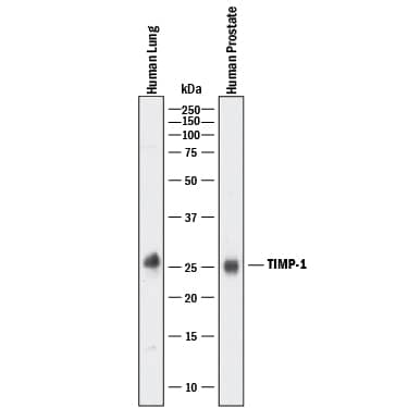
HAF017). A specific band was detected for TIMP-1 at approximately 25 kDa (as indicated). This experiment was conducted under reducing conditions and using
Immunoblot Buffer Group 1." alt="Western blot shows lysates of human lung tissue and human prostate tissue. PVDF membrane was probed with 1 µg/mL of Goat Anti-Human TIMP-1 Antigen Affinity-purified Polyclonal Antibody (Catalog # AF970) followed by HRP-conjugated Anti-Goat IgG Secondary Antibody (Catalog #
HAF017). A specific band was detected for TIMP-1 at approximately 25 kDa (as indicated). This experiment was conducted under reducing conditions and using
Immunoblot Buffer Group 1." class="big_thumb" />
HAF017). A specific band was detected for TIMP-1 at approximately 26 kDa (as indicated). This experiment was conducted under reducing conditions and using
Immunoblot Buffer Group 1." class="big_lightbox">
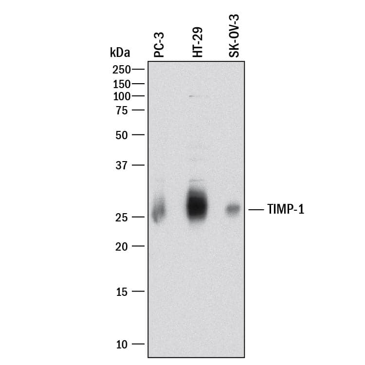
HAF017). A specific band was detected for TIMP-1 at approximately 26 kDa (as indicated). This experiment was conducted under reducing conditions and using
Immunoblot Buffer Group 1." class="big_thumb" />
VC004). Before incubation with the primary antibody, tissue was subjected to heat-induced epitope retrieval using Antigen Retrieval Reagent-Basic (Catalog #
CTS013). Tissue was stained using DAB (brown) and counterstained with hematoxylin (blue). Specific staining was localized to cytoplasm and extracellular space. View our protocol for
IHC Staining with VisUCyte HRP Polymer Detection Reagents." class="big_lightbox">
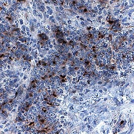
VC004). Before incubation with the primary antibody, tissue was subjected to heat-induced epitope retrieval using Antigen Retrieval Reagent-Basic (Catalog #
CTS013). Tissue was stained using DAB (brown) and counterstained with hematoxylin (blue). Specific staining was localized to cytoplasm and extracellular space. View our protocol for
IHC Staining with VisUCyte HRP Polymer Detection Reagents." class="big_thumb" />
HAF109). This experiment was conducted under reducing conditions and using the 12-230 kDa separation system." class="big_lightbox">
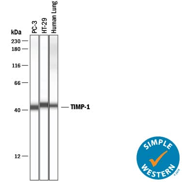
HAF109). This experiment was conducted under reducing conditions and using the 12-230 kDa separation system." class="big_thumb" />
HAF017). A specific band was detected for TIMP-1 at approximately 25 kDa (as indicated) in the parental SK-OV-3 cell line, but is not detectable in knockout SK-OV-3 cell line. GAPDH (Catalog #
AF5718) is shown as a loading control. This experiment was conducted under reducing conditions and using
Immunoblot Buffer Group 1." class="big_lightbox">
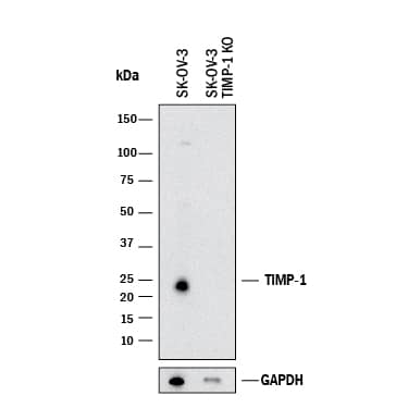
HAF017). A specific band was detected for TIMP-1 at approximately 25 kDa (as indicated) in the parental SK-OV-3 cell line, but is not detectable in knockout SK-OV-3 cell line. GAPDH (Catalog #
AF5718) is shown as a loading control. This experiment was conducted under reducing conditions and using
Immunoblot Buffer Group 1." class="big_thumb" />
902-MP) activity is measured in the presence of Recombinant Human TIMP-1 (0.1 µg/mL, Catalog #
970-TM) that has been preincubated with increasing concentrations of Goat Anti-Human TIMP-1 Antigen Affinity-purified Polyclonal Antibody (Catalog # AF970). The ND
50 is typically 1 µg/mL." class="big_lightbox">
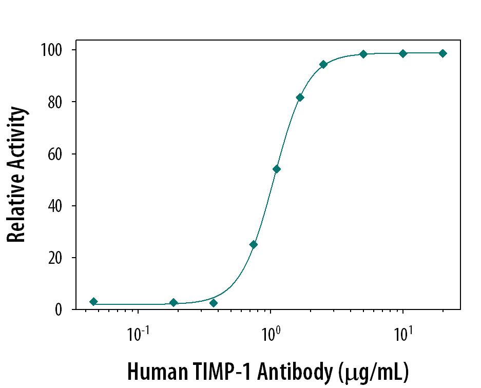
902-MP) activity is measured in the presence of Recombinant Human TIMP-1 (0.1 µg/mL, Catalog #
970-TM) that has been preincubated with increasing concentrations of Goat Anti-Human TIMP-1 Antigen Affinity-purified Polyclonal Antibody (Catalog # AF970). The ND
50 is typically 1 µg/mL." class="big_thumb" />


970-TM) was serially diluted and captured by Mouse Anti-Human TIMP‑1 Monoclonal Antibody (Catalog #
MAB970) coated on a Clear Polystyrene Microplate (Catalog #
DY990). Goat Anti-Human TIMP‑1 Antigen Affinity-purified Polyclonal Antibody (Catalog # AF970) was biotinylated and incubated with the protein captured on the plate. Detection of the standard curve was achieved by incubating Streptavidin-HRP (Catalog #
DY998)" class="big_lightbox">

970-TM) was serially diluted and captured by Mouse Anti-Human TIMP‑1 Monoclonal Antibody (Catalog #
MAB970) coated on a Clear Polystyrene Microplate (Catalog #
DY990). Goat Anti-Human TIMP‑1 Antigen Affinity-purified Polyclonal Antibody (Catalog # AF970) was biotinylated and incubated with the protein captured on the plate. Detection of the standard curve was achieved by incubating Streptavidin-HRP (Catalog #
DY998)" class="big_thumb" />
 HAF017). A specific band was detected for TIMP-1 at approximately 25 kDa (as indicated). This experiment was conducted under reducing conditions and using Immunoblot Buffer Group 1." alt="Western blot shows lysates of human lung tissue and human prostate tissue. PVDF membrane was probed with 1 µg/mL of Goat Anti-Human TIMP-1 Antigen Affinity-purified Polyclonal Antibody (Catalog # AF970) followed by HRP-conjugated Anti-Goat IgG Secondary Antibody (Catalog # HAF017). A specific band was detected for TIMP-1 at approximately 25 kDa (as indicated). This experiment was conducted under reducing conditions and using Immunoblot Buffer Group 1." class="big_thumb" />
 NL007) and counterstained with DAPI (blue). Specific staining was localized to cytoplasm and endoplasmic reticulum. View our protocol for Fluorescent ICC Staining of Cells on Coverslips." alt="TIMP-1 was detected in immersion fixed Wi-38 human lung fibroblast cell line using Mouse Anti-Human TIMP-1 Monoclonal Antibody (Catalog # MAB970) at 3 µg/mL for 3 hours at room temperature. Cells were stained using the NorthernLights™ 557-conjugated Anti-Mouse IgG Secondary Antibody (red; Catalog # NL007) and counterstained with DAPI (blue). Specific staining was localized to cytoplasm and endoplasmic reticulum. View our protocol for Fluorescent ICC Staining of Cells on Coverslips." class="big_thumb" />
 VC004). Before incubation with the primary antibody, tissue was subjected to heat-induced epitope retrieval using VisUCyte Antigen Retrieval Reagent-Basic (Catalog # VCTS021). Tissue was stained using DAB (brown) and counterstained with hematoxylin (blue). Specific staining was localized to neuronal cell bodies. View our protocol for IHC Staining with VisUCyte HRP Polymer Detection Reagents." alt="TIMP‑1 was detected in perfusion fixed paraffin-embedded sections of Rat Brain using Goat Anti-Rat TIMP‑1 Antigen Affinity-purified Polyclonal Antibody (Catalog # AF580) at 5 µg/mL for 1 hour at room temperature followed by incubation with the Anti-Goat IgG VisUCyte™ HRP Polymer Antibody (Catalog # VC004). Before incubation with the primary antibody, tissue was subjected to heat-induced epitope retrieval using VisUCyte Antigen Retrieval Reagent-Basic (Catalog # VCTS021). Tissue was stained using DAB (brown) and counterstained with hematoxylin (blue). Specific staining was localized to neuronal cell bodies. View our protocol for IHC Staining with VisUCyte HRP Polymer Detection Reagents." class="big_thumb" />


![N/A TIMP-1 [HRP]](http://images.novusbio.com/fullsize/elisa/DATA_TIMP1_DTM100_ELISA_847.jpg "N/A TIMP-1 [HRP]")
![N/A TIMP-1 [HRP]](http://images.novusbio.com/fullsize/elisa/TIMP-1_DTM100_ELISA_194.jpg)
![N/A TIMP-1 [Biotin]](http://images.novusbio.com/fullsize/elisa/DATA_TIMP1_DY970_ELISA_2284.jpg "N/A TIMP-1 [Biotin]")
 HAF017). A specific band was detected for TIMP-1 at approximately 26 kDa (as indicated). This experiment was conducted under reducing conditions and using
  VC004). Before incubation with the primary antibody, tissue was subjected to heat-induced epitope retrieval using Antigen Retrieval Reagent-Basic (Catalog #
  HAF109). This experiment was conducted under reducing conditions and using the 12-230 kDa separation system." class="big_thumb" />
 HAF017). A specific band was detected for TIMP-1 at approximately 25 kDa (as indicated) in the parental SK-OV-3 cell line, but is not detectable in knockout SK-OV-3 cell line. GAPDH (Catalog #
  902-MP) activity is measured in the presence of Recombinant Human TIMP-1 (0.1 µg/mL, Catalog #


 970-TM) was serially diluted and captured by Mouse Anti-Human TIMP‑1 Monoclonal Antibody (Catalog #

 980-MT) was serially diluted and captured by Rat Anti-Mouse TIMP‑1 Monoclonal Antibody (Catalog #
 ![N/A TIMP-1 [HRP]](http://images.novusbio.com/fullsize/elisa/DATA_TIMP1_MTM100_ELISA_1041.jpg "N/A TIMP-1 [HRP]")
![N/A TIMP-1 [HRP]](http://images.novusbio.com/fullsize/elisa/TIMP-1_MTM100_ELISA_328.jpg)
 CTS013). Tissue was stained using the Anti-Mouse HRP-DAB Cell & Tissue Staining Kit (brown; Catalog #
 970-TM) was serially diluted and captured by Mouse Anti-Human TIMP‑1 Monoclonal Antibody (Catalog # MAB970) coated on a Clear Polystyrene Microplate (Catalog #
 ![Western Blot: TIMP-1 Antibody (F31 P2 A5) [NB100-74551] - Analysis was performed on whole cell extracts (30 ug lysate) of A431 (Lane 1), HeLa (Lane 2), Jurkat (Lane 3), HT29 (Lane 4) and COS-7 (Lane 5). The blots were probed with Anti-TIMP1 Mouse .](http://images.novusbio.com/fullsize/TIMP-1-Antibody-F31-P2-A5-Western-Blot-NB100-74551-img0009.jpg "Western Blot: TIMP-1 Antibody (F31 P2 A5) [NB100-74551] - Analysis was performed on whole cell extracts (30 ug lysate) of A431 (Lane 1), HeLa (Lane 2), Jurkat (Lane 3), HT29 (Lane 4) and COS-7 (Lane 5). The blots were probed with Anti-TIMP1 Mouse .")
![Immunocytochemistry/Immunofluorescence: TIMP-1 Antibody (F31 P2 A5) [NB100-74551] - Formalin-fixed cells were permeabilized with 0.1% Triton X-100 in TBS for 5-10 minutes at room temperature and blocked with 3% BSA-PBS for 30 minutes at room temperature. Cells were probed with a TIMP1 Monclonal Antibody (F31 P2 A5) at a dilution of 1:20 and incubated overnight in a humidified chamber. Cells were washed with PBST and incubated with a DyLight-conjugated secondary antibody for 45 minutes at room temperature in the dark. F-actin (red) was stained with a fluorescent phalloidin and nuclei (blue) were stained with DAPI.](http://images.novusbio.com/fullsize/TIMP-1-Antibody-F31-P2-A5-Immunocytochemistry-Immunofluorescence-NB100-74551-img0005.jpg)
![Immunohistochemistry-Paraffin: TIMP-1 Antibody (F31 P2 A5) [NB100-74551] - Normal biopsies of deparaffinized Human prostate tissue at a dilution of 1:20.](http://images.novusbio.com/fullsize/TIMP-1-Antibody-F31-P2-A5-Immunohistochemistry-Paraffin-NB100-74551-img0006.jpg)
![Immunocytochemistry/Immunofluorescence: TIMP-1 Antibody (F31 P2 A5) [NB100-74551] - Analysis of TIMP1 (green) in Hela cells. Formalin-fixed cells were permeabilized with 0.1% Triton X-100 in TBS for 5-10 minutes at room temperature and blocked with 3% BSA-PBS for 30 minutes at room temperature. Cells were probed with a TIMP1 Monclonal Antibody (F31 P2 A5) at a dilution of 1:100 and incubated overnight in a humidified chamber. Cells were washed with PBST and incubated with a DyLight-conjugated secondary antibody for 45 minutes at room temperature in the dark. F-actin (red) was stained with a fluorescent phalloidin and nuclei (blue) were stained with DAPI. Images were taken at a 60X magnification.](http://images.novusbio.com/fullsize/TIMP-1-Antibody-F31-P2-A5-Immunocytochemistry-Immunofluorescence-NB100-74551-img0004.jpg)
![Immunohistochemistry-Paraffin: TIMP-1 Antibody (F31 P2 A5) [NB100-74551] - Cancer biopsies of deparaffinized Human breast carcinoma tissue at a dilution of 1:20.](http://images.novusbio.com/fullsize/TIMP-1-Antibody-F31-P2-A5-Immunohistochemistry-Paraffin-NB100-74551-img0007.jpg)


 580-RT) was serially diluted and captured by Mouse Anti-Rat TIMP‑1 Monoclonal Antibody (Catalog #
 ![N/A TIMP-1 [Biotin]](http://images.novusbio.com/fullsize/elisa/DATA_TIMP1_DY980_ELISA_2289.jpg "N/A TIMP-1 [Biotin]")
![N/A TIMP-1 [HRP]](http://images.novusbio.com/fullsize/elisa/DATA_TIMP1_RTM100_ELISA_1100.jpg "N/A TIMP-1 [HRP]")
![N/A TIMP-1 [HRP]](http://images.novusbio.com/fullsize/elisa/TIMP-1_PRTM100_ELISA_366.jpg)


