HAF017). A specific band was detected for ACE-2 at approximately 100 and 110 kDa (as indicated). This experiment was conducted under reducing conditions and using Western Blot Buffer Group 1." class="big_lightbox">
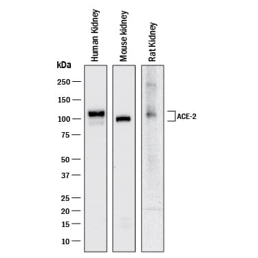
HAF017). A specific band was detected for ACE-2 at approximately 100 and 110 kDa (as indicated). This experiment was conducted under reducing conditions and using Western Blot Buffer Group 1." alt="Western blot shows lysates of human kidney tissue, mouse kidney tissue, and rat kidney tissue. PVDF membrane was probed with 1 µg/mL of Goat Anti-Human/Mouse/Rat/Hamster ACE-2 Antigen Affinity-purified Polyclonal Antibody (Catalog # AF933) followed by HRP-conjugated Anti-Goat IgG Secondary Antibody (Catalog #
HAF017). A specific band was detected for ACE-2 at approximately 100 and 110 kDa (as indicated). This experiment was conducted under reducing conditions and using Western Blot Buffer Group 1." class="big_thumb" />
F0108). Quadrant markers were set based on Goat IgG control antibody (
AB-108-C, data not shown). Staining was performed using our Staining Membrane-associated Proteins protocol. " class="big_lightbox">
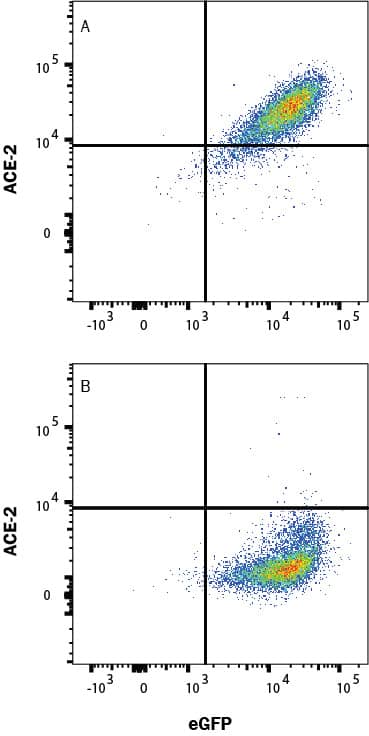
F0108). Quadrant markers were set based on Goat IgG control antibody (
AB-108-C, data not shown). Staining was performed using our Staining Membrane-associated Proteins protocol. " class="big_thumb" />
CTS008) and counterstained with hematoxylin (blue). Lower panel shows a lack of labeling if primary antibodies are omitted and tissue is stained only with secondary antibody followed by incubation with detection reagents. View our protocol for
Chromogenic IHC Staining of Paraffin-embedded Tissue Sections." class="big_lightbox">
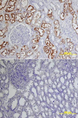
CTS008) and counterstained with hematoxylin (blue). Lower panel shows a lack of labeling if primary antibodies are omitted and tissue is stained only with secondary antibody followed by incubation with detection reagents. View our protocol for
Chromogenic IHC Staining of Paraffin-embedded Tissue Sections." class="big_thumb" />
VC004). Before incubation with the primary antibody, tissue was subjected to heat-induced epitope retrieval using Antigen Retrieval Reagent-Basic (
CTS013). Tissue was stained using DAB (brown) and counterstained with hematoxylin (blue). Specific staining was localized to respiratory bronchioles. Staining was performed using our protocol for IHC Staining with VisUCyte HRP Polymer Detection Reagents." class="big_lightbox">
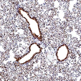
VC004). Before incubation with the primary antibody, tissue was subjected to heat-induced epitope retrieval using Antigen Retrieval Reagent-Basic (
CTS013). Tissue was stained using DAB (brown) and counterstained with hematoxylin (blue). Specific staining was localized to respiratory bronchioles. Staining was performed using our protocol for IHC Staining with VisUCyte HRP Polymer Detection Reagents." class="big_thumb" />
VC004). Before incubation with the primary antibody, tissue was subjected to heat-induced epitope retrieval using Antigen Retrieval Reagent-Basic (
CTS013). Tissue was stained using DAB (brown) and counterstained with hematoxylin (blue). Specific staining was localized to cell surface in eputhelial cells in bronchioles. Staining was performed using our protocol for IHC Staining with VisUCyte HRP Polymer Detection Reagents." class="big_lightbox">
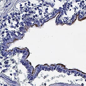
VC004). Before incubation with the primary antibody, tissue was subjected to heat-induced epitope retrieval using Antigen Retrieval Reagent-Basic (
CTS013). Tissue was stained using DAB (brown) and counterstained with hematoxylin (blue). Specific staining was localized to cell surface in eputhelial cells in bronchioles. Staining was performed using our protocol for IHC Staining with VisUCyte HRP Polymer Detection Reagents." class="big_thumb" />
VC004). Before incubation with the primary antibody, tissue was subjected to heat-induced epitope retrieval using Antigen Retrieval Reagent-Basic (
CTS013). Tissue was stained using DAB (brown) and counterstained with hematoxylin (blue). Staining was performed using our protocol for IHC Staining with VisUCyte HRP Polymer Detection Reagents." class="big_lightbox">
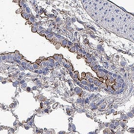
VC004). Before incubation with the primary antibody, tissue was subjected to heat-induced epitope retrieval using Antigen Retrieval Reagent-Basic (
CTS013). Tissue was stained using DAB (brown) and counterstained with hematoxylin (blue). Staining was performed using our protocol for IHC Staining with VisUCyte HRP Polymer Detection Reagents." class="big_thumb" />
VC004). Before incubation with the primary antibody, tissue was subjected to heat-induced epitope retrieval using Antigen Retrieval Reagent-Basic (
CTS013). Tissue was stained using DAB (brown) and counterstained with hematoxylin (blue). Specific staining was localized to cell surface in convoluted tubules. Staining was performed using our protocol for IHC Staining with VisUCyte HRP Polymer Detection Reagents." class="big_lightbox">
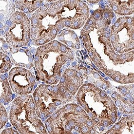
VC004). Before incubation with the primary antibody, tissue was subjected to heat-induced epitope retrieval using Antigen Retrieval Reagent-Basic (
CTS013). Tissue was stained using DAB (brown) and counterstained with hematoxylin (blue). Specific staining was localized to cell surface in convoluted tubules. Staining was performed using our protocol for IHC Staining with VisUCyte HRP Polymer Detection Reagents." class="big_thumb" />
HAF109). This experiment was conducted under reducing conditions and using the 12-230 kDa separation system." class="big_lightbox">
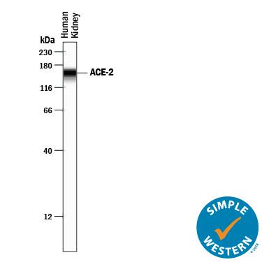
HAF109). This experiment was conducted under reducing conditions and using the 12-230 kDa separation system." class="big_thumb" />
 HAF017). A specific band was detected for ACE-2 at approximately 100 and 110 kDa (as indicated). This experiment was conducted under reducing conditions and using Western Blot Buffer Group 1." alt="Western blot shows lysates of human kidney tissue, mouse kidney tissue, and rat kidney tissue. PVDF membrane was probed with 1 µg/mL of Goat Anti-Human/Mouse/Rat/Hamster ACE-2 Antigen Affinity-purified Polyclonal Antibody (Catalog # AF933) followed by HRP-conjugated Anti-Goat IgG Secondary Antibody (Catalog # HAF017). A specific band was detected for ACE-2 at approximately 100 and 110 kDa (as indicated). This experiment was conducted under reducing conditions and using Western Blot Buffer Group 1." class="big_thumb" />
 HAF007). A specific band was detected for ACE-2 at approximately 110 kDa (as indicated). This experiment was conducted under reducing conditions and using Immunoblot Buffer Group 1." alt="Western blot shows lysates of NS0 mouse myeloma cell line and human kidney tissue. PVDF membrane was probed with 2 µg/mL of Mouse Anti-Human ACE-2 Monoclonal Antibody (Catalog # MAB933) followed by HRP-conjugated Anti-Mouse IgG Secondary Antibody (Catalog # HAF007). A specific band was detected for ACE-2 at approximately 110 kDa (as indicated). This experiment was conducted under reducing conditions and using Immunoblot Buffer Group 1." class="big_thumb" />
 HAF019). A specific band was detected for ACE-2 at approximately 95 kDa (as indicated). This experiment was conducted under reducing conditions and using Immunoblot Buffer Group 1." alt="Western blot shows lysates of mouse kidney tissue. PVDF membrane was probed with 0.25 µg/mL of Goat Anti-Mouse ACE-2 Antigen Affinity-purified Polyclonal Antibody (Catalog # AF3437) followed by HRP-conjugated Anti-Goat IgG Secondary Antibody (Catalog # HAF019). A specific band was detected for ACE-2 at approximately 95 kDa (as indicated). This experiment was conducted under reducing conditions and using Immunoblot Buffer Group 1." class="big_thumb" />
 HAF018). A specific band was detected for ACE‑2 at approximately 120 kDa (as indicated). This experiment was conducted under reducing conditions and using Western Blot Buffer Group 1." alt="Western blot shows lysates of human kidney and human testis. PVDF membrane was probed with 2 µg/mL of Mouse Anti-Human/Rat/Hamster ACE‑2 Monoclonal Antibody (Catalog # MAB9332) followed by HRP-conjugated Anti-Mouse IgG Secondary Antibody (HAF018). A specific band was detected for ACE‑2 at approximately 120 kDa (as indicated). This experiment was conducted under reducing conditions and using Western Blot Buffer Group 1." class="big_thumb" />
 HAF007). A specific band was detected for ACE-2 at approximately 110 kDa (as indicated). This experiment was conducted under reducing conditions and using Immunoblot Buffer Group 1." alt="Western blot shows lysates of NS0 mouse myeloma cell line and human kidney tissue. PVDF membrane was probed with 2 µg/mL of Mouse Anti-Human ACE-2 Monoclonal Antibody (Catalog # MAB9331) followed by HRP-conjugated Anti-Mouse IgG Secondary Antibody (Catalog # HAF007). A specific band was detected for ACE-2 at approximately 110 kDa (as indicated). This experiment was conducted under reducing conditions and using Immunoblot Buffer Group 1." class="big_thumb" />
 HAF005). A specific band was detected for ACE‑2 at approximately 120 kDa (as indicated). This experiment was conducted under reducing conditions and using Western Blot Buffer Group 1." alt="Western blot shows lysates of human kidney. PVDF membrane was probed with 0.5 µg/mL of Rat Anti-Human/Mouse/Rat/Hamster ACE‑2 Monoclonal Antibody (Catalog # MAB3437) followed by HRP-conjugated Anti-Rat IgG Secondary Antibody (HAF005). A specific band was detected for ACE‑2 at approximately 120 kDa (as indicated). This experiment was conducted under reducing conditions and using Western Blot Buffer Group 1." class="big_thumb" />


 F0108). Quadrant markers were set based on Goat IgG control antibody (
 CTS008) and counterstained with hematoxylin (blue). View our protocol for
  CTS008) and counterstained with hematoxylin (blue). Lower panel shows a lack of labeling if primary antibodies are omitted and tissue is stained only with secondary antibody followed by incubation with detection reagents. View our protocol for
  VC004). Before incubation with the primary antibody, tissue was subjected to heat-induced epitope retrieval using Antigen Retrieval Reagent-Basic (
 VC004). Before incubation with the primary antibody, tissue was subjected to heat-induced epitope retrieval using Antigen Retrieval Reagent-Basic (
 VC004). Before incubation with the primary antibody, tissue was subjected to heat-induced epitope retrieval using Antigen Retrieval Reagent-Basic (
 VC004). Before incubation with the primary antibody, tissue was subjected to heat-induced epitope retrieval using Antigen Retrieval Reagent-Basic (
 HAF109). This experiment was conducted under reducing conditions and using the 12-230 kDa separation system." class="big_thumb" />


![Simple Western: ACE-2 Antibody [Unconjugated] [AF933] - Design for a membrane-localized ACE2 expression system. (A) Our ACE2 construct is driven by a CMV promoter followed by the first 25 residues of ACE2 containing the leader sequence that direct ACE2 to the plasma membrane. This is followed by a 3xHA tag linked to the remainder of ACE2 (20-805) and a C-terminal sfGFP. Both 3xHA and sfGFP fusions are separated from ACE2 by flexible 3xGGGGS linkers. (B) The ACE2 fusion protein is designed to be embedded in the plasma membrane where it can perform extracellular carboxypeptidase-mediated metabolism and its levels can be detected by cell staining with antibodies to HA. (C) Lysates from untransfected or 3xHA-ACE2-sfGFP-transfected HEK293 cells were analyzed by automated Jess capillary immunoassay using antibodies to HA, GFP, and two ACE2 antibodies. (D) Confocal fluorescence microscopy of HEK cells transfected with 3xHA-ACE2-sfGFP and stained with HA and the nuclear stain DAPI. Image collected and cropped by CiteAb from the following publication (//pubmed.ncbi.nlm.nih.gov/37644110), licensed under a CC-BY license. Not internally tested by R&D Systems.](http://images.novusbio.com/fullsize/af933_human-ace-2-ectodomain-affinity-purified-polyclonal-ab-30820241044556.jpg)


 CTS013). Tissue was stained using the Anti-Mouse HRP-DAB Cell & Tissue Staining Kit (brown; Catalog #
  VC001). Before incubation with the primary antibody, tissue was subjected to heat-induced epitope retrieval using Antigen Retrieval Reagent-Basic (


 933-ZN were tested in a functional ELISA for binding to recombinant SARS-CoV-2 Spike RBD Fc Chimera Protein
  10499-CV) was immobilized on a Biacore Sensor Chip CM5, and binding to recombinant human ACE-2 (
 ES007)." class="big_thumb" />

 F0108). Quadrant markers were set based on Goat IgG control antibody (
 Fluorescent IHC Staining of Frozen Tissue Sections." class="big_thumb" />
 CTS008) and counterstained with hematoxylin (blue). Lower panel shows a lack of labeling if primary antibodies are omitted and tissue is stained only with secondary antibody followed by incubation with detection reagents. View our protocol for
  HAF109). This experiment was conducted under reducing conditions and using the 12-230 kDa separation system." class="big_thumb" />
 MAB34371) coated on a Clear Polystyrene Microplate (

 3437-ZN) was serially diluted and captured by Rat Anti-Mouse ACE‑2 Monoclonal Antibody (Catalog #
 ![Immunohistochemistry-Paraffin: ACE-2 Antibody (SN0754) [NBP2-67692] - ACE-2 staining in immersion fixed paraffin-embedded sections of human lung normal (left) and lung cancer tissue (right) using Mouse Anti-Human ACE-2 (SN0754) Monoclonal Antibody (Catalog # NBP2-67692) at 10 ug/ml for 1 hour at room temperature followed by incubation with Goat anti-Mouse IgG Secondary Antibody.](http://images.novusbio.com/fullsize/ACE-2-Antibody-SN0754-Immunohistochemistry-Paraffin-NBP2-67692-img0009.jpg "Immunohistochemistry-Paraffin: ACE-2 Antibody (SN0754) [NBP2-67692] - ACE-2 staining in immersion fixed paraffin-embedded sections of human lung normal (left) and lung cancer tissue (right) using Mouse Anti-Human ACE-2 (SN0754) Monoclonal Antibody (Catalog # NBP2-67692) at 10 ug/ml for 1 hour at room temperature followed by incubation with Goat anti-Mouse IgG Secondary Antibody.")
![Western Blot: ACE-2 Antibody (SN0754) [NBP2-67692] - Analysis of ACE2 on different lysates. Proteins were transferred to a PVDF membrane and blocked with 5% BSA in PBS for 1 hour at room temperature. The primary antibody (1/500) was used in 5% BSA at room temperature for 2 hours. Goat Anti-Rabbit IgG - HRP Secondary Antibody at 1:5,000 dilution was used for 1 hour at room temperature. Positive control: Lane 1: human kidney tissue lysateLane 2: human small intestine tissue lysate](http://images.novusbio.com/fullsize/ACE-2-Antibody-SN0754-Western-Blot-NBP2-67692-img0011.jpg)
![Immunocytochemistry/Immunofluorescence: ACE-2 Antibody (SN0754) [NBP2-67692] - Staining ACE2 in MCF-7 cells (green). The nuclear counter stain is DAPI (blue). Cells were fixed in paraformaldehyde, permeabilised with 0.25% Triton X100/PBS.](http://images.novusbio.com/fullsize/ACE-2-Antibody-SN0754-Immunocytochemistry-Immunofluorescence-NBP2-67692-img0003.jpg)
![Immunohistochemistry-Paraffin: ACE-2 Antibody (SN0754) [NBP2-67692] - ACE-2 staining in paraffin-embedded sections of hamster lung tissue using Rabbit Anti-Human ACE-2 (SN0754) Monoclonal Antibody (Catalog # NBP2-67692) at 3 ug/ml overnight at 4 degrees C. Detection kit: VC003. Counterstain: Hematoxylin. Localization: respiratory bronchioles.](http://images.novusbio.com/fullsize/ACE-2-Antibody-SN0754-Immunohistochemistry-Paraffin-NBP2-67692-img0010.jpg)
![Western Blot: ACE-2 Antibody (SN0754) [NBP2-67692] - Western blot analysis of ACE-2 on Hamster testis (1) and stomach (2) tissue lysates using anti-ACE-2 antibody at 1/1,000 dilution.](http://images.novusbio.com/fullsize/ACE-2-Antibody-SN0754-Western-Blot-NBP2-67692-img0008.jpg)
![Immunocytochemistry/Immunofluorescence: ACE-2 Antibody (SN0754) [NBP2-67692] - Staining ACE2 in 293 cells (green). The nuclear counter stain is DAPI (blue). Cells were fixed in paraformaldehyde, permeabilised with 0.25% Triton X100/PBS.](http://images.novusbio.com/fullsize/ACE-2-Antibody-SN0754-Immunocytochemistry-Immunofluorescence-NBP2-67692-img0001.jpg)
![Immunocytochemistry/Immunofluorescence: ACE-2 Antibody (SN0754) [NBP2-67692] - Staining ACE2 in HepG2 cells (green). The nuclear counter stain is DAPI (blue). Cells were fixed in paraformaldehyde, permeabilised with 0.25% Triton X100/PBS.](http://images.novusbio.com/fullsize/ACE-2-Antibody-SN0754-Immunocytochemistry-Immunofluorescence-NBP2-67692-img0002.jpg)
![Immunohistochemistry-Paraffin: ACE-2 Antibody (SN0754) [NBP2-67692] - Analysis of paraffin-embedded human breast carcinoma tissue using anti-ACE2 antibody. Counter stained with hematoxylin.](http://images.novusbio.com/fullsize/ACE-2-Antibody-SN0754-Immunohistochemistry-Paraffin-NBP2-67692-img0004.jpg)
![Immunohistochemistry-Paraffin: ACE-2 Antibody (SN0754) [NBP2-67692] - Analysis of paraffin-embedded human kidney tissue using anti-ACE2 antibody. Counter stained with hematoxylin.](http://images.novusbio.com/fullsize/ACE-2-Antibody-SN0754-Immunohistochemistry-Paraffin-NBP2-67692-img0005.jpg)
![Immunohistochemistry-Paraffin: ACE-2 Antibody (SN0754) [NBP2-67692] - Analysis of paraffin-embedded mouse kidney tissue using anti-ACE2 antibody. Counter stained with hematoxylin.](http://images.novusbio.com/fullsize/ACE-2-Antibody-SN0754-Immunohistochemistry-Paraffin-NBP2-67692-img0006.jpg)
![Western blot analysis on different lysates with NBP2-67692 at 1/2,000 dilution. Lane 1: Caco-2 cell lysate (20 µg/Lane) Lane 2: HepG2 cell lysate (20 µg/Lane) Lane 3: 293T cell lysate (20 µg/Lane) Lane 4: Mouse testis tissue lysate (40 µg/Lane) Lane 5: Mouse lung tissue lysate (40 µg/Lane) Lane 6: Rat testis tissue lysate (40 µg/Lane) Lane 7: Rat lung tissue lysate (40 µg/Lane) Predicted band size: 92 kDa Observed band size: 130 kDa Exposure time: 3 minutes; 4-20% SDS-PAGE gel. Proteins were transferred to a PVDF membrane and blocked with 5% NFDM/TBST for 1 hour at room temperature. NBP2-67692 at 1/2,000 dilution was used in 5% NFDM/TBST at 4℃ overnight. Goat Anti-Rabbit IgG [HRP] Secondary Antibody at 1/50,000 dilution was used for 1 hour at room temperature.](http://images.novusbio.com/fullsize/nbp2-67692_-western-blot-639204586672234702.jpg)
![Western Blot: ACE-2 Antibody [NBP1-76614] - Independent Antibody Validation (IAV) via Protein Expression Profile in Human TissuesLoading: 15 ug of lysates per lane.Antibodies: ACE2, (2 ug/mL), ACE2, (2 ug/mL), ACE2, (2 ug/mL) and beta-actin (1 ug/mL), 1h incubation at RT in 5% NFDM/TBST.Secondary: Goat anti-rabbit IgG HRP conjugate at 1:10000 dilution.](http://images.novusbio.com/fullsize/ACE-2-Antibody-Western-Blot-NBP1-76614-img0009.jpg "Western Blot: ACE-2 Antibody [NBP1-76614] - Independent Antibody Validation (IAV) via Protein Expression Profile in Human TissuesLoading: 15 ug of lysates per lane.Antibodies: ACE2, (2 ug/mL), ACE2, (2 ug/mL), ACE2, (2 ug/mL) and beta-actin (1 ug/mL), 1h incubation at RT in 5% NFDM/TBST.Secondary: Goat anti-rabbit IgG HRP conjugate at 1:10000 dilution.")
![Immunohistochemistry-Paraffin: ACE-2 Antibody [NBP1-76614] - Analysis of paraffin-embedded human kidney tissue using anti-ACE2 antibody 2 ug/ml. Tissue was fixed with formaldehyde and blocked with 10% serum for 1 h at RT; antigen retrieval was by heat mediation with a citrate buffer (pH6). Samples were incubated with primary antibody overnight at 4C. A goat anti-rabbit IgG H&L (HRP) at 1/250 was used as secondary. Counter stained with Hematoxylin.](http://images.novusbio.com/fullsize/ACE-2-Antibody-Immunohistochemistry-Paraffin-NBP1-76614-img0005.jpg)
![Western Blot: ACE-2 Antibody [NBP1-76614] - Validation in Rat Skin Tissue Loading: 15 ug of lysates per lane.Antibodies: ACE2 (2 ug/mL), 1h incubation at RT in 5% NFDM/TBST.Secondary: Goat anti-rabbit IgG HRP conjugate at 1:10000 dilution.](http://images.novusbio.com/fullsize/ACE-2-Antibody-Western-Blot-NBP1-76614-img0006.jpg)
![Western Blot: ACE-2 Antibody [NBP1-76614] - Loading: 15 ug of lysates per lane.Antibodies: ACE2, (2 ug/mL), 1h incubation at RT in 5% NFDM/TBST.Secondary: Goat anti-rabbit IgG HRP conjugate at 1:10000 dilution.](http://images.novusbio.com/fullsize/ACE-2-Antibody-Western-Blot-NBP1-76614-img0007.jpg)
![Western Blot: ACE-2 Antibody [NBP1-76614] - Validation in Human TissuesLoading: 15 ug of lysates per lane.Antibodies: ACE2, (2 ug/mL), 1h incubation at RT in 5% NFDM/TBST.Secondary: Goat anti-rabbit IgG HRP conjugate at 1:10000 dilution.](http://images.novusbio.com/fullsize/ACE-2-Antibody-Western-Blot-NBP1-76614-img0008.jpg)
![Immunocytochemistry/Immunofluorescence: ACE-2 Antibody [NBP1-76614] - Analysis of 4% paraformaldehyde-fixed Caco2 cells labeling ACE2 at 20 mg/mL, followed by goat anti-rabbit IgG secondary antibody at 1/500 dilution (green) and DAPI staining (blue). Image showing membrane staining on Caco2 cells.](http://images.novusbio.com/fullsize/ACE-2-Antibody-Immunocytochemistry-Immunofluorescence-NBP1-76614-img0001.jpg)
![Immunocytochemistry/Immunofluorescence: ACE-2 Antibody [NBP1-76614] - Analysis of 4% paraformaldehyde-fixed human lung tissue labeling ACE-2 at 20 mg/mL, followed by goat anti-rabbit IgG secondary antibody at 1/500 dilution (green) and DAPI staining (blue).](http://images.novusbio.com/fullsize/ACE-2-Antibody-Immunocytochemistry-Immunofluorescence-NBP1-76614-img0003.jpg)
![Immunocytochemistry/ Immunofluorescence: ACE-2 Antibody - BSA Free [NBP1-76614] - Validation of ACE-2 in Rat Lung Tissue. Immunofluorescent analysis of 4% paraformaldehyde-fixed rat lung tissue labeling ACE-2 with at 20 ug/mL, followed by goat anti-rabbit IgG secondary antibody at 1/500 dilution (green) and DAPI staining (blue).](http://images.novusbio.com/fullsize/nbp1-76614_rabbit-polyclonal-ace-2-antibody-2711202420175165.gif)
![Immunocytochemistry/ Immunofluorescence: ACE-2 Antibody - BSA Free [NBP1-76614] - Validation of ACE-2 in Human Testis Tissue. Immunofluorescent analysis of 4% paraformaldehyde-fixed human testis tissue labeling ACE-2 with at 20 ug/mL, followed by goat anti-rabbit IgG secondary antibody at 1/500 dilution (green) and DAPI staining (blue).](http://images.novusbio.com/fullsize/nbp1-76614_rabbit-polyclonal-ace-2-antibody-2711202419514040.gif)
![Block/Neutralize: ACE-2 Antibody (AC384) [NBP2-80038] - In a functional flow cytometry assay using a HEK293 human embryonic kidney cell line transfected with human ACE-2, 1ug/mL Fc-tagged recombinant human Spike S1 subunit RBD is shown to bind Ace-2 (black dotted line) detected by Human IgG Fc-APC Secondary Antibody (FAB110A). Binding is completely blocked (orange histogram) by 25 ug/mL of Mouse Anti-Human ACE-2 Monoclonal Antibody (NBP2-80038). 25 ug/mL Mouse Anti-Human DC-SIGN/CD209 Monoclonal Antibody (MAB161) was used as an irrelevant control (blue line).](http://images.novusbio.com/fullsize/ACE-2-Antibody-AC384-Functional-NBP2-80038-img0003.jpg "Block/Neutralize: ACE-2 Antibody (AC384) [NBP2-80038] - In a functional flow cytometry assay using a HEK293 human embryonic kidney cell line transfected with human ACE-2, 1ug/mL Fc-tagged recombinant human Spike S1 subunit RBD is shown to bind Ace-2 (black dotted line) detected by Human IgG Fc-APC Secondary Antibody (FAB110A). Binding is completely blocked (orange histogram) by 25 ug/mL of Mouse Anti-Human ACE-2 Monoclonal Antibody (NBP2-80038). 25 ug/mL Mouse Anti-Human DC-SIGN/CD209 Monoclonal Antibody (MAB161) was used as an irrelevant control (blue line).")
![Block/Neutralize: ACE-2 Antibody (AC384) [NBP2-80038] - In a functional flow cytometry assay using a HEK293 human embryonic kidney cell line transfected with human ACE-2, 0.5 ug/mL sheep Fc-tagged recombinant human Spike S1 is shown to bind ACE-2 (black dotted line) detected by Donkey anti-Sheep APC Secondary Antibody (Catalog # F0127). Binding is completely blocked (orange histogram) by 25 ug/mL of Mouse Anti-Human ACE-2 Monoclonal Antibody (NBP2-80038). 25 ug/mL Mouse anti-Human DC-SIGN/CD209 Monoclonal Antibody (MAB161) was used as an irrelevant control (blue line).](http://images.novusbio.com/fullsize/ACE-2-Antibody-AC384-Functional-NBP2-80038-img0004.jpg)
![Western Blot: ACE-2 Antibody (AC384) [NBP2-80038] - 1: Human ACE2 Fc-fusion protein. 2: Human ACE2 (Ecto domain) (FLAG®-tagged). 3: HepG2 cell lysate. 4: Other Fc-fusion (hGITR-Fc) proteins (negative control). 1:2'000 dilution.](http://images.novusbio.com/fullsize/ACE-2-Antibody-AC384-Western-Blot-NBP2-80038-img0005.jpg)
![Immunohistochemistry-Paraffin: ACE-2 Antibody (AC384) [NBP2-80038] - ACE-2 staining in paraffin-embedded sections of hamster lung tissue using Mouse Anti-Human ACE-2 (AC384) Monoclonal Antibody (Catalog # NBP2-80038) at 10 ug/ml overnight at 4 degrees C. Detection kit: VC001. Counterstain: Hematoxylin. Localization: respiratory bronchioles.](http://images.novusbio.com/fullsize/ACE-2-Antibody-AC384-Immunohistochemistry-Paraffin-NBP2-80038-img0007.jpg)
![Flow Cytometry: ACE-2 Antibody (AC384) [NBP2-80038] - Detection of ACE-2 in HEK293 Human Cell Line Transfected with Human ACE-2 and eGFP by Flow Cytometry. HEK293 human embryonic kidney cell line transfected with (A) human ACE-2 or (B) irrelevant protein, and eGFP was stained with Mouse Anti-Human ACE-2 Monoclonal Antibody (Catalog # NBP2-80038) followed by Allophycocyanin-conjugated Anti-Mouse IgG Secondary Antibody (Catalog # F0101B). Quadrant markers were set based on Mouse IgG1 control antibody staining (Catalog # MAB002, data not shown). View our protocol for Staining Membrane-associated Proteins.](http://images.novusbio.com/fullsize/ACE-2-Antibody-AC384-Flow-Cytometry-NBP2-80038-img0002.jpg)
![Immunohistochemistry-Paraffin: ACE-2 Antibody (AC384) [NBP2-80038] - ACE-2 was detected in immersion fixed paraffin-embedded sections of human lung normal (left) and lung cancer tissue (right) using Mouse Anti-HumanACE-2 (AC18F) Monoclonal Antibody (Catalog # NBP2-80038) at 15ug/mL for 1 hour at room temperature followed by incubation with the Goat anti-Mouse IgG Secondary Antibody.](http://images.novusbio.com/fullsize/ACE-2-Antibody-AC384-Immunohistochemistry-Paraffin-NBP2-80038-img0006.jpg)
![Functional: ACE-2 Antibody (AC384) [NBP2-80038] - Binding of ACE2 (human) to the Spike protein of SARS-CoV-2 is inhibited by the antibody anti-ACE2 (human), mAb (blocking) (AC384). Spike (SARS-CoV-2):Fc (human) (RBD) (rec.) is coated on an ELISA plate at 1ug/ml. ACE2 (human), mAb (blocking) (AC384) or an unrelated mAb Control are added (starting at 40 ug/mL with a two-fold serial dilution) together with 500 ng/uL of ACE2 (human). After incubation for 1h at RT, the binding was detected using an anti-FLAG antibody (HRP).](http://images.novusbio.com/fullsize/ACE-2-Antibody-AC384-Functional-NBP2-80038-img0001.jpg)
 F0101B). Quadrant markers were set based on Mouse IgG2A Isotype Control (
 CTS013). Tissue was stained using the NorthernLights™ 557-conjugated Anti-Mouse IgG Secondary Antibody (red;
  VC001). Before incubation with the primary antibody, tissue was subjected to heat-induced epitope retrieval using Antigen Retrieval Reagent-Basic (
![N/A ACE-2 [Biotin]](http://images.novusbio.com/fullsize/dy933-05_human-ace-2-duoset-elisa-5-plate-26102020101415.jpg "N/A ACE-2 [Biotin]")
 CTS013). Tissue was stained using the NorthernLights™ 557-conjugated Anti-Mouse IgG Secondary Antibody (red;
  VC001). Before incubation with the primary antibody, tissue was subjected to heat-induced epitope retrieval using Antigen Retrieval Reagent-Basic (
 HAF005). A specific band was detected for ACE‑2 at approximately 120 kDa (as indicated). This experiment was conducted under reducing conditions and using Western Blot Buffer Group 1." class="big_thumb" />

![Western Blot: ACE-2 Antibody (AC18F) [NBP2-80035] - Western blot analysis using anti-ACE2 (human), mAb (AC18F) at 1: 2,000 dilution. Lane 1. hACE2 (Fc protein). Lane 2. hACE2 (Ecto domain) (FLAG(R)-tagged). Lane 3. Other hGITR (Fc protein) (control).](http://images.novusbio.com/fullsize/ACE-2-Antibody-AC18F-Western-Blot-NBP2-80035-img0003.jpg "Western Blot: ACE-2 Antibody (AC18F) [NBP2-80035] - Western blot analysis using anti-ACE2 (human), mAb (AC18F) at 1: 2,000 dilution. Lane 1. hACE2 (Fc protein). Lane 2. hACE2 (Ecto domain) (FLAG(R)-tagged). Lane 3. Other hGITR (Fc protein) (control).")
![Immunohistochemistry-Paraffin: ACE-2 Antibody (AC18F) [NBP2-80035] - ACE-2 staining in paraffin-embedded sections of hamster lung tissue using Mouse Anti-Human ACE-2 (AC18F) Monoclonal Antibody (Catalog # NBP2-80035) at 10 ug/ml overnight at 4 degrees C. Detection kit: VC001. Counterstain: Hematoxylin. Localization: respiratory bronchioles.](http://images.novusbio.com/fullsize/ACE-2-Antibody-AC18F-Immunohistochemistry-Paraffin-NBP2-80035-img0006.jpg)
![<b>Biological Strategies Validation. </b>Flow Cytometry: ACE-2 Antibody (AC18F) [NBP2-80035] - Detection of endogenous human ACE2 by anti-ACE2 (h), mAb (AC18F). HepG2 cell line treated with CellTase solution (Abeomics) before analysis and then stained with anti-ACE2 (h), mAb (AC18F) (red line) or an isotype control, mouse IgG1 (green line) at 1ug/10E6 cells each, revealed with a PE-conjugated goat anti-mouse IgG and then analyzed by flow cytometry.](http://images.novusbio.com/fullsize/ACE-2-Antibody-AC18F-Flow-Cytometry-NBP2-80035-img0005.jpg)
![Flow Cytometry: ACE-2 Antibody (AC18F) [NBP2-80035] - HepG2 cells were stained significantly using anti-ACE2 (human), mAb (AC18F).](http://images.novusbio.com/fullsize/ACE-2-Antibody-AC18F-Flow-Cytometry-NBP2-80035-img0001.jpg)


