MAB1259) at 10 µg/mL, for 3 hours at room temperature. Cells were stained using the NorthernLights™ 557-conjugated Anti-Goat IgG Secondary Antibody (red; Catalog #
NL001), NorthernLights™ 493-conjugated Anti-Mouse IgG Secondary Antibody (green; Catalog #
NL009) and counterstained with DAPI (blue). Specific staining for HOXD10 was localized to nucleus, while Nestin co-staining was cytoplasmic. View our protocol for
Fluorescent ICC Staining of Cells on Coverslips." class="big_lightbox">
MAB1259) at 10 µg/mL, for 3 hours at room temperature. Cells were stained using the NorthernLights™ 557-conjugated Anti-Goat IgG Secondary Antibody (red; Catalog #
NL001), NorthernLights™ 493-conjugated Anti-Mouse IgG Secondary Antibody (green; Catalog #
NL009) and counterstained with DAPI (blue). Specific staining for HOXD10 was localized to nucleus, while Nestin co-staining was cytoplasmic. View our protocol for
Fluorescent ICC Staining of Cells on Coverslips." alt="HOXD10 was detected in immersion fixed human induced pluripotent stem (iPS) cells differentiated to neural progenitor cells (NPC) using Goat Anti-Human HOXD10 Antigen Affinity-purified Polyclonal Antibody (Catalog # AF10413) at 10 µg/mL, and co-stained with Mouse Anti-Human Nestin Monoclonal Antibody (Catalog #
MAB1259) at 10 µg/mL, for 3 hours at room temperature. Cells were stained using the NorthernLights™ 557-conjugated Anti-Goat IgG Secondary Antibody (red; Catalog #
NL001), NorthernLights™ 493-conjugated Anti-Mouse IgG Secondary Antibody (green; Catalog #
NL009) and counterstained with DAPI (blue). Specific staining for HOXD10 was localized to nucleus, while Nestin co-staining was cytoplasmic. View our protocol for
Fluorescent ICC Staining of Cells on Coverslips." class="big_thumb" />
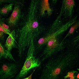
 MAB1259) at 10 µg/mL, for 3 hours at room temperature. Cells were stained using the NorthernLights™ 557-conjugated Anti-Goat IgG Secondary Antibody (red; Catalog # NL001), NorthernLights™ 493-conjugated Anti-Mouse IgG Secondary Antibody (green; Catalog # NL009) and counterstained with DAPI (blue). Specific staining for HOXD10 was localized to nucleus, while Nestin co-staining was cytoplasmic. View our protocol for Fluorescent ICC Staining of Cells on Coverslips." alt="HOXD10 was detected in immersion fixed human induced pluripotent stem (iPS) cells differentiated to neural progenitor cells (NPC) using Goat Anti-Human HOXD10 Antigen Affinity-purified Polyclonal Antibody (Catalog # AF10413) at 10 µg/mL, and co-stained with Mouse Anti-Human Nestin Monoclonal Antibody (Catalog # MAB1259) at 10 µg/mL, for 3 hours at room temperature. Cells were stained using the NorthernLights™ 557-conjugated Anti-Goat IgG Secondary Antibody (red; Catalog # NL001), NorthernLights™ 493-conjugated Anti-Mouse IgG Secondary Antibody (green; Catalog # NL009) and counterstained with DAPI (blue). Specific staining for HOXD10 was localized to nucleus, while Nestin co-staining was cytoplasmic. View our protocol for Fluorescent ICC Staining of Cells on Coverslips." class="big_thumb" />


![Western Blot: HOXD10 Antibody (1D11) [NBP2-45744] - Analysis of HEK293T cells were transfected with the pCMV6-ENTRY control (Left lane) or pCMV6-ENTRY HOXD10.](http://images.novusbio.com/fullsize/HOXD10-Antibody-1D11-Western-Blot-NBP2-45744-img0008.jpg "Western Blot: HOXD10 Antibody (1D11) [NBP2-45744] - Analysis of HEK293T cells were transfected with the pCMV6-ENTRY control (Left lane) or pCMV6-ENTRY HOXD10.")
![Immunohistochemistry: HOXD10 Antibody (1D11) [NBP2-45744] - Analysis of Carcinoma of Human bladder tissue. (Heat-induced epitope retrieval by 10mM citric buffer, pH6.0, 120C for 3min)](http://images.novusbio.com/fullsize/HOXD10-Antibody-1D11-Immunohistochemistry-NBP2-45744-img0007.jpg)
![Immunohistochemistry: HOXD10 Antibody (1D11) [NBP2-45744] - Analysis of Carcinoma of Human pancreas tissue. (Heat-induced epitope retrieval by 10mM citric buffer, pH6.0, 120C for 3min)](http://images.novusbio.com/fullsize/HOXD10-Antibody-1D11-Immunohistochemistry-NBP2-45744-img0001.jpg)
![Immunohistochemistry: HOXD10 Antibody (1D11) [NBP2-45744] - Analysis of Adenocarcinoma of Human ovary tissue. (Heat-induced epitope retrieval by 10mM citric buffer, pH6.0, 120C for 3min)](http://images.novusbio.com/fullsize/HOXD10-Antibody-1D11-Immunohistochemistry-NBP2-45744-img0002.jpg)
![Immunohistochemistry: HOXD10 Antibody (1D11) [NBP2-45744] - Analysis of Human pancreas tissue. (Heat-induced epitope retrieval by 10mM citric buffer, pH6.0, 120C for 3min)](http://images.novusbio.com/fullsize/HOXD10-Antibody-1D11-Immunohistochemistry-NBP2-45744-img0003.jpg)
![Immunohistochemistry: HOXD10 Antibody (1D11) [NBP2-45744] - Analysis of Carcinoma of Human thyroid tissue. (Heat-induced epitope retrieval by 10mM citric buffer, pH6.0, 120C for 3min)](http://images.novusbio.com/fullsize/HOXD10-Antibody-1D11-Immunohistochemistry-NBP2-45744-img0004.jpg)
![Immunohistochemistry: HOXD10 Antibody (1D11) [NBP2-45744] - Analysis of Human bladder tissue. (Heat-induced epitope retrieval by 10mM citric buffer, pH6.0, 120C for 3min)](http://images.novusbio.com/fullsize/HOXD10-Antibody-1D11-Immunohistochemistry-NBP2-45744-img0005.jpg)
![Immunohistochemistry: HOXD10 Antibody (1D11) [NBP2-45744] - Analysis of Adenocarcinoma of Human endometrium tissue. (Heat-induced epitope retrieval by 10mM citric buffer, pH6.0, 120C for 3min)](http://images.novusbio.com/fullsize/HOXD10-Antibody-1D11-Immunohistochemistry-NBP2-45744-img0006.jpg)
![Immunohistochemistry: HOXD10 Antibody [NBP1-28476] - (3.8ug/ml) staining of paraffin embedded Human Kidney. Steamed antigen retrieval with citrate buffer pH 6, AP-staining.](http://images.novusbio.com/fullsize/HOXD10-Antibody-Immunohistochemistry-NBP1-28476-img0003.jpg "Immunohistochemistry: HOXD10 Antibody [NBP1-28476] - (3.8ug/ml) staining of paraffin embedded Human Kidney. Steamed antigen retrieval with citrate buffer pH 6, AP-staining.")
![Flow Cytometry: HOXD10 Antibody [NBP1-28476] - Flow cytometric analysis of paraformaldehyde fixed HEK293 cells (blue line), permeabilized with 0.5% Triton. Primary incubation 1hr (10ug/ml) followed by Alexa Fluor 488 secondary antibody (1ug/ml). IgG control: Unimmunized goat IgG (black line) followed by Alexa Fluor 488 secondary antibody.](http://images.novusbio.com/fullsize/HOXD10-Antibody-Flow-Cytometry-NBP1-28476-img0002.jpg)
![Immunocytochemistry/Immunofluorescence: HOXD10 Antibody [NBP2-57634] - Staining of human cell line HEK 293 shows localization to nucleoplasm, cytosol & cytoplasmic bodies.](http://images.novusbio.com/fullsize/HOXD10-Antibody-Immunocytochemistry-Immunofluorescence-NBP2-57634-img0001.jpg "Immunocytochemistry/Immunofluorescence: HOXD10 Antibody [NBP2-57634] - Staining of human cell line HEK 293 shows localization to nucleoplasm, cytosol & cytoplasmic bodies.")

![Western Blot: HOXD10 Antibody - BSA Free [NBP3-04565] - Western blot analysis of Mouse kidney, using HOXD10 antibody (A7686) at 1:800 dilution.<br>Secondary antibody: HRP Goat Anti-Rabbit IgG (H+L) (AS014) at 1:10000 dilution.<br>Lysates/proteins: 25μg per lane.<br>Blocking buffer: 3% nonfat dry milk in TBST.<br>Detection: ECL Basic Kit (RM00020).<br>Exposure time: 60s.](http://images.novusbio.com/fullsize/nbp3-04565_rabbit-polyclonal-hoxd10-antibody-5220251931395.jpg "Western Blot: HOXD10 Antibody - BSA Free [NBP3-04565] - Western blot analysis of Mouse kidney, using HOXD10 antibody (A7686) at 1:800 dilution.<br>Secondary antibody: HRP Goat Anti-Rabbit IgG (H+L) (AS014) at 1:10000 dilution.<br>Lysates/proteins: 25μg per lane.<br>Blocking buffer: 3% nonfat dry milk in TBST.<br>Detection: ECL Basic Kit (RM00020).<br>Exposure time: 60s.")
![Western Blot: HOXD10 Antibody (5H5S6) [NBP3-33466] - Western blot analysis of lysates from Mouse kidney, using HOXD10 Rabbit mAb at 1:1000 dilution.<br/>Secondary antibody: HRP-conjugated Goat anti-Rabbit IgG (H+L) at 1:10000 dilution.<br/>Lysates/proteins: 25ug per lane.<br/>Blocking buffer: 3% nonfat dry milk in TBST.<br/>Detection: ECL Basic Kit.<br/>Exposure time: 10s.](http://images.novusbio.com/fullsize/nbp3-33466_rabbit-hoxd10-mab-5h5s6-1012202411343392.jpg "Western Blot: HOXD10 Antibody (5H5S6) [NBP3-33466] - Western blot analysis of lysates from Mouse kidney, using HOXD10 Rabbit mAb at 1:1000 dilution.<br/>Secondary antibody: HRP-conjugated Goat anti-Rabbit IgG (H+L) at 1:10000 dilution.<br/>Lysates/proteins: 25ug per lane.<br/>Blocking buffer: 3% nonfat dry milk in TBST.<br/>Detection: ECL Basic Kit.<br/>Exposure time: 10s.")
![Western Blot: HOXD10 Overexpression Lysate (Adult Normal) [NBL1-11682] Left-Empty vector transfected control cell lysate (HEK293 cell lysate); Right -Over-expression Lysate for HOXD10.](http://images.novusbio.com/fullsize/HOXD10-Overexpression-Lysate-Adult-Normal-Western-Blot-NBL1-11682-img0002.jpg "Western Blot: HOXD10 Overexpression Lysate (Adult Normal) [NBL1-11682] Left-Empty vector transfected control cell lysate (HEK293 cell lysate); Right -Over-expression Lysate for HOXD10.")
