After August 17, 2026, Novus Biologicals products and services will no longer be available on this website; you will access all products and services on rndsystems.com. Create your R&D Systems online account today.
251-KG) stimulates proliferation in the 4MBr‑5 rhesus monkey epithelial cell line in a dose-dependent manner (orange line). Proliferation elicited by Recombinant Human KGF/FGF‑7 (125 ng/mL) is neutralized (green line) by increasing concentrations of Goat Anti-Human KGF/FGF‑7 Antigen Affinity-purified Polyclonal Antibody (Catalog # AF-251-NA). The ND50 is typically 6-12 µg/mL." class="big_lightbox" target="_blank">
CTS013). Tissue was stained using the Anti-Goat HRP-DAB Cell & Tissue Staining Kit (brown; Catalog # CTS008) and counterstained with hematoxylin (blue). Specific staining was localized to trophoblast cells in chorionic villi. View our protocol for Chromogenic IHC Staining of Paraffin-embedded Tissue Sections." class="big_lightbox" target="_blank">
251-KG) was serially diluted and captured by Mouse Anti-Human KGF/FGF‑7 Monoclonal Antibody (Catalog # MAB251) coated on a Clear Polystyrene Microplate (Catalog # DY990). Goat Anti-Human KGF/FGF‑7 Antigen Affinity-purified Polyclonal Antibody (Catalog # AF-251-NA) was biotinylated and incubated with the protein captured on the plate. Detection of the standard curve was achieved by incubating Streptavidin-HRP (Catalog # DY998)" class="big_lightbox" target="_blank">
251-KG) stimulates proliferation in the 4MBr-5 rhesus monkey epithelial cell line in a dose-dependent manner (orange line), as measured by Resazurin (Catalog # AR002). Proliferation elicited by Recombinant Human KGF/FGF-7 (100 ng/mL) is neutralized (green line) by increasing concentrations of Mouse Anti-Human KGF/FGF-7 Monoclonal Antibody (Catalog # MAB251). The ND50 is typically 0.1-0.5 µg/mL." class="big_lightbox" target="_blank">
251-KG) was serially diluted and captured by Mouse Anti-Human KGF/FGF‑7 Monoclonal Antibody (Catalog # MAB251) coated on a Clear Polystyrene Microplate (Catalog # DY990). Goat Anti-Human KGF/FGF‑7 Antigen Affinity-purified Polyclonal Antibody (Catalog # AF-251-NA) was biotinylated and incubated with the protein captured on the plate. Detection of the standard curve was achieved by incubating Streptavidin-HRP (Catalog # DY998)" class="big_lightbox" target="_blank">
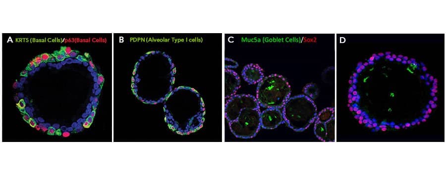
345-FG), Recombinant Human Noggin (6057-NG), and Recombinant Human R-Spondin 1 (4645-RS), along with the other reagents listed in the lung organoid expansion medium recipe in the lung organoid culture protocol. Lung organoids were able to differentiate and exhibit markers for various cell types of the lung. Lung organoids were stained with (A) a Rabbit Anti-Human Cytokeratin 5 (KRT5) Monoclonal Antibody (Novus Biologicals, Catalog # NB110-56916; green) and a Goat Anti-Human p63/TP73L Polyclonal Antibody (AF1916; red) to visualize basal cells, (B) a Hamster Anti-Mouse Podoplanin (PDPN) Monoclonal Antibody (Novus Biologicals, Catalog # NB600-1015; green) to visualize alveolar type I cells and a Goat Anti-Human p63/TP73L Polyclonal Antibody (AF1916; red) to visualize basal cells, and (C, D) a Mouse Anti-MUC5AC Monoclonal Antibody (Novus Biologicals, Catalog # NBP2-15196; green) to visualize Goblet cells and a Mouse Anti-Human/Mouse/Rat SOX2 Monoclonal Antibody (MAB2018; red). All samples were counterstained with DAPI (5748; blue). " class="big_lightbox" target="_blank">
BT-KGF-AFL) grades of RecombinantHuman KGF/FGF-7 measured in a cell proliferationassay using Ba/F3 mouse pro Bcells transfected with human FGFRIIb (orange and green, respectively)." class="big_lightbox" target="_blank">
BT-KGF, BT-KGF-AFL, and BT-KGF-GMP). The iPSC cells (1210a cell line) were cultured in a 24-well plate and differentiated into pancreatic progenitor cells using different lots of KGF and other differentiation factors. Expression of pancreatic progenitor markers was analyzed via flow cytometry with anti-PDX-1 (Catalog # MAB2419) and anti-NKX6.1 antibodies. The PDX-1+/NKX6.1+ double positive cell population was quantified as the pancreatic progenitor cells. The data is represented as the average of three technical replicates." class="big_lightbox" target="_blank">
MAB14171) and the anti-somatostatin antibody (Catalog # MAB2358), followed by the secondary antibody staining (anti-mouse IgG NL493-conjugated antibody: Catalog # NL009; anti-rat IgG NL557-conjugated antibody: Catalog # NL013). A Leica THUNDER Imaging System was utilized for the imaging of C-peptide and somatostatin expression in the beta islets. GMP KGF successfully differentiates the iPSCs into functional beta islets capable of expressing these characteristic markers. " class="big_lightbox" target="_blank">
Keratinocyte Growth Factor (KGF, FGF-7, HBGF-7) is a member of the FGF family of 23 related mitogenic proteins. These proteins play a central role during embryonic development and postnatal growth and tissue repair, by promoting cellular proliferation and differentiation. KGF specifically stimulates epithelial cells and keratinocytes, it is the most potent growth factor identified thus far for skin keratinocytes. KGF takes part in the development of kidney and lung, angiogenesis and has a key role in wound healing following skin injuries. It shows considerable species cross-reactivity (mouse, monkey, porcine cells) as the other members of the FGF family.
![Neutralization KGF/FGF-7 Antibody [Unconjugated]](http://images.novusbio.com/images/antibody/KGF_AF-251-NA_Block_Neutralize_8667.jpg)

![Neutralization KGF/FGF-7 Antibody (29522) [Unconjugated]](http://images.novusbio.com/images/antibody/KGF_MAB251_Block_Neutralize_16999.jpg)
![N/A KGF/FGF-7 [HRP]](http://images.novusbio.com/fullsize/elisa/DATA_KGF_DKG00_ELISA_725.jpg)
![N/A KGF/FGF-7 [Biotin]](http://images.novusbio.com/fullsize/elisa/DATA_KGF_DY251_ELISA_1838.jpg)


![Bioactivity KGF/FGF-7 [Unconjugated]](http://images.novusbio.com/fullsize/protein/KGF_251KG_2860.jpg)
![Bioactivity KGF/FGF-7 [Unconjugated]](http://images.novusbio.com/fullsize/bt-kgf-gmp_recombinant-human-kgf-fgf-7-gmp-protein-cf-bioactivity-42202514259.jpg)

![Immunohistochemistry KGF/FGF-7 Antibody [Unconjugated]](http://images.novusbio.com/images/antibody/KGF_AF-251-NA_Immunohistochemistry_10185.jpg)
![Immunohistochemistry-Paraffin KGF/FGF-7 Antibody [Unconjugated]](http://images.novusbio.com/fullsize/af-251-na_human-kgf-fgf-7-affinity-purified-polyclonal-ab-8220249505911.jpg)
![Immunohistochemistry-Paraffin KGF/FGF-7 Antibody [Unconjugated]](http://images.novusbio.com/fullsize/af-251-na_human-kgf-fgf-7-affinity-purified-polyclonal-ab-44202415425045.jpg)
![Western Blot KGF/FGF-7 Antibody [Unconjugated]](http://images.novusbio.com/fullsize/af-251-na_human-kgf-fgf-7-affinity-purified-polyclonal-ab-8120255541760.jpg)
![ELISA KGF/FGF-7 Antibody [Unconjugated]](http://images.novusbio.com/images/af-251-na_human-kgf-fgf-7-affinity-purified-polyclonal-ab-elisa-639201854768098860.jpg)


![ELISA KGF/FGF-7 Antibody (29522) [Unconjugated]](http://images.novusbio.com/images/mab251_human-kgf-fgf-7-mab-clone-29522-elisa-639201776849790128.jpg)

![N/A KGF/FGF-7 [HRP]](http://images.novusbio.com/fullsize/elisa/DATA_KGF_DKG00_ELISA_726.jpg)
![N/A KGF/FGF-7 [HRP]](http://images.novusbio.com/fullsize/elisa/KGF_DKG00_ELISA_122.jpg)
![Bioactivity KGF/FGF-7 [Unconjugated]](http://images.novusbio.com/images/251-kg_recombinant-human-kgf-fgf-7-protein-bioactivity-2520239811.jpg)
![Bioactivity KGF/FGF-7 [Unconjugated]](http://images.novusbio.com/images/bt-kgf-gmp_recombinant-human-kgf-fgf-7-gmp-protein-cf-bioactivity-422025141227..jpg)
![SDS-Page KGF/FGF-7 [Unconjugated]](http://images.novusbio.com/fullsize/bt-kgf-gmp_recombinant-human-kgf-fgf-7-gmp-protein-cf-sds-page-422025141534.jpg)
![Data KGF/FGF-7 [Unconjugated]](http://images.novusbio.com/images/bt-kgf-gmp_recombinant-human-kgf-fgf-7-gmp-protein-cf-data-155202593924.jpg)
![Data KGF/FGF-7 [Unconjugated]](http://images.novusbio.com/fullsize/bt-kgf-gmp_recombinant-human-kgf-fgf-7-gmp-protein-cf-data-1552025101340.jpg)
![Data KGF/FGF-7 [Unconjugated]](http://images.novusbio.com/images/bt-kgf-gmp_recombinant-human-kgf-fgf-7-gmp-protein-cf-data-155202513169.jpg)
![Data KGF/FGF-7 [Unconjugated]](http://images.novusbio.com/fullsize/bt-kgf-gmp_recombinant-human-kgf-fgf-7-gmp-protein-cf-data-1552025132534.jpg)