DR647RB) and counterstained with DAPI (blue; Lunaphore Catalog #
DR100). Specific staining was localized to the neuronal nuclei. Protocol available in
COMET™ Panel Builder." class="big_lightbox">

DR647RB) and counterstained with DAPI (blue; Lunaphore Catalog #
DR100). Specific staining was localized to the neuronal nuclei. Protocol available in
COMET™ Panel Builder." alt="RBFOX3 was detected in immersion fixed paraffin-embedded sections of human brain Cortex using Rabbit Anti-Human RBFOX3, Monoclonal Antibody (Catalog # MAB11690) at 3ug/mL at 37° Celsius for 4 minutes. Before incubation with the primary antibody, tissue underwent an all-in-one dewaxing and antigen retrieval preprocessing using PreTreatment Module (PT Module) and Dewax and HIER Buffer H (pH 9; Epredia Catalog # TA-999-DHBH). Tissue was stained using the Alexa Fluor™ Plus 647 Goat anti-Rabbit IgG Secondary Antibody at 1:200 at 37 ° Celsius for 2 minutes. (Yellow; Lunaphore Catalog #
DR647RB) and counterstained with DAPI (blue; Lunaphore Catalog #
DR100). Specific staining was localized to the neuronal nuclei. Protocol available in
COMET™ Panel Builder." class="big_thumb" />
DR555RB) and counterstained with DAPI (blue; Lunaphore Catalog #
DR100). Specific staining was localized to the cytoplasm and nucleus. Protocol available in
COMET™ Panel Builder." class="big_lightbox">
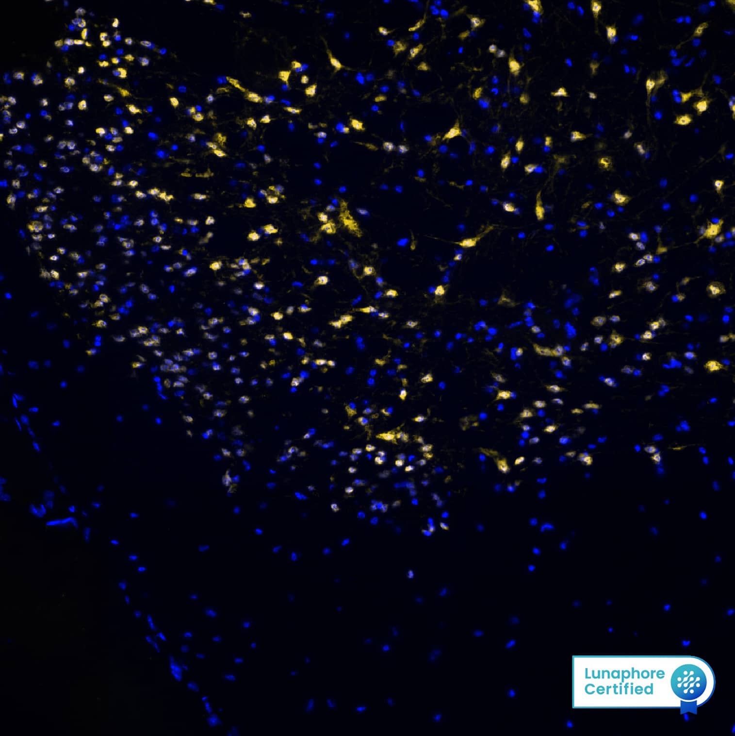
DR555RB) and counterstained with DAPI (blue; Lunaphore Catalog #
DR100). Specific staining was localized to the cytoplasm and nucleus. Protocol available in
COMET™ Panel Builder." class="big_thumb" />
DR555RB) and counterstained with DAPI (blue; Lunaphore Catalog #
DR100). Specific staining was localized to the cytoplasm and nucleus. Protocol available in
COMET™ Panel Builder." class="big_lightbox">
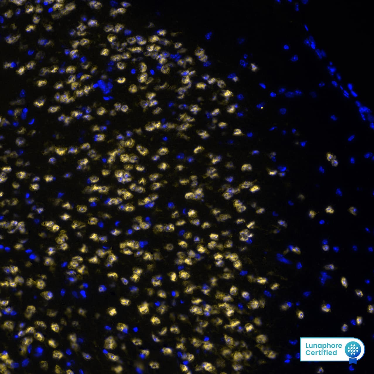
DR555RB) and counterstained with DAPI (blue; Lunaphore Catalog #
DR100). Specific staining was localized to the cytoplasm and nucleus. Protocol available in
COMET™ Panel Builder." class="big_thumb" />
DR555RB) and counterstained with DAPI (blue; Lunaphore Catalog #
DR100). Specific staining was localized to the cytoplasm and nucleus. Protocol available in
COMET™ Panel Builder." class="big_lightbox">
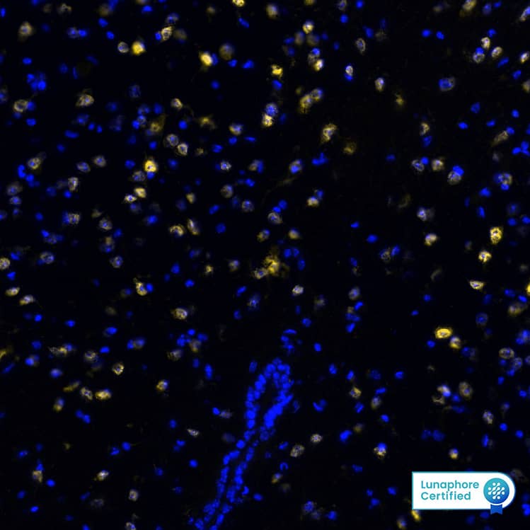
DR555RB) and counterstained with DAPI (blue; Lunaphore Catalog #
DR100). Specific staining was localized to the cytoplasm and nucleus. Protocol available in
COMET™ Panel Builder." class="big_thumb" />
VC003) or the HRP-conjugated Anti-Rabbit IgG Secondary Antibody (
HAF008). Before incubation with the primary antibody, tissue was subjected to heat-induced epitope retrieval using VisUCyte Antigen Retrieval Reagent-Basic (
VCTS021). Tissue was stained using DAB (brown) and counterstained with hematoxylin (blue). Specific staining was localized to the nucleus. View our protocol for
IHC Staining with VisUCyte HRP Polymer Detection Reagents." class="big_lightbox">

VC003) or the HRP-conjugated Anti-Rabbit IgG Secondary Antibody (
HAF008). Before incubation with the primary antibody, tissue was subjected to heat-induced epitope retrieval using VisUCyte Antigen Retrieval Reagent-Basic (
VCTS021). Tissue was stained using DAB (brown) and counterstained with hematoxylin (blue). Specific staining was localized to the nucleus. View our protocol for
IHC Staining with VisUCyte HRP Polymer Detection Reagents." class="big_thumb" />
HAF008). Specific bands were detected for RBFOX3/NeuN at approximately 40 and 50 kDa (as indicated). This experiment was conducted under reducing conditions and using
Western Blot Buffer Group 1." class="big_lightbox">

HAF008). Specific bands were detected for RBFOX3/NeuN at approximately 40 and 50 kDa (as indicated). This experiment was conducted under reducing conditions and using
Western Blot Buffer Group 1." class="big_thumb" />
 DR647RB) and counterstained with DAPI (blue; Lunaphore Catalog # DR100). Specific staining was localized to the neuronal nuclei. Protocol available in COMET™ Panel Builder." alt="RBFOX3 was detected in immersion fixed paraffin-embedded sections of human brain Cortex using Rabbit Anti-Human RBFOX3, Monoclonal Antibody (Catalog # MAB11690) at 3ug/mL at 37° Celsius for 4 minutes. Before incubation with the primary antibody, tissue underwent an all-in-one dewaxing and antigen retrieval preprocessing using PreTreatment Module (PT Module) and Dewax and HIER Buffer H (pH 9; Epredia Catalog # TA-999-DHBH). Tissue was stained using the Alexa Fluor™ Plus 647 Goat anti-Rabbit IgG Secondary Antibody at 1:200 at 37 ° Celsius for 2 minutes. (Yellow; Lunaphore Catalog # DR647RB) and counterstained with DAPI (blue; Lunaphore Catalog # DR100). Specific staining was localized to the neuronal nuclei. Protocol available in COMET™ Panel Builder." class="big_thumb" />
 DR647MS) and counterstained with DAPI (blue; Lunaphore Catalog # DR100). Specific staining was localized to the neuronal nuclei. Protocol available in COMET™ Panel Builder." alt="RBFOX3 was detected in immersion fixed paraffin-embedded sections of mouse Brain Cortex using Anti-Mouse RBFOX3, Monoclonal Antibody (Catalog #NBP3-21203) at 1:1000 dilution at 37 ° Celsius for 4 minutes. Before incubation with the primary antibody, tissue underwent an all-in-one dewaxing and antigen retrieval preprocessing using PreTreatment Module (PT Module) and Dewax and HIER Buffer H (pH 9; Epredia Catalog # TA-999-DHBH). Tissue was stained using the Alexa Fluor™ 647 Goat anti-Mouse IgG Secondary Antibody at 1:200 at 37 ° Celsius for 2 minutes. (Yellow; Lunaphore Catalog # DR647MS) and counterstained with DAPI (blue; Lunaphore Catalog # DR100). Specific staining was localized to the neuronal nuclei. Protocol available in COMET™ Panel Builder." class="big_thumb" />
 DR647MS) and counterstained with DAPI (blue; Lunaphore Catalog # DR100). Specific staining was localized to the neuronal nuclei. Protocol available in COMET™ Panel Builder." alt="RBFOX3 was detected in immersion fixed paraffin-embedded sections of human Cerebellum using Mouse Anti-Human RBFOX3, Monoclonal Antibody (Catalog #MAB11665) at 25ug/mL at 37 ° Celsius for 8 minutes. Before incubation with the primary antibody, tissue underwent an all-in-one dewaxing and antigen retrieval preprocessing using PreTreatment Module (PT Module) and Dewax and HIER Buffer H (pH 9; Epredia Catalog # TA-999-DHBH). Tissue was stained using the Alexa Fluor™ 647 Goat anti-Mouse IgG Secondary Antibody at 1:200 at 37 ° Celsius for 8 minutes. (Yellow; Lunaphore Catalog # DR647MS) and counterstained with DAPI (blue; Lunaphore Catalog # DR100). Specific staining was localized to the neuronal nuclei. Protocol available in COMET™ Panel Builder." class="big_thumb" />


![Analysis of whole brain tissue lysates using mouse mAb to FOX3/NeuN NBP1-92693, dilution 1:1,000 in green: [1] protein standard (red), [2] adult rat brain, [3] embryonic E20 rat brain, [4] adult mouse brain. Note the strong twin bands corresponding to the two alternate transcripts of FOX3/NeuN protein with apparent SDS-PAGE molecular weights of 46 and 48kDa. As with other FOX3/NeuN antibodies, an additional band at ~70kDa is revealed in some lysates.](http://images.novusbio.com/fullsize/RBFOX3-NeuN-Antibody-1B7-Western-Blot-NBP1-92693-img0020.jpg)


![Immunocytochemistry/ Immunofluorescence: RBFOX3/NeuN Antibody (1B7) [NBP1-92693] - NF kappa B localizes to both neuronal & glial nuclei in superior colliculus. Representative confocal images of NF kappa B in the superior colliculus (SC) of vehicle- & HE3286-treated rats with all nuclei indicated (DAPI). Sections were also labeled for astrocytes (A, GFAP), microglia (B, Iba1), or neurons using antibodies against either NeuN (C) or phosphorylated neurofilament heavy (pNFH; D). Representative nuclei from each cell class demonstrated NF kappa B localization (arrows). Insets (solid white, B) show region contained within dashed box with red channel removed to better visualize nuclear localization of NF kappa B. Scale: 10 μm. Image collected & cropped by CiteAb from the following publication (//journal.frontiersin.org/article/10.3389/fnins.2017.00045/full), licensed under a CC-BY license. Not internally tested by Novus Biologicals.](http://images.novusbio.com/fullsize/nbp1-92693_mouse-monoclonal-rbfox3-neun-antibody-1b7-310202415382419.jpg)
![Immunohistochemistry-Paraffin: RBFOX3/NeuN Antibody [NBP1-77686] - IHC analysis of formalin fixed paraffin embedded tissue section from hippocampus of mouse brain with NeuN antibody at 1:200 dilution. The antibody generated a strong nuclear with weak cytoplasmic staining in NeuN reactive cells.](http://images.novusbio.com/fullsize/RBFOX3-NeuN-Antibody-Immunohistochemistry-Paraffin-NBP1-77686-img0007.jpg "Immunohistochemistry-Paraffin: RBFOX3/NeuN Antibody [NBP1-77686] - IHC analysis of formalin fixed paraffin embedded tissue section from hippocampus of mouse brain with NeuN antibody at 1:200 dilution. The antibody generated a strong nuclear with weak cytoplasmic staining in NeuN reactive cells.")

![Immunocytochemistry/Immunofluorescence: RBFOX3/NeuN Antibody [NBP1-77686] - Neuro2a cells were fixed in 4% paraformaldehyde for 10 minutes and permeabilized in 0.5% Triton X-100 in PBS for 5 minutes. The cells were incubated with anti-RBFOX3/NeuN Antibody NBP1-77686 at 1 ug/ml overnight at 4C and detected with an anti-rabbit Dylight 488 (Green) at a 1:1000 dilution for 60 minutes. Nuclei were counterstained with DAPI (Blue). Cells were imaged using a 40X objective.](http://images.novusbio.com/fullsize/RBFOX3-NeuN-Antibody-Immunocytochemistry-Immunofluorescence-NBP1-77686-img0013.jpg)
![Western Blot: RBFOX3/NeuN Antibody [NBP1-77686] - Western blot image submitted by a verified customer review.](http://images.novusbio.com/fullsize/RBFOX3-NeuN-Antibody-Western-Blot-NBP1-77686-img0012.jpg)
![Flow Cytometry: RBFOX3/NeuN Antibody [NBP1-77686] - An intracellular stain was performed on Hek293 cells with NBP1-77686AF647 (blue) and a matched isotype control (orange). Cells were fixed with 4% PFA and then permeabilized with 0.1% saponin. Cells were incubated in an antibody dilution of 2.5 ug/mL for 30 minutes at room temperature. Both antibodies were conjugated to Alexa Fluor 647.](http://images.novusbio.com/fullsize/RBFOX3-NeuN-Antibody-Flow-Cytometry-NBP1-77686-img0010.jpg)
![Immunocytochemistry/Immunofluorescence: RBFOX3/NeuN Antibody [NBP1-77686] - NeuN antibody was tested in U2OS cells with DyLight 488 (green). Nuclei and alpha-tubulin were counterstained with DAPI (blue) and DyLight 550 (red).](http://images.novusbio.com/fullsize/RBFOX3-NeuN-Antibody-Immunocytochemistry-Immunofluorescence-NBP1-77686-img0005.jpg)
![Immunohistochemistry: RBFOX3/NeuN Antibody [NBP1-77686] - Analysis of NeuN in mouse brain using DAB with hematoxylin counterstain.](http://images.novusbio.com/fullsize/RBFOX3-NeuN-Antibody-Immunohistochemistry-NBP1-77686-img0008.jpg)
![Flow Cytometry: RBFOX3/NeuN Antibody [NBP1-77686] - An intracellular stain was performed on U2OS cells with RBFOX3/NeuN Antibody NBP1-77686AF647 (blue) and a matched isotype control (orange). Cells were fixed with 4% PFA and then permeabilized with 0.1% saponin. Cells were incubated in an antibody dilution of 5 ug/mL for 30 minutes at room temperature. Both antibodies were conjugated to Alexa Fluor 647.](http://images.novusbio.com/fullsize/RBFOX3-NeuN-Antibody-Flow-Cytometry-NBP1-77686-img0009.jpg)


![Immunocytochemistry/ Immunofluorescence: RBFOX3/NeuN Antibody - BSA Free [NBP1-77686] - Double immunofluorescence indicates that NF-kappa B & CCL5 were co-localized with microglia, astrocytes, & neurons in the ipsilateral L4–5 spinal cord on day 7 after CCI.NF-kappa B (green) colocalizes with Iba-1, GFAP, & NeuN (red) in laminae II–III of the superficial dorsal horn (A). CCL5 (red) co-localizes with Iba-1, GFAP, & NeuN (green) in the medial superficial dorsal horn (laminae II–III) (B). NF-kappa B (green) co-localizes with CCL5 (red) in laminae II–IV of the superficial dorsal horn following CCI on day 7 in the sham & CCI groups in the ipsilateral L4–5 spinal cord (C). Two single stained images (yellow) were merged. Scale = 100 μm. Image collected & cropped by CiteAb from the following publication (//dx.plos.org/10.1371/journal.pone.0115120), licensed under a CC-BY license. Not internally tested by Novus Biologicals.](http://images.novusbio.com/fullsize/nbp1-77686_rabbit-polyclonal-rbfox3-neun-antibody-31020241538241.jpg)
![Immunocytochemistry/ Immunofluorescence: RBFOX3/NeuN Antibody - BSA Free [NBP1-77686] - (A) Representative Western blots depicting phosphorylated JAK3 (pJAK3), total JAK3, & corresponding beta -actin levels in the ipsilateral (CXI) versus the contralateral (CXC) cortex at 24 h in sham & mice subjected to permanent middle cerebral artery occlusion (pMCAO). (B,C) Quantified pJAK3 & JAK3 values normalized to beta -actin in the cortex of sham & mice subjected to stroke. Both pJAK3 & JAK3 are significantly increased in the ipsilateral cortex of stroked animals (*P < 0.05 & **P < 0.01 with respect to sham ipsilateral/contralateral & stroke contralateral; sham, n = 5; stroke, n = 13). Data were analyzed using a two-way ANOVA (ipsilateral/contralateral or sham/stroke) with Bonferroni post-test. (D) Diagram showing peri-infarct brain region from which images were taken (n = 3). Scale bar is 10 µm, & magnification is 60×. (E) JAK3 colocalized with microglia/macrophages labeled with Iba-1. (F) JAK3 colocalized with neurons labeled with NeuN. (G) JAK3 colocalized with endothelial cells labeled with CD31. (H) JAK3 is not colocalized with astrocytes labeled with GFAP. Image collected & cropped by CiteAb from the following publication (//pubmed.ncbi.nlm.nih.gov/28790974), licensed under a CC-BY license. Not internally tested by Novus Biologicals.](http://images.novusbio.com/fullsize/nbp1-77686_rabbit-polyclonal-rbfox3-neun-antibody-310202416175267.jpg)


![Western Blot: RBFOX3/NeuN Antibody [NBP3-05554] - Western blot analysis of whole brain lysates using RBFOX3/NeuN Antibody at a dilution of 1:1,000 in green: [1] protein standard (red), [2] mouse brain, [3] rat brain. Bands at 46k and 48 kDa correspond to protein isotypes of RBFOX3/NeuN.](http://images.novusbio.com/fullsize/RBFOX3-NeuN-Antibody-Western-Blot-NBP3-05554-img0002.jpg "Western Blot: RBFOX3/NeuN Antibody [NBP3-05554] - Western blot analysis of whole brain lysates using RBFOX3/NeuN Antibody at a dilution of 1:1,000 in green: [1] protein standard (red), [2] mouse brain, [3] rat brain. Bands at 46k and 48 kDa correspond to protein isotypes of RBFOX3/NeuN.")
![Immunocytochemistry/Immunofluorescence: RBFOX3/NeuN Antibody [NBP3-05554] - Immunofluorescent analysis of a section of adult rat hippocampus stained with RBFOX3/NeuN Antibody at a dilution of 1:2,000 in red, costained with mouse monoclonal antibdy to MAP2 at a dilution of 1:2,000, in green. Nuclear DNA was revealed in blue using the DAPI stain. Following transcardial perfusion of mouse with 4% paraformaldehyde, brain was post fixed for 24 hours, cut to 45uM, and free-floating sections were stained with the above antibodies. The RBFOX3/NeuN antibody stains the nuclei of neurons in the hippocampus while the MAP2 antibody stains the dendritic processes of neurons.](http://images.novusbio.com/fullsize/RBFOX3-NeuN-Antibody-Immunocytochemistry-Immunofluorescence-NBP3-05554-img0001.jpg)


![Simple Western: RBFOX3/NeuN Antibody [NBP1-92716] - Simple Western lane view shows a specific band for RBFOX3/NeuN in 0.5 mg/ml of Human Brain lysate. This experiment was performed under reducing conditions using the 12-230 kDa separation system.](http://images.novusbio.com/fullsize/RBFOX3-NeuN-Antibody-Simple-Western-NBP1-92716-img0004.jpg "Simple Western: RBFOX3/NeuN Antibody [NBP1-92716] - Simple Western lane view shows a specific band for RBFOX3/NeuN in 0.5 mg/ml of Human Brain lysate. This experiment was performed under reducing conditions using the 12-230 kDa separation system.")
![Immunohistochemistry Free-Floating: RBFOX3/NeuN Antibody [NBP1-92716] - Analysis of a section of adult mouse cerebellum stained with rabbit pAb to FOX3/NeuN, NBP1-92716, dilution 1:5,000 in red, costained with chicken pAb to GFAP, dilution 1:5,000, in green. Following transcardial perfusion of mouse with 4% paraformaldehyde, brain was post fixed for 24 hours, cut to 45uM, and free-floating sections were stained with above antibodies. The FOX3/NeuN antibody stains the nuclei of neurons in the cerebellar granule layer. The GFAP antibody stains the processes of Bergmann glia in the molecular layer and astroglia in the granule and white matter layers.](http://images.novusbio.com/fullsize/RBFOX3-NeuN-Antibody-Immunocytochemistry-Immunofluorescence-NBP1-92716-img0005.jpg)
![Western Blot: RBFOX3/NeuN Antibody [NBP1-92716] - Analysis of whole brain lysates using rabbit pAb to Fox3/NeuN, NBP1-92716, dilution 1:1,000 in green: [1] protein standard (red), [2] mouse brain, [3] rat brain. Bands at 46-48kDa correspond to protein isotypes of Fox3/NeuN.](http://images.novusbio.com/fullsize/RBFOX3-NeuN-Antibody-Western-Blot-NBP1-92716-img0006.jpg)
![Immunohistochemistry-Paraffin: RBFOX3/NeuN Antibody (SR45-07) [NBP2-67314] - Human brain tissue using anti-NeuN antibody. The section was pre-treated using heat mediated antigen retrieval with Tris-EDTA buffer (pH 8.0-8.4) for 20 minutes.The tissues were blocked in 5% BSA for 30 minutes at room temperature, washed with ddH2O and PBS, and then probed with the primary antibody (1/200) for 30 minutes at room temperature. The detection was performed using an HRP conjugated compact polymer system. DAB was used as the chromogen. Tissues were counterstained with hematoxylin and mounted with DPX.](http://images.novusbio.com/fullsize/RBFOX3-NeuN-Antibody-SR45-07-Immunohistochemistry-Paraffin-NBP2-67314-img0008.jpg "<b>Biological Strategies Validation. </b>Immunohistochemistry-Paraffin: RBFOX3/NeuN Antibody (SR45-07) [NBP2-67314] - Human brain tissue using anti-NeuN antibody. The section was pre-treated using heat mediated antigen retrieval with Tris-EDTA buffer (pH 8.0-8.4) for 20 minutes.The tissues were blocked in 5% BSA for 30 minutes at room temperature, washed with ddH2O and PBS, and then probed with the primary antibody (1/200) for 30 minutes at room temperature. The detection was performed using an HRP conjugated compact polymer system. DAB was used as the chromogen. Tissues were counterstained with hematoxylin and mounted with DPX.")
![<b>Biological Strategies Validation. </b>Immunohistochemistry-Paraffin: RBFOX3/NeuN Antibody (SR45-07) [NBP2-67314] - Analysis of paraffin-embedded mouse cerebellum tissue using anti-NeuN antibody. Counter stained with hematoxylin.](http://images.novusbio.com/fullsize/RBFOX3-NeuN-Antibody-SR45-07-Immunohistochemistry-Paraffin-NBP2-67314-img0004.jpg)
![Western Blot: RBFOX3/NeuN Antibody (SR45-07) [NBP2-67314] - Analysis of RBFOX/NeuN on different lysates with Rabbit anti-RBFOX/NeuN antibody at 1/500 dilution. Lane 1: Rat cerebellum tissue lysate Lane 2: Mouse cerebellum tissue lysate Lysates/proteins at 20 ug/Lane. Predicted band size: 34 kDa Observed band size: 45/50 kDa Exposure time: 2 minutes; 12% SDS-PAGE gel. Proteins were transferred to a PVDF membrane and blocked with 5% NFDM/TBST for 1 hour at room temperature. The primary antibody at 1/500 dilution was used in 5% NFDM/TBST at room temperature for 2 hours. Goat Anti-Rabbit IgG - HRP Secondary Antibody at 1:300,000 dilution was used for 1 hour at room temperature.](http://images.novusbio.com/fullsize/RBFOX3-NeuN-Antibody-SR45-07-Western-Blot-NBP2-67314-img0010.jpg)
![Immunocytochemistry/Immunofluorescence: RBFOX3/NeuN Antibody (SR45-07) [NBP2-67314] - Analysis of SHG-44 cells labeling RBFOX3/NeuN with Rabbit anti-RBFOX3/NeuN antibody (permeabilized with 0.05% Triton X-100 in PBS for 20 minutes, and then blocked with 2% negative goat serum for 30 minutes at room temperature. Cells were then incubated with Rabbit anti-NeuN antibody was used as the secondary antibody at 1/1,000 dilution. PBS instead of the primary antibody was used as the secondary antibody only control. Nuclear DNA was labelled in blue with DAPI.](http://images.novusbio.com/fullsize/RBFOX3-NeuN-Antibody-SR45-07-Immunocytochemistry-Immunofluorescence-NBP2-67314-img0011.jpg)
![Immunohistochemistry-Paraffin: RBFOX3/NeuN Antibody (SR45-07) [NBP2-67314] - Immunofluorescence analysis of paraffin-embedded Rat cerebellum tissue labeling NeuN ( washed with PBS. And then probed with the primary antibodies NeuN ( Green) at 1/50 dilution and GFAP (Red) at 1/500 dilution overnight at 4 , washed with PBS. iFluor(TM) 488 conjugate-Goat anti-Rabbit IgG and iFluor(TM) 594 conjugate-Goat anti-Mouse IgG were used as the secondary antibody at 1/1,000 dilution. DAPI was used as nuclear counterstain.](http://images.novusbio.com/fullsize/RBFOX3-NeuN-Antibody-SR45-07-Immunohistochemistry-Paraffin-NBP2-67314-img0016.jpg)
![Flow Cytometry: RBFOX3/NeuN Antibody (SR45-07) [NBP2-67314] - Analysis of SH-SY-5Y cells with NeuN antibody at 1/50 dilution (blue) compared with an unlabelled control (cells without incubation with primary antibody; red). Alexa Fluor 488-conjugated goat anti rabbit IgG was used as the secondary antibody.](http://images.novusbio.com/fullsize/RBFOX3-NeuN-Antibody-SR45-07-Flow-Cytometry-NBP2-67314-img0001.jpg)
![Western Blot: RBFOX3/NeuN Antibody (SR45-07) [NBP2-67314] - Analysis of NeuN on different lysates. Proteins were transferred to a PVDF membrane and blocked with 5% BSA in PBS for 1 hour at room temperature. The primary antibody (1/500) was used in 5% BSA at room temperature for 2 hours. Goat Anti-Rabbit IgG - HRP Secondary Antibody at 1:5,000 dilution was used for 1 hour at room temperature. Positive control: Lane 1: human brain tissue lysateLane 2: rat brain tissue lysate Lane 2: mouse brain tissue lysate](http://images.novusbio.com/fullsize/RBFOX3-NeuN-Antibody-SR45-07-Western-Blot-NBP2-67314-img0005.jpg)
![Immunocytochemistry/Immunofluorescence: RBFOX3/NeuN Antibody (SR45-07) [NBP2-67314] - Staining NeuN in SH-SY-5Y cells (green). The nuclear counter stain is DAPI (blue). Cells were fixed in paraformaldehyde, permeabilised with 0.25% Triton X100/PBS.](http://images.novusbio.com/fullsize/RBFOX3-NeuN-Antibody-SR45-07-Immunocytochemistry-Immunofluorescence-NBP2-67314-img0002.jpg)
![<b>Biological Strategies Validation. </b>Immunohistochemistry-Paraffin: RBFOX3/NeuN Antibody (SR45-07) [NBP2-67314] - Mouse brain tissue using anti-NeuN antibody. The section was pre-treated using heat mediated antigen retrieval with Tris-EDTA buffer (pH 8.0-8.4) for 20 minutes.The tissues were blocked in 5% BSA for 30 minutes at room temperature, washed with ddH2O and PBS, and then probed with the primary antibody (1/200) for 30 minutes at room temperature. The detection was performed using an HRP conjugated compact polymer system. DAB was used as the chromogen. Tissues were counterstained with hematoxylin and mounted with DPX.](http://images.novusbio.com/fullsize/RBFOX3-NeuN-Antibody-SR45-07-Immunohistochemistry-Paraffin-NBP2-67314-img0006.jpg)
![<b>Biological Strategies Validation. </b>Immunohistochemistry-Paraffin: RBFOX3/NeuN Antibody (SR45-07) [NBP2-67314] - Rat brain tissue using anti-NeuN antibody. The section was pre-treated using heat mediated antigen retrieval with Tris-EDTA buffer (pH 8.0-8.4) for 20 minutes.The tissues were blocked in 5% BSA for 30 minutes at room temperature, washed with ddH2O and PBS, and then probed with the primary antibody (1/200) for 30 minutes at room temperature. The detection was performed using an HRP conjugated compact polymer system. DAB was used as the chromogen. Tissues were counterstained with hematoxylin and mounted with DPX.](http://images.novusbio.com/fullsize/RBFOX3-NeuN-Antibody-SR45-07-Immunohistochemistry-Paraffin-NBP2-67314-img0007.jpg)
![<b>Biological Strategies Validation. </b>Immunohistochemistry-Paraffin: RBFOX3/NeuN Antibody (SR45-07) [NBP2-67314] - Immunohistochemical analysis of paraffin-embedded mouse cerebellum tissue using anti-RBFOX3/NeuN antibody. The section was pre-treated using heat mediated antigen retrieval with Tris-EDTA buffer (pH 8.0-8.4) for 20 minutes. The tissues were blocked in 5%](http://images.novusbio.com/fullsize/RBFOX3-NeuN-Antibody-SR45-07-Immunohistochemistry-Paraffin-NBP2-67314-img0009.jpg)
![Immunohistochemistry-Paraffin: RBFOX3/NeuN Antibody (SR45-07) [NBP2-67314] - Analysis of paraffin-embedded mouse brain tissue with Rabbit anti-ROBX3/NeuN antibody washed with ddH2O and PBS, and then probed with the primary antibody at 1/500 dilution for 1 hour at room temperature. The detection was performed using an HRP conjugated compact polymer system. DAB was used as the chromogen. Tissues were counterstained with hematoxylin and mounted with DPX.](http://images.novusbio.com/fullsize/RBFOX3-NeuN-Antibody-SR45-07-Immunohistochemistry-Paraffin-NBP2-67314-img0012.jpg)
![Immunohistochemistry-Paraffin: RBFOX3/NeuN Antibody (SR45-07) [NBP2-67314] - Analysis of paraffin-embedded rat brain tissue with Rabbit anti-RBFOX3/NeuN antibody washed with ddH2O and PBS, and then probed with the primary antibody at 1/500 dilution for 1 hour at room temperature. The detection was performed using an HRP conjugated compact polymer system. DAB was used as the chromogen. Tissues were counterstained with hematoxylin and mounted with DPX.](http://images.novusbio.com/fullsize/RBFOX3-NeuN-Antibody-SR45-07-Immunohistochemistry-Paraffin-NBP2-67314-img0013.jpg)
![Immunohistochemistry-Paraffin: RBFOX3/NeuN Antibody (SR45-07) [NBP2-67314] - Analysis of paraffin-embedded mouse hippocampus tissue with Rabbit anti-RBFOX3/NeuN antibody washed with ddH2O and PBS, and then probed with the primary antibody at 1/500 dilution for 1 hour at room temperature. The detection was performed using an HRP conjugated compact polymer system. DAB was used as the chromogen. Tissues were counterstained with hematoxylin and mounted with DPX.](http://images.novusbio.com/fullsize/RBFOX3-NeuN-Antibody-SR45-07-Immunohistochemistry-Paraffin-NBP2-67314-img0014.jpg)
![Immunohistochemistry-Paraffin: RBFOX3/NeuN Antibody (SR45-07) [NBP2-67314] - Analysis of paraffin-embedded rat hippocampus tissue with Rabbit anti-RBFOX/NeuN antibody washed with ddH2O and PBS, and then probed with the primary antibody at 1/500 dilution for 1 hour at room temperature. The detection was performed using an HRP conjugated compact polymer system. DAB was used as the chromogen. Tissues were counterstained with hematoxylin and mounted with DPX.](http://images.novusbio.com/fullsize/RBFOX3-NeuN-Antibody-SR45-07-Immunohistochemistry-Paraffin-NBP2-67314-img0015.jpg)
![Western blot analysis on different lysates with NBP2-67314 at 1/5,000 dilution. Lane 1: Mouse brain tissue lysate Lane 2: Rat brain tissue lysate Lane 3: Mouse cerebellum tissue lysate Lane 4: Rat cerebellum tissue lysate Lysates/proteins at 20 µg/Lane. Predicted band size: 34 kDa Observed band size: 45/50 kDa Exposure time: 43 seconds; 4-20% SDS-PAGE gel. Proteins were transferred to a PVDF membrane and blocked with 5% NFDM/TBST for 1 hour at room temperature. NBP2-67314 at 1/5,000 dilution was used in 5% NFDM/TBST at room temperature for 2 hours. Goat Anti-Rabbit IgG [HRP] Secondary Antibody at 1/100,000 dilution was used for 1 hour at room temperature.](http://images.novusbio.com/fullsize/nbp2-67314_-western-blot-639204586674911646.jpg)

![<b>Orthogonal Strategies Validation. </b>Immunohistochemistry-Paraffin: RBFOX3/NeuN Antibody [NBP1-89821] - Analysis in human cerebral cortex and tonsil tissues. Corresponding RBFOX3/NeuN RNA-seq data are presented for the same tissues.](http://images.novusbio.com/fullsize/RBFOX3-NeuN-Antibody-Immunohistochemistry-Paraffin-NBP1-89821-img0013.jpg)

![Immunohistochemistry-Paraffin: RBFOX3/NeuN Antibody [NBP1-89821] - Staining of human tonsil shows no positivity in germinal center cells as expected.](http://images.novusbio.com/fullsize/RBFOX3-NeuN-Antibody-Immunohistochemistry-Paraffin-NBP1-89821-img0018.jpg)
![Immunohistochemistry-Paraffin: RBFOX3/NeuN Antibody [NBP1-89821] - Staining of human cerebellum shows strong nuclear positivity in cells in granular layer.](http://images.novusbio.com/fullsize/RBFOX3-NeuN-Antibody-Immunohistochemistry-Paraffin-NBP1-89821-img0014.jpg)

![Immunohistochemistry-Paraffin: RBFOX3/NeuN Antibody [NBP1-89821] - Staining of human hippocampus shows strong nuclear and cytoplasmic positivity in pyramidal cells.](http://images.novusbio.com/fullsize/RBFOX3-NeuN-Antibody-Immunohistochemistry-Paraffin-NBP1-89821-img0016.jpg)

 DR555RB) and counterstained with DAPI (blue; Lunaphore Catalog #
  DR555RB) and counterstained with DAPI (blue; Lunaphore Catalog #
  DR555RB) and counterstained with DAPI (blue; Lunaphore Catalog #
  VC003) or the HRP-conjugated Anti-Rabbit IgG Secondary Antibody (
 HAF008). Specific bands were detected for RBFOX3/NeuN at approximately 40 and 50 kDa (as indicated). This experiment was conducted under reducing conditions and using
  VCTS021). Tissue was stained using the HRP-conjugated Anti-Rabbit IgG Secondary Antibody (
![Western Blot: RBFOX3/NeuN Antibody (2F9F0) [RBFOX3/NeuN] - Western blot analysis of various lysates using RBFOX3/NeuN Rabbit mAb at1:1000 dilution.<br>Secondary antibody: HRP-conjugated Goat anti-Rabbit IgG (H+L) at1:10000 dilution.<br>Lysates/proteins: 25ug per lane.<br>Blocking buffer: 3% nonfat dry milk in TBST.<br>Detection: ECL Basic Kit .<br>Exposure time: 10s.](http://images.novusbio.com/fullsize/nbp3-15656_rabbit-rbfox3-neun-mab-2f9f0-52202517344212.jpg "Western Blot: RBFOX3/NeuN Antibody (2F9F0) [RBFOX3/NeuN] - Western blot analysis of various lysates using RBFOX3/NeuN Rabbit mAb at1:1000 dilution.<br>Secondary antibody: HRP-conjugated Goat anti-Rabbit IgG (H+L) at1:10000 dilution.<br>Lysates/proteins: 25ug per lane.<br>Blocking buffer: 3% nonfat dry milk in TBST.<br>Detection: ECL Basic Kit .<br>Exposure time: 10s.")
![Immunohistochemistry-Paraffin: RBFOX3/NeuN Antibody (2F9F0) [NBP3-15656] - Immunohistochemistry of paraffin-embedded mouse brain using RBFOX3/NeuN Rabbit mAb (NBP3-15656) at dilution of 1:100 (40x lens).Perform microwave antigen retrieval with 10 mM PBS buffer pH 7.2 before commencing with IHC staining protocol.](http://images.novusbio.com/fullsize/RBFOX3-NeuN-Antibody-2F9F0-Immunohistochemistry-Paraffin-NBP3-15656-img0003.jpg)
![Immunohistochemistry: RBFOX3/NeuN Antibody (2F9F0) [RBFOX3/NeuN] - Immunohistochemistry analysis of paraffin-embedded Human brain tissue using RBFOX3/NeuN Rabbit mAb at a dilution of 1:2000 (40x lens). High pressure antigen retrieval performed with 0.01M Tris-EDTA Buffer(pH 9.0) prior to IHC staining.](http://images.novusbio.com/fullsize/nbp3-15656_rabbit-rbfox3-neun-mab-2f9f0-52202517333816.jpg)
![Immunocytochemistry/ Immunofluorescence: RBFOX3/NeuN Antibody (2F9F0) [RBFOX3/NeuN] - Confocal imaging of paraffin-embedded human brain tissue using RBFOX3/NeuN Rabbit mAb followed by a further incubation with Cy3 Goat Anti-Rabbit IgG (H+L) . DAPI was used for nuclear staining (Blue). High pressure antigen retrieval performed with 0.01M Citrate Buffer (pH 6.0) prior to IF staining. Objective: 40x.](http://images.novusbio.com/fullsize/nbp3-15656_rabbit-rbfox3-neun-mab-2f9f0-52202517361261.jpg)
![Immunocytochemistry/ Immunofluorescence: RBFOX3/NeuN Antibody (2F9F0) [RBFOX3/NeuN] - Confocal imaging of paraffin-embedded rat brain tissue using RBFOX3/NeuN Rabbit mAb followed by a further incubation with Cy3 Goat Anti-Rabbit IgG (H+L) . DAPI was used for nuclear staining (Blue). High pressure antigen retrieval performed with 0.01M Citrate Buffer (pH 6.0) prior to IF staining. Objective: 40x.](http://images.novusbio.com/fullsize/nbp3-15656_rabbit-rbfox3-neun-mab-2f9f0-52202517315573.jpg)
![Immunocytochemistry/ Immunofluorescence: RBFOX3/NeuN Antibody (2F9F0) [RBFOX3/NeuN] - Confocal imaging of paraffin-embedded mouse brain tissue using RBFOX3/NeuN Rabbit mAb followed by a further incubation with Cy3 Goat Anti-Rabbit IgG (H+L) . DAPI was used for nuclear staining (Blue). High pressure antigen retrieval performed with 0.01M Citrate Buffer (pH 6.0) prior to IF staining. Objective: 40x.](http://images.novusbio.com/fullsize/nbp3-15656_rabbit-rbfox3-neun-mab-2f9f0-5220251734517.jpg)
![Immunohistochemistry: RBFOX3/NeuN Antibody (2F9F0) [RBFOX3/NeuN] - Immunohistochemistry analysis of paraffin-embedded Mouse brain tissue using RBFOX3/NeuN Rabbit mAb at a dilution of 1:2000 (40x lens). High pressure antigen retrieval performed with 0.01M Tris-EDTA Buffer(pH 9.0) prior to IHC staining.](http://images.novusbio.com/fullsize/nbp3-15656_rabbit-rbfox3-neun-mab-2f9f0-5220251734529.jpg)
![Immunohistochemistry: RBFOX3/NeuN Antibody (2F9F0) [RBFOX3/NeuN] - Immunohistochemistry analysis of paraffin-embedded Rat brain tissue using RBFOX3/NeuN Rabbit mAb at a dilution of 1:2000 (40x lens). High pressure antigen retrieval performed with 0.01M Tris-EDTA Buffer(pH 9.0) prior to IHC staining.](http://images.novusbio.com/fullsize/nbp3-15656_rabbit-rbfox3-neun-mab-2f9f0-5220251734591.jpg)


![Western Blot: RBFOX3/NeuN Antibody (NeuN/288R) [NBP3-13857] - Western blot analysis of HeLa cell lysate using RBFOX3/NeuN antibody (NeuN/288R).](http://images.novusbio.com/fullsize/RBFOX3-NeuN-Antibody-NeuN-288R-Western-Blot-NBP3-13857-img0002.jpg "Western Blot: RBFOX3/NeuN Antibody (NeuN/288R) [NBP3-13857] - Western blot analysis of HeLa cell lysate using RBFOX3/NeuN antibody (NeuN/288R).")
![Immunohistochemistry-Paraffin: RBFOX3/NeuN Antibody (NeuN/288R) [NBP3-13857] - Formalin-fixed, paraffin-embedded human cerebrum stained with RBFOX3/NeuN antibody (NeuN/288R). HIER: Tris/EDTA, pH9.0, 45min. HRP-polymer, 30min. DAB, 5min.](http://images.novusbio.com/fullsize/RBFOX3-NeuN-Antibody-NeuN-288R-Immunohistochemistry-Paraffin-NBP3-13857-img0003.jpg)
![Western Blot: RBFOX3/NeuN Antibody (NeuN/288R) [NBP3-13857] - Western Blot Analysis of human brain tissue lysate using RBFOX3/NeuN antibody (NeuN/288R).](http://images.novusbio.com/fullsize/RBFOX3-NeuN-Antibody-NeuN-288R-Western-Blot-NBP3-13857-img0001.jpg)
![Immunohistochemistry-Paraffin: RBFOX3/NeuN Antibody (NeuN/288R) [NBP3-13857] - Formalin-fixed, paraffin-embedded human cerebellum stained with RBFOX3/NeuN antibody (NeuN/288R). Inset: PBS instead of primary antibody, secondary negative control.](http://images.novusbio.com/fullsize/RBFOX3-NeuN-Antibody-NeuN-288R-Immunohistochemistry-Paraffin-NBP3-13857-img0004.jpg)
![Immunohistochemistry-Frozen: RBFOX3/NeuN Antibody (PD01-45) [NBP3-32644] - Immunofluorescence analysis of frozen mouse hippocampus tissue labeling RBFOX3/NeuN with Mouse anti-RBFOX3/NeuN antibody (NBP3-32644).<br/><br/>The tissues were blocked in 3% BSA for 30 minutes at room temperature, washed with PBS, and then probed with the primary antibody (NBP3-32644, green) at 1/50 dilution overnight at 4℃, washed with PBS. Goat Anti-Mouse IgG H&L (Alexa Fluor® 488) was used as the secondary antibody at 1/200 dilution. Nuclei were counterstained with DAPI (blue).](http://images.novusbio.com/fullsize/nbp3-32644_mouse-rbfox3-neun-mab-pd01-45-2682024159391.jpg "Immunohistochemistry-Frozen: RBFOX3/NeuN Antibody (PD01-45) [NBP3-32644] - Immunofluorescence analysis of frozen mouse hippocampus tissue labeling RBFOX3/NeuN with Mouse anti-RBFOX3/NeuN antibody (NBP3-32644).<br/><br/>The tissues were blocked in 3% BSA for 30 minutes at room temperature, washed with PBS, and then probed with the primary antibody (NBP3-32644, green) at 1/50 dilution overnight at 4℃, washed with PBS. Goat Anti-Mouse IgG H&L (Alexa Fluor® 488) was used as the secondary antibody at 1/200 dilution. Nuclei were counterstained with DAPI (blue).")
![Western Blot: RBFOX3/NeuN Antibody (PD01-45) [NBP3-32644] - Western blot analysis of RBFOX3/NeuN on mouse cerebellum tissue lysates with Mouse anti-RBFOX3/NeuN antibody (NBP3-32644) at 1/1,000 dilution.<br/><br/> Lysates/proteins at 20 ug/Lane.<br/><br/> Predicted band size: 34 kDa<br/> Observed band size: 45/50 kDa<br/><br/> Exposure time: 2 minutes;<br/><br/> 10% SDS-PAGE gel.<br/><br/> Proteins were transferred to a PVDF membrane and blocked with 5% NFDM/TBST for 1 hour at room temperature. The primary antibody (NBP3-32644) at 1/1,000 dilution was used in 5% NFDM/TBST at room temperature for 2 hours. Goat Anti-Mouse IgG - HRP Secondary Antibody at 1:150,000 dilution was used for 1 hour at room temperature.](http://images.novusbio.com/fullsize/nbp3-32644_mouse-rbfox3-neun-mab-pd01-45-27820241633121.jpg)
![Immunohistochemistry: RBFOX3/NeuN Antibody (PD01-45) [NBP3-32644] - Immunohistochemical analysis of paraffin-embedded human brain tissue with Mouse anti-RBFOX3/NeuN antibody (NBP3-32644) at 1/1,000 dilution.<br/><br/> The section was pre-treated using heat mediated antigen retrieval with Tris-EDTA buffer (pH 9.0) for 20 minutes. The tissues were blocked in 1% BSA for 20 minutes at room temperature, washed with ddH2O and PBS, and then probed with the primary antibody (NBP3-32644) at 1/1,000 dilution for 1 hour at room temperature. The detection was performed using an HRP conjugated compact polymer system. DAB was used as the chromogen. Tissues were counterstained with hematoxylin and mounted with DPX.](http://images.novusbio.com/fullsize/nbp3-32644_mouse-rbfox3-neun-mab-pd01-45-298202414115415.jpg)

![Western blot analysis on different lysates with NBP3-32644 at 1/500 dilution. Lane 1: SH-SY5Y cell lysate Lane 2: SHG44 cell lysate Lysates/proteins at 10 µg/Lane. Predicted band size: 34 kDa Observed band size: 50 kDa Exposure time: 2 minutes; 10% SDS-PAGE gel. Proteins were transferred to a PVDF membrane and blocked with 5% NFDM/TBST for 1 hour at room temperature. NBP3-32644 at 1/500 dilution was used in 5% NFDM/TBST at room temperature for 2 hours. Goat anti-Mouse IgG [HRP] at 1:150,000 dilution was used for 1 hour at room temperature.](http://images.novusbio.com/fullsize/nbp3-32644_-western-blot-639204597792802624.png)
 VC001) or the HRP-conjugated Anti-Mouse IgG Secondary Antibody (Catalog #
  HAF018). Specific bands were detected for RBFOX3/NeuN at approximately 40 and 50 kDa (as indicated). This experiment was conducted under reducing conditions and using


