MAB003, open histogram) followed by Allophycocyanin-conjugated Anti-Mouse IgG Secondary Antibody (Catalog #
F0101B). To facilitate intracellular staining, cells were fixed with Flow Cytometry Fixation Buffer (Catalog #
FC004) and permeabilized with Saponin. View our protocol for
Staining Intracellular Molecules." class="big_lightbox">
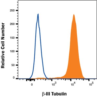
MAB003, open histogram) followed by Allophycocyanin-conjugated Anti-Mouse IgG Secondary Antibody (Catalog #
F0101B). To facilitate intracellular staining, cells were fixed with Flow Cytometry Fixation Buffer (Catalog #
FC004) and permeabilized with Saponin. View our protocol for
Staining Intracellular Molecules." alt="A172 cells were stained with Mouse Anti-Neuron-specific beta ‑III Tubulin Monoclonal Antibody (Catalog # MAB1195, filled histogram) or isotype control antibody (Catalog #
MAB003, open histogram) followed by Allophycocyanin-conjugated Anti-Mouse IgG Secondary Antibody (Catalog #
F0101B). To facilitate intracellular staining, cells were fixed with Flow Cytometry Fixation Buffer (Catalog #
FC004) and permeabilized with Saponin. View our protocol for
Staining Intracellular Molecules." class="big_thumb" />
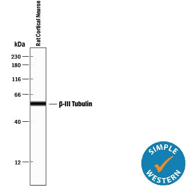
HAF018). A specific band was detected for beta -III Tubulin at approximately 55 kDa (as indicated). This experiment was conducted under reducing conditions and using
Immunoblot Buffer Group 1." class="big_lightbox">
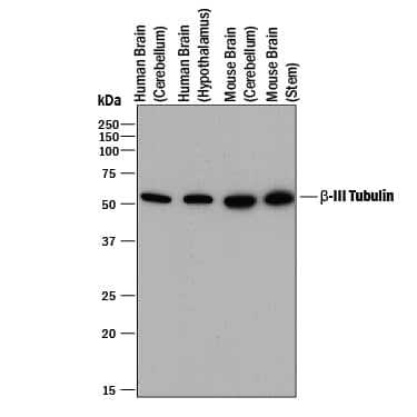
HAF018). A specific band was detected for beta -III Tubulin at approximately 55 kDa (as indicated). This experiment was conducted under reducing conditions and using
Immunoblot Buffer Group 1." class="big_thumb" />
AF2594). Cells were incubated with primary antibodies for 3 hours at room temperature. Cells were stained for beta-III Tubulin using the NorthernLights™ 557-conjugated Anti-Mouse IgG Secondary Antibody (red;
NL007) and for GFAP using the NorthernLights 493-conjugated Anti-Sheep IgG Secondary Antibody (green;
NL012). View our protocol for
Fluorescent ICC Staining of Cells on Coverslips." class="big_lightbox">
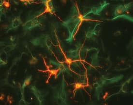
AF2594). Cells were incubated with primary antibodies for 3 hours at room temperature. Cells were stained for beta-III Tubulin using the NorthernLights™ 557-conjugated Anti-Mouse IgG Secondary Antibody (red;
NL007) and for GFAP using the NorthernLights 493-conjugated Anti-Sheep IgG Secondary Antibody (green;
NL012). View our protocol for
Fluorescent ICC Staining of Cells on Coverslips." class="big_thumb" />
NSC001) using 5 µg/mL neuron-specific Mouse Anti-Neuron-specific beta -III Tubulin Monoclonal Antibody (Catalog # MAB1195) and 10 µg/mL Rat Nestin Antigen Affinity-purified Polyclonal Antibody (
AF2736). Cells were incubated with primary antibodies for 3 hours at room temperature. Cells were stained for beta-III Tubulin using the NorthernLights™ 557-conjugated Anti-Mouse IgG Secondary Antibody (red;
NL007) and for Nestin using the NorthernLights 493-conjugated Anti-Goat IgG Secondary Antibody (green;
NL003). Tissue was counterstained with DAPI (blue). View our protocol for
Fluorescent ICC Staining of Cells on Coverslips." class="big_lightbox">
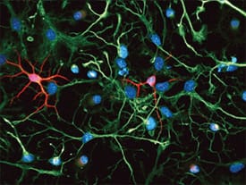
NSC001) using 5 µg/mL neuron-specific Mouse Anti-Neuron-specific beta -III Tubulin Monoclonal Antibody (Catalog # MAB1195) and 10 µg/mL Rat Nestin Antigen Affinity-purified Polyclonal Antibody (
AF2736). Cells were incubated with primary antibodies for 3 hours at room temperature. Cells were stained for beta-III Tubulin using the NorthernLights™ 557-conjugated Anti-Mouse IgG Secondary Antibody (red;
NL007) and for Nestin using the NorthernLights 493-conjugated Anti-Goat IgG Secondary Antibody (green;
NL003). Tissue was counterstained with DAPI (blue). View our protocol for
Fluorescent ICC Staining of Cells on Coverslips." class="big_thumb" />
VC001). Before incubation with the primary antibody, tissue was subjected to heat-induced epitope retrieval using VisUCyte Antigen Retrieval Reagent-Basic (Catalog #
VCTS021). Tissue was stained using DAB (brown) and counterstained with hematoxylin (blue). Specific staining was localized to Purkinje neurons. View our protocol for
IHC Staining with VisUCyte HRP Polymer Detection Reagents." class="big_lightbox">

VC001). Before incubation with the primary antibody, tissue was subjected to heat-induced epitope retrieval using VisUCyte Antigen Retrieval Reagent-Basic (Catalog #
VCTS021). Tissue was stained using DAB (brown) and counterstained with hematoxylin (blue). Specific staining was localized to Purkinje neurons. View our protocol for
IHC Staining with VisUCyte HRP Polymer Detection Reagents." class="big_thumb" />
 MAB003, open histogram) followed by Allophycocyanin-conjugated Anti-Mouse IgG Secondary Antibody (Catalog # F0101B). To facilitate intracellular staining, cells were fixed with Flow Cytometry Fixation Buffer (Catalog # FC004) and permeabilized with Saponin. View our protocol for Staining Intracellular Molecules." alt="A172 cells were stained with Mouse Anti-Neuron-specific beta ‑III Tubulin Monoclonal Antibody (Catalog # MAB1195, filled histogram) or isotype control antibody (Catalog # MAB003, open histogram) followed by Allophycocyanin-conjugated Anti-Mouse IgG Secondary Antibody (Catalog # F0101B). To facilitate intracellular staining, cells were fixed with Flow Cytometry Fixation Buffer (Catalog # FC004) and permeabilized with Saponin. View our protocol for Staining Intracellular Molecules." class="big_thumb" />


 HAF018). A specific band was detected for beta -III Tubulin at approximately 55 kDa (as indicated). This experiment was conducted under reducing conditions and using
  AF2594). Cells were incubated with primary antibodies for 3 hours at room temperature. Cells were stained for beta-III Tubulin using the NorthernLights™ 557-conjugated Anti-Mouse IgG Secondary Antibody (red;
  Fluorescent ICC Staining of Cells on Coverslips." class="big_thumb" />
 NSC001) using 5 µg/mL neuron-specific Mouse Anti-Neuron-specific beta -III Tubulin Monoclonal Antibody (Catalog # MAB1195) and 10 µg/mL Rat Nestin Antigen Affinity-purified Polyclonal Antibody (
 VC001). Tissue was stained using DAB (brown) and counterstained with hematoxylin (blue). Specific staining was localized to Purkinje neurons. View our protocol for
 VC001). Before incubation with the primary antibody, tissue was subjected to heat-induced epitope retrieval using VisUCyte Antigen Retrieval Reagent-Basic (Catalog #
  NL007) and counterstained with DAPI (blue). Specific staining was localized to cell cytoplasm. View our protocol for


![Immunocytochemistry/Immunofluorescence: beta Tubulin Antibody [NB600-936] - MCF7 cells were fixed in 4% paraformaldehyde for 10 minutes and permeabilized in 0.05% Triton X-100 in PBS for 5 minutes. The cells were incubated with anti- (NB600-936) at 2 ug/ml overnight at 4C and detected with an anti-rabbit Dylight 488 (Green) at a 1:1000 dilution for 60 minutes. Nuclei were counterstained with DAPI (Blue). Cells were imaged using a 100X objective and digitally deconvolved.](http://images.novusbio.com/fullsize/beta-Tubulin-Antibody-Immunocytochemistry-Immunofluorescence-NB600-936-img0021.jpg "Immunocytochemistry/Immunofluorescence: beta Tubulin Antibody [NB600-936] - MCF7 cells were fixed in 4% paraformaldehyde for 10 minutes and permeabilized in 0.05% Triton X-100 in PBS for 5 minutes. The cells were incubated with anti- (NB600-936) at 2 ug/ml overnight at 4C and detected with an anti-rabbit Dylight 488 (Green) at a 1:1000 dilution for 60 minutes. Nuclei were counterstained with DAPI (Blue). Cells were imaged using a 100X objective and digitally deconvolved.")
![Simple Western: beta Tubulin Antibody [NB600-936] - Lane view shows a specific band for beta Tubulin in 0.05 mg/ml of MCF-7 lysate. This experiment was performed under reducing conditions using the 12-230 kDa separation system.](http://images.novusbio.com/fullsize/beta-Tubulin-Antibody-Simple-Western-NB600-936-img0016.jpg)
![Western Blot: beta Tubulin Antibody [NB600-936] - Analysis of extracts from HeLa, A549 and Jurkat cells using NB600-936 Beta-tubulin antibody at 1:1000](http://images.novusbio.com/fullsize/beta-Tubulin-Antibody-Western-Blot-NB600-936-img0006.jpg)

![Immunocytochemistry/Immunofluorescence: beta Tubulin Antibody [NB600-936] - PC12 cells were fixed in 4% paraformaldehyde for 10 minutes and permeabilized in 0.05% Triton X-100 in PBS for 5 minutes. The cells were incubated with anti-beta Tubulin Antibody (NB600-936) at 2 ug/ml overnight at 4C and detected with an anti-rabbit Dylight 488 (Green) at a 1:1000 dilution for 60 minutes. Nuclei were counterstained with DAPI (Blue). Cells were imaged using a 100X objective and digitally deconvolved.](http://images.novusbio.com/fullsize/beta-Tubulin-Antibody-Immunocytochemistry-Immunofluorescence-NB600-936-img0023.jpg)
![Immunohistochemistry-Paraffin: beta Tubulin Antibody [NB600-936] - Analysis of FFPE tissue section of normal human skin using 1:2000 dilution of beta Tubulin antibody. Intense cytoplasmic staining of beta Tubulin (TUBB) protein was observed in various cells of the epidermal as well as the dermal cells [10X Magnification].](http://images.novusbio.com/fullsize/beta-Tubulin-Antibody-Immunohistochemistry-Paraffin-NB600-936-img0011.jpg)
![Western Blot: beta Tubulin Antibody [NB600-936] - Analysis in HeLa whole cell lysate at a 1:1,000 dilution.](http://images.novusbio.com/fullsize/beta-Tubulin-Antibody-Western-Blot-NB600-936-img0004.jpg)

![Immunocytochemistry/Immunofluorescence: beta Tubulin Antibody [NB600-936] - Confocal immunofluorescent analysis of C2C12 cells using beta Tubulin antibody (NB600-936, 1:5). An Alexa Fluor 488-conjugated Goat to rabbit IgG was used as secondary antibody (green). Actin filaments were labeled with Alexa Fluor 568 phalloidin (red). DAPI was used to stain the cell nuclei (blue).](http://images.novusbio.com/fullsize/beta-Tubulin-Antibody-Immunocytochemistry-Immunofluorescence-NB600-936-img0015.jpg)
![Immunocytochemistry/Immunofluorescence: beta Tubulin Antibody [NB600-936] - Analysis of beta Tubulin in mouse hippocampal primary culture. Image courtesy of product review by Lin Yi-Wen.](http://images.novusbio.com/fullsize/beta-Tubulin-Antibody-Immunocytochemistry-Immunofluorescence-NB600-936-img0001.jpg)
![Immunocytochemistry/Immunofluorescence: beta Tubulin Antibody [NB600-936] - NIH3T3 cells were fixed in 4% paraformaldehyde for 10 minutes and permeabilized in 0.05% Triton X-100 in PBS for 5 minutes. The cells were incubated with anti-beta Tubulin Antibody (NB600-936) at 2 ug/ml overnight at 4C and detected with an anti-rabbit Dylight 488 (Green) at a 1:1000 dilution for 60 minutes. Nuclei were counterstained with DAPI (Blue). Cells were imaged using a 100X objective and digitally deconvolved.](http://images.novusbio.com/fullsize/beta-Tubulin-Antibody-Immunocytochemistry-Immunofluorescence-NB600-936-img0022.jpg)
![Immunohistochemistry-Paraffin: beta Tubulin Antibody [NB600-936] - Analysis of FFPE tissue section of human esophageal squamous cell carcinoma (SCC) using 1:2000 dilution of beta Tubulin antibody. Strong cytoplasmic immuno-positivity of beta Tubulin (TUBB) was observed in SCC cells as well as the associated tumor stromal cells [10X Magnification].](http://images.novusbio.com/fullsize/beta-Tubulin-Antibody-Immunohistochemistry-Paraffin-NB600-936-img0014.jpg)
![Immunohistochemistry-Paraffin: beta Tubulin Antibody [NB600-936] - Analysis of FFPE tissue section of human esophageal squamous cell carcinoma (SCC) using 1:2000 dilution of beta Tubulin antibody. This representative image shows a cytoplasm specific staining of beta Tubulin (TUBB) in SCC cells [60X Magnification].](http://images.novusbio.com/fullsize/beta-Tubulin-Antibody-Immunohistochemistry-Paraffin-NB600-936-img0013.jpg)
![Immunohistochemistry-Paraffin: beta Tubulin Antibody [NB600-936] - Analysis of FFPE tissue section of normal human brain using 1:2000 dilution of beta Tubulin antibody. The various brain cells depicted strong cytoplasmic immunoreactivity of beta Tubulin (TUBB) protein [10X Magnification].](http://images.novusbio.com/fullsize/beta-Tubulin-Antibody-Immunohistochemistry-Paraffin-NB600-936-img0012.jpg)


![Western Blot: beta Tubulin Antibody - BSA Free [NB600-936] - Dual pathway inhibition regulates TK1 protein levels & results in greater p27 protein levels than single agents alone.Single agent PLX4032 resulted in activation of p-AKT Ser473 following 24 hours of exposure at two concentrations (100 nM, 1 µM). The addition of the dual PI3K/mTOR inhibitor BEZ235 blocks p-AKT Ser473 activation & resulted in a greater increase in p27 protein levels & diminished TK1 protein levels. Image collected & cropped by CiteAb from the following publication (//pubmed.ncbi.nlm.nih.gov/25247710), licensed under a CC-BY license. Not internally tested by Novus Biologicals.](http://images.novusbio.com/fullsize/nb600-936_rabbit-polyclonal-beta-tubulin-antibody-310202415175297.jpg)
![Western Blot: beta Tubulin Antibody - BSA Free [NB600-936] - Expression of pluripotency related markers & a sphere culture of BEAS-2B under normoxia.(a) HIF-2 alpha expression was detected in the nucleus of BEAS-2B under normoxia by western blotting. Beta tubulin & TATA binding protein (TBP) were used as protein markers for the cytosol & nucleus fraction, respectively. (b) Western blotting for Oct-4. (c) For knockdown of HIF-2 alpha , BEAS-2B cells were transfected with shRNA targeting HIF-2 alpha & sh-Luc was used as control. (d) Western blotting of Nanog & a culture of BEAS-2B containing floating spheres. Representative result from two or three experiments was presented. Image collected & cropped by CiteAb from the following publication (//pubmed.ncbi.nlm.nih.gov/27373565), licensed under a CC-BY license. Not internally tested by Novus Biologicals.](http://images.novusbio.com/fullsize/nb600-936_rabbit-polyclonal-beta-tubulin-antibody-310202415334929.jpg)
![Western Blot: beta Tubulin Antibody - BSA Free [NB600-936] - Expression of AAV transgene before & after experimental autoimmune uveoretinitis (EAU) induction. (A) B10.RIII mice were evaluated by funduscopy one month after an intravitreal injection of AAV delivering sGFP-TatM013v5 or secreted GFP (sGFP). As a control we evaluated mice that received an intravitreal injection of saline. Diffuse fluorescence indicates the secretion of sGFP & sGFP-TatM013v5 in the retina. (B) Western blot from retinas harvested 14 days after IRBP immunization. Membrane was probed with anti-GFP & anti-Tubulin antibodies. Image shows expression of sGFP on both sGFP & sGFP-TatM013v5 retina lysates. (n = 2–3 retina samples from different mice per group). Image collected & cropped by CiteAb from the following publication (//pubmed.ncbi.nlm.nih.gov/31795515), licensed under a CC-BY license. Not internally tested by Novus Biologicals.](http://images.novusbio.com/fullsize/nb600-936_rabbit-polyclonal-beta-tubulin-antibody-310202415384143.jpg)
![Western Blot: beta Tubulin Antibody - BSA Free [NB600-936] - Combined V600EBRAF & mTOR inhibition results in transcriptional control of TK1 protein levels in COLO 205 cells.(A) Western blot of COLO 205 cells treated with PP242 (250 nM) & increasing PLX4032. Similar to single agent PLX4032, p-MEK, but not p-ERK, was inhibited in a PLX4032-dependent manner. Consistent with mTORC1/mTORC2 inhibition, p-AKT Ser473, but not p-AKT Thr308, was inhibited. Unlike single agent PLX4032, which resulted in concentration-dependent activation of p-rpS6, combined treatment maintained p-rpS6 levels at essentially baseline levels except at the highest PLX4032 concentration. Similarly, DUSP6 levels were inversely related to p-ERK protein levels. With combined mTOR & V600EBRAF blockade, p27 & TK1 protein levels were inversely correlated & dramatically affected by PLX4032 exposure. (B) Similarly, TK1 mRNA was significantly reduced at PLX4032 concentrations as low as 10 nM. (C) Despite elevated p27 protein levels, p27 mRNA was unaffected by combined mTOR-V600EBRAF inhibition. Image collected & cropped by CiteAb from the following publication (//pubmed.ncbi.nlm.nih.gov/25247710), licensed under a CC-BY license. Not internally tested by Novus Biologicals.](http://images.novusbio.com/fullsize/nb600-936_rabbit-polyclonal-beta-tubulin-antibody-310202416171469.jpg)
![Western Blot: beta Tubulin Antibody - BSA Free [NB600-936] - [18F]-FLT PET reflects BEZ235-dependent inhibition of PI3K/mTOR activity in PLX4720 treated COLO 205 xenografts. Xenograft-bearing mice were imaged with [18F]-FLT PET on treatment day 4. (A) [18F]-FLT uptake was diminished in the combination treatment cohort relative to vehicle (p = 0.0087), but not single agent PLX4720- or BEZ235-treated cohorts. (B) Western blot of xenograft tissue harvested immediately following imaging illustrated elevated p-ERK & p-rpS6 levels in PLX4720-treated mice. Combining PLX4032 with BEZ235 resulted in reduced p-ERK & p-rpS6 protein levels. (C) TK1 levels, as measured by IHC, were reduced only in the combination treatment group in agreement with [18F]-FLT PET. (D) Consistent with in vitro studies, diminished TK1 levels, & consequently [18F]-FLT PET, correlated with elevated p27 that was elevated only in the combination treated group. Image collected & cropped by CiteAb from the following publication (//pubmed.ncbi.nlm.nih.gov/25247710), licensed under a CC-BY license. Not internally tested by Novus Biologicals.](http://images.novusbio.com/fullsize/nb600-936_rabbit-polyclonal-beta-tubulin-antibody-31020241617144.jpg)
![Western Blot: beta Tubulin Antibody - BSA Free [NB600-936] - PLX4720 exposure does not affect [18F]-FLT PET in COLO 205 xenografts, despite evidence of target inhibition & diminished [18F]-FDG uptake.(A) Representative transverse [18F]-FLT & [18F]-FDG PET images acquired after three daily treatments with vehicle or 60 mg/kg PLX4720 (tumor indicated by arrowhead). (B) Quantification of PET data illustrated similar [18F]-FLT uptake in vehicle-treated & PLX4720-treated tumors. Unlike [18F]-FLT PET, PLX4720 exposure elicited a significant reduction in [18F]-FDG uptake (p = 0.0006). (C) Western blot analysis of vehicle- & PLX4720-treated tumor tissue confirmed that PLX4720 had no effect on TK1 protein levels in agreement with [18F]-FLT PET. Target inhibition as measured by p-MEK levels was observed. However, similar to in vitro studies, PLX4720-treated COLO 205 xenografts exhibited elevated p-ERK & p-rpS6 protein levels relative to vehicle controls. Image collected & cropped by CiteAb from the following publication (//pubmed.ncbi.nlm.nih.gov/25247710), licensed under a CC-BY license. Not internally tested by Novus Biologicals.](http://images.novusbio.com/fullsize/nb600-936_rabbit-polyclonal-beta-tubulin-antibody-310202416171490.jpg)
![Western Blot: beta Tubulin Antibody - BSA Free [NB600-936] - Deletion of Phb1 affects lipid metabolism.Western blot (a) & quantification (b) of Acetyl-CoA carboxylase (ACC) & phosphorylated ACC (p-ACC) expression at P20. N = 6–7 animals per genotype. Unpaired two-tailed t-test [p-ACC (t = 0.4627, df = 11, p = 0.021), ACC (t = 1.355, df = 11, p = 0.26)]. Western blot (c) & quantification (d) of ACC & p-ACC expression at P40. N = 6–8 animals per genotype [p-ACC (t = 0.5447, df = 12, p = 0.42), ACC (t = 1.153, df = 12, p = 0.17)]. Unpaired two-tailed t-test. By RT-qPCR, we identified a significant downregulation of many enzymes involved with lipid biosynthesis at both P20 (e) & P40 (f): sterol regulatory element-binding protein 1 (Srebp1), 3-hydroxy-3-methylglutaryl-CoA reductase (Hmgcr), ATP citrate lyase (Acly), fatty acid synthase (FASN), acetyl-CoA carboxylase 2 (ACC2), N = 5 animals per genotype. Unpaired two-tailed t-test P20 [Srebp1 (t = 3.26, df = 8, p = 0.012), Hmgcr (t = 7.63, df = 8, p = 0.000061), Acly (t = 4.418, df = 8, 0.0022), FASN (t = 4.109, df = 8, p = 0.0034), ACC2 (t = 3.408, df = 8, p = 0.0092)]; P40 [Srebp1 (t = 7.551, df = 8, p = 0.000066), Hmgcr (t = 5.091, df = 8, p = 0.00094), Acly (t = 4.934, df = 8, p = 0.0011), FASN (t = 7.186, df = 8, p = 0.000094), ACC2 (t = 2.697, df = 8, p = 0.027)]. Data are presented as mean ± SEM. *p < 0.05; **p < 0.01; ***p < 0.001. n.s. non-significant. Image collected & cropped by CiteAb from the following publication (//pubmed.ncbi.nlm.nih.gov/34078899), licensed under a CC-BY license. Not internally tested by Novus Biologicals.](http://images.novusbio.com/fullsize/nb600-936_rabbit-polyclonal-beta-tubulin-antibody-31020241616030.jpg)
![Western Blot: beta Tubulin Antibody - BSA Free [NB600-936] - Deletion of Phb1 affects lipid metabolism.Western blot (a) & quantification (b) of Acetyl-CoA carboxylase (ACC) & phosphorylated ACC (p-ACC) expression at P20. N = 6–7 animals per genotype. Unpaired two-tailed t-test [p-ACC (t = 0.4627, df = 11, p = 0.021), ACC (t = 1.355, df = 11, p = 0.26)]. Western blot (c) & quantification (d) of ACC & p-ACC expression at P40. N = 6–8 animals per genotype [p-ACC (t = 0.5447, df = 12, p = 0.42), ACC (t = 1.153, df = 12, p = 0.17)]. Unpaired two-tailed t-test. By RT-qPCR, we identified a significant downregulation of many enzymes involved with lipid biosynthesis at both P20 (e) & P40 (f): sterol regulatory element-binding protein 1 (Srebp1), 3-hydroxy-3-methylglutaryl-CoA reductase (Hmgcr), ATP citrate lyase (Acly), fatty acid synthase (FASN), acetyl-CoA carboxylase 2 (ACC2), N = 5 animals per genotype. Unpaired two-tailed t-test P20 [Srebp1 (t = 3.26, df = 8, p = 0.012), Hmgcr (t = 7.63, df = 8, p = 0.000061), Acly (t = 4.418, df = 8, 0.0022), FASN (t = 4.109, df = 8, p = 0.0034), ACC2 (t = 3.408, df = 8, p = 0.0092)]; P40 [Srebp1 (t = 7.551, df = 8, p = 0.000066), Hmgcr (t = 5.091, df = 8, p = 0.00094), Acly (t = 4.934, df = 8, p = 0.0011), FASN (t = 7.186, df = 8, p = 0.000094), ACC2 (t = 2.697, df = 8, p = 0.027)]. Data are presented as mean ± SEM. *p < 0.05; **p < 0.01; ***p < 0.001. n.s. non-significant. Image collected & cropped by CiteAb from the following publication (//pubmed.ncbi.nlm.nih.gov/34078899), licensed under a CC-BY license. Not internally tested by Novus Biologicals.](http://images.novusbio.com/fullsize/nb600-936_rabbit-polyclonal-beta-tubulin-antibody-31020241616031.jpg)
![Western Blot: beta Tubulin Antibody - BSA Free [NB600-936] - TK1 protein levels do not reflect p-ERK attenuation following inhibition of V600EBRAF inhibition in COLO 205 cells.COLO 205 cells were collected 48 hours of PLX 4032 exposure at 10 nM, 100 nM, 500 nM, 1 µM, or 5 µM. (A) Western blot analysis demonstrated target inhibition of p-MEK despite increased p-ERK levels. PI3K-mTOR signaling was elevated in a PLX 4032-dependent manner as exhibited by a steady rise in p-rpS6 levels. The ERK-phosphatase DUSP6 decreased in conjunction with mTOR signaling & was inversely proportional to p-ERK levels. A slight increase in p27 levels were observed concomitantly with only modest changes in TK1 levels, except at the highest dose of PLX4032. (B) Decreased TK1 mRNA levels were observed at all drug concentrations above 10 nM (p<0.05). Image collected & cropped by CiteAb from the following publication (//pubmed.ncbi.nlm.nih.gov/25247710), licensed under a CC-BY license. Not internally tested by Novus Biologicals.](http://images.novusbio.com/fullsize/nb600-936_rabbit-polyclonal-beta-tubulin-antibody-310202416175297.jpg)

![Western Blot: beta Tubulin Antibody [NB100-56459] - Analysis of beta-Tubulin in A: human brain, B: mouse brain and C: rat brain tissue lysates using the this antibody at 25 ug/mL.](http://images.novusbio.com/fullsize/beta-Tubulin-Antibody-Western-Blot-NB100-56459-img0004.jpg "Western Blot: beta Tubulin Antibody [NB100-56459] - Analysis of beta-Tubulin in A: human brain, B: mouse brain and C: rat brain tissue lysates using the this antibody at 25 ug/mL.")
![Immunohistochemistry-Paraffin: beta Tubulin Antibody [NB100-56459] - Human kidney tubules stained with beta-Tubulin antibody at 10 ug/mL. Staining of formalin-fixed tissues is enhanced by boiling tissue sections in 10 mM sodium citrate buffer, pH 6.0 for 10-20 min followed by cooling at RT for 20 min.](http://images.novusbio.com/fullsize/beta-Tubulin-Antibody-Immunohistochemistry-Paraffin-NB100-56459-img0005.jpg)
![Simple Western: beta Tubulin Antibody [NB100-56459] - Image shows a specific band for beta Tubulin in 0.5 mg/mL of Human Brain lysate. This experiment was performed under reducing conditions using the 12-230 kDa separation system.](http://images.novusbio.com/fullsize/beta-Tubulin-Antibody-Simple-Western-NB100-56459-img0006.jpg)
![Western Blot: beta Tubulin Antibody (BT7R) [NBP2-37830] - Analysis of 20ug of THP-1 whole cell lysate per well onto a SDS-PAGE gel.](http://images.novusbio.com/fullsize/beta-Tubulin-Antibody-BT7R-Western-Blot-NBP2-37830-img0017.jpg "Western Blot: beta Tubulin Antibody (BT7R) [NBP2-37830] - Analysis of 20ug of THP-1 whole cell lysate per well onto a SDS-PAGE gel.")
![Immunocytochemistry/Immunofluorescence: beta Tubulin Antibody (BT7R) [NBP2-37830] - Analysis of Beta-Tubulin (green) showing staining in the in the cytoskeleton of A431 cells (right) compared to a negative control without primary antibody (left).](http://images.novusbio.com/fullsize/beta-Tubulin-Antibody-BT7R-Immunocytochemistry-Immunofluorescence-NBP2-37830-img0011.jpg)
![Immunohistochemistry-Paraffin: beta Tubulin Antibody (BT7R) [NBP2-37830] - Analysis showing staining in the cytoskeleton of human colon carcinoma (right) compared with a negative control without primary antibody (left).](http://images.novusbio.com/fullsize/beta-Tubulin-Antibody-BT7R-Immunohistochemistry-Paraffin-NBP2-37830-img0014.jpg)
![Flow (Intracellular): beta Tubulin Antibody (BT7R) [NBP2-37830] - An intracellular stain was performed on HeLa cells with beta Tubulin Antibody (BT7R) NBP2-37830PCP (blue) and a matched isotype control (orange). Cells were fixed with 4% PFA and then permeabilized with 0.1% saponin. Cells were incubated in an antibody dilution of 5 ug/mL for 30 minutes at room temperature. Both antibodies were conjugated to PerCP.](http://images.novusbio.com/fullsize/beta-Tubulin-Antibody-BT7R-Flow-Intracellular-NBP2-37830-img0018.jpg)
![Western Blot: beta Tubulin Antibody (BT7R) [NBP2-37830] - Analysis of mouse brain tissue lysate loaded at 10 ug/lane and detected with 0.25 ug/ml of NBP2-37830.](http://images.novusbio.com/fullsize/beta-Tubulin-Antibody-BT7R-Western-Blot-NBP2-37830-img0015.jpg)
![Western Blot: beta Tubulin Antibody (BT7R) [NBP2-37830] - Analysis of 8ug of mouse embryonic stem cell lysate onto an SDS-PAGE gel.](http://images.novusbio.com/fullsize/beta-Tubulin-Antibody-BT7R-Western-Blot-NBP2-37830-img0016.jpg)
![Immunocytochemistry/Immunofluorescence: beta Tubulin Antibody (BT7R) [NBP2-37830] - Analysis of Beta-Tubulin (red) in HEK293T cells. Cells fixed in 4% formaldehyde were permeabilized and blocked with 1X PBS containing 5% BSA and 0.3% Triton X-100 for 1 hour at room temperature. Cells were probed with a Beta-Tubulin monoclonal antibody at a dilution of 1:100 overnight at 4C in 1X PBS containing 1% BSA and 0.3% Triton X-100, washed with 1X PBS, and incubated with a fluorophore-conjugated goat anti-mouse IgG secondary antibody at a dilution of 1:200 for 1 hour at room temperature. Nuclei (blue) were stained with DAPI. Images were taken on a Leica DM1000 microscope at 40X magnification. Data courtesy of the Innovators Program.](http://images.novusbio.com/fullsize/beta-Tubulin-Antibody-BT7R-Immunocytochemistry-Immunofluorescence-NBP2-37830-img0005.jpg)
![Immunocytochemistry/Immunofluorescence: beta Tubulin Antibody (BT7R) [NBP2-37830] - Analysis of Beta-Tubulin (green) showing staining in the in the cytoskeleton of Hela cells (right) compared to a negative control without primary antibody (left).](http://images.novusbio.com/fullsize/beta-Tubulin-Antibody-BT7R-Immunocytochemistry-Immunofluorescence-NBP2-37830-img0009.jpg)
![Immunocytochemistry/Immunofluorescence: beta Tubulin Antibody (BT7R) [NBP2-37830] - Analysis of Beta-Tubulin (green) showing staining in the in the cytoskeleton of C2C12 cells (right) compared to a negative control without primary antibody (left). Formalin-fixed cells were permeabilized with 0.1% Triton X-100 in TBS for 5-10 minutes and blocked with 3% BSA-PBS for 30 minutes at room temperature. Cells were probed with a Beta-Tubulin loading control antibody in 3% BSA-PBS at a dilution of 1:150 and incubated overnight at 4C in a humidified chamber. Cells were washed with PBST and incubated with a DyLight-conjugated secondary antibody in PBS at room temperature in the dark. Nuclei (blue) were stained with Hoechst or DAPI. Images were taken at a magnification of 60x.](http://images.novusbio.com/fullsize/beta-Tubulin-Antibody-BT7R-Immunocytochemistry-Immunofluorescence-NBP2-37830-img0010.jpg)
![Immunohistochemistry-Paraffin: beta Tubulin Antibody (BT7R) [NBP2-37830] - Analysis of Beta-Tubulin showing staining in the cytoskeleton of paraffin-embedded mouse colon tissue (right) compared with a negative control without primary antibody (left). To expose target proteins, antigen retrieval was performed using 10mM sodium citrate (pH 6.0), microwaved for 8-15 min. Following antigen retrieval, tissues were blocked in 3% H2O2-methanol for 15 min at room temperature, washed with ddH2O and PBS, and then probed with a Beta-Tubulin loading control antibody diluted in 3% BSA-PBS at a dilution of 1:20 overnight at 4C in a humidified chamber. Tissues were washed extensively in PBST and detection was performed using an HRP-conjugated secondary antibody followed by colorimetric detection using a DAB kit. Tissues were counterstained with hematoxylin and dehydrated with ethanol and xylene to prep for mounting.](http://images.novusbio.com/fullsize/beta-Tubulin-Antibody-BT7R-Immunohistochemistry-Paraffin-NBP2-37830-img0012.jpg)
![Immunohistochemistry-Paraffin: beta Tubulin Antibody (BT7R) [NBP2-37830] - Analysis showing staining in the cytoskeleton of paraffin-embedded human lung adenocarcinoma (right) compared with a negative control without primary antibody (left).](http://images.novusbio.com/fullsize/beta-Tubulin-Antibody-BT7R-Immunohistochemistry-Paraffin-NBP2-37830-img0013.jpg)
![Flow Cytometry: beta Tubulin Antibody (BT7R) [NBP2-37830] - Analysis of Beta Tubulin in NIH-3T3 cells (green) compared to an isotype control (blue).](http://images.novusbio.com/fullsize/beta-Tubulin-Antibody-BT7R-Flow-Cytometry-NBP2-37830-img0006.jpg)
![Flow Cytometry: beta Tubulin Antibody (BT7R) [NBP2-37830] - Analysis of Beta Tubulin in Hela cells (green) compared to an isotype control (blue).](http://images.novusbio.com/fullsize/beta-Tubulin-Antibody-BT7R-Flow-Cytometry-NBP2-37830-img0007.jpg)
![Flow Cytometry: beta Tubulin Antibody (BT7R) [NBP2-37830] - Analysis of Beta Tubulin in CEM cells (green) compared to an isotype control (blue).](http://images.novusbio.com/fullsize/beta-Tubulin-Antibody-BT7R-Flow-Cytometry-NBP2-37830-img0008.jpg)
![Western Blot: beta Tubulin Antibody (SAP.4G5) [NB120-11312] - Cell line lysates were separated on SDS-PAGE and probed with Monoclonal Anti-beta-Tubulin I antibody Clone: SAP.4G5. The antibody was developed using Goat Anti-Mouse IgG-Peroxidase and a chemiluminescent substrate. Lanes: 1.) Chicken Fibroblasts, 2.) Mouse brain extract I, 3.) Mouse brain extract II, 4.) Rat brain, 5.) Jurkat, 6.) Daudi, 7.) SK-Mel-28, 8.) G361, 9.) HeLa](http://images.novusbio.com/fullsize/beta-Tubulin-Antibody-SAP-4G5-Western-Blot-NB120-11312-img0004.jpg "Western Blot: beta Tubulin Antibody (SAP.4G5) [NB120-11312] - Cell line lysates were separated on SDS-PAGE and probed with Monoclonal Anti-beta-Tubulin I antibody Clone: SAP.4G5. The antibody was developed using Goat Anti-Mouse IgG-Peroxidase and a chemiluminescent substrate. Lanes: 1.) Chicken Fibroblasts, 2.) Mouse brain extract I, 3.) Mouse brain extract II, 4.) Rat brain, 5.) Jurkat, 6.) Daudi, 7.) SK-Mel-28, 8.) G361, 9.) HeLa")
![Immunocytochemistry/Immunofluorescence: beta Tubulin Antibody (SAP.4G5) [NB120-11312] - HeLa cells were fixed and permeabilized with 4% paraformaldehyde followed by 0.5% Triton(TM) X-100. Fixed cells were stained with Monoclonal Anti-beta-Tubulin I antibody Clone: SAP.4G5. The antibody was developed using Goat Anti-Mouse IgG, Atto-488 conjugate. Cells were counterstained with DAPI (blue) to stain nuclei.](http://images.novusbio.com/fullsize/beta-Tubulin-Antibody-SAP-4G5-Immunofluorescence-NB120-11312-img0003.jpg)
![Immunohistochemistry-Paraffin: beta Tubulin Antibody (SAP.4G5) [NB120-11312] - Staining of formalin-fixed, paraffin-embedded human testis sections with Monoclonal Anti-beta-Tubulin I, Clone: SAP.4G5 using biotin/ExtrAvidin(R)-Peroxidase.](http://images.novusbio.com/fullsize/beta-Tubulin-Antibody-SAP-4G5-Immunohistochemistry-Paraffin-NB120-11312-img0002.jpg)
![Immunohistochemistry-Paraffin: beta Tubulin Antibody (SAP.4G5) [NB120-11312] - Staining of formalin-fixed, paraffin-embedded human fallopian tube sections with Monoclonal Anti-beta-Tubulin I, Clone: SAP.4G5 using biotin/ExtrAvidin(R)-Peroxidase.](http://images.novusbio.com/fullsize/beta-Tubulin-Antibody-SAP-4G5-Immunohistochemistry-Paraffin-NB120-11312-img0001.jpg)


![Western Blot: beta Tubulin Antibody (TU-06) [NB120-7792] - Analysis (reducing conditions) of HPB-ALL human peripheral blood leukemia cell line. Lane 1: negative control. Lane 2,3,4,5,6: immunostaining with anti-beta-tubulin (TU-06; dilution 0,5 ug/ml, 1 ug/ml, 2 ug/ml, 4 ug/ml, 5 ug/ml).](http://images.novusbio.com/fullsize/beta-Tubulin-Antibody-TU-06-Western-Blot-NB120-7792-img0002.jpg "Western Blot: beta Tubulin Antibody (TU-06) [NB120-7792] - Analysis (reducing conditions) of HPB-ALL human peripheral blood leukemia cell line. Lane 1: negative control. Lane 2,3,4,5,6: immunostaining with anti-beta-tubulin (TU-06; dilution 0,5 ug/ml, 1 ug/ml, 2 ug/ml, 4 ug/ml, 5 ug/ml).")
![Immunocytochemistry/Immunofluorescence: beta Tubulin Antibody (TU-06) [NB120-7792] - Staining of 3T3 mouse embryonal fibroblast cell line using anti-beta-tubulin (TU-06) (detection by Goat anti-mouse IgM Cy(R)5). Nucleus is stained with DAPI (blue).](http://images.novusbio.com/fullsize/beta-Tubulin-Antibody-TU-06-Immunocytochemistry-Immunofluorescence-NB120-7792-img0003.jpg)
![Western Blot: beta Tubulin Antibody (S11B) [NBP2-81064] - Western Blot using anti-Beta-Tubulin antibody (S11B) [NBP2-81064]. HeLa (A), A431 (B), HEK293 (C) and MCF-7 (D) cell lysate samples (35ug protein in RIPA buffer) were resolved on a 10% SDS PAGE gel and blots probed with the chimeric rabbit version of S11B [NBP2-81064] at 0.01 ug/ml before detection using an anti-rabbit secondary antibody. A primary incubation of 1h was used and protein was detected by chemiluminescence. The expected band size for Beta-Tubulin is ~54kDa. NBP2-81064 successfully detected human Beta-Tubulin in HeLa, A431, HEK293 and MCF-7 cell lysate samples.](http://images.novusbio.com/fullsize/beta-Tubulin-Antibody-S11B-Western-Blot-NBP2-81064-img0004.jpg "Western Blot: beta Tubulin Antibody (S11B) [NBP2-81064] - Western Blot using anti-Beta-Tubulin antibody (S11B) [NBP2-81064]. HeLa (A), A431 (B), HEK293 (C) and MCF-7 (D) cell lysate samples (35ug protein in RIPA buffer) were resolved on a 10% SDS PAGE gel and blots probed with the chimeric rabbit version of S11B [NBP2-81064] at 0.01 ug/ml before detection using an anti-rabbit secondary antibody. A primary incubation of 1h was used and protein was detected by chemiluminescence. The expected band size for Beta-Tubulin is ~54kDa. NBP2-81064 successfully detected human Beta-Tubulin in HeLa, A431, HEK293 and MCF-7 cell lysate samples.")
![Immunocytochemistry/Immunofluorescence: beta Tubulin Antibody (S11B) [NBP2-81064] - Immunofluoresence staining of fixed HeLa cells with anti-Beta-tubulin antibody (S11B) [NBP2-81064]. Immunofluorescence analysis of paraformaldehyde fixed HeLa cells, permeabilized with 0.15% Triton stained with the chimeric rabbit IgG version of S11B [NBP2-81064] at 10 ug/ml for 1h followed by Alexa Fluor 488 secondary antibody (1 ug/ml), showing cytoplasmic staining. The nuclear stain is DAPI (blue). Panels show from left-right, top-bottom NBP2-81064, DAPI, merged channels and a negative control. The negative control was stained with unimmunized rabbit IgG followed by Alexa Fluor 488 secondary antibody.](http://images.novusbio.com/fullsize/beta-Tubulin-Antibody-S11B-Immunocytochemistry-Immunofluorescence-NBP2-81064-img0003.jpg)
![Immunohistochemistry-Paraffin: beta Tubulin Antibody (S11B) [NBP2-81064] - Immunohistochemical staining of human cerebral cortex tissue using [NBP2-81064]. Anti-Beta Tubulin staining of paraffin embedded human cerebral cortex tissue using the rabbit-chimeric version of S11B [NBP2-81064]. Antigen retreival was acheived by microwaving in citrate buffer (pH6), followed by blocking with protein block serum-free buffer. Primary antibody incubation with NBP2-81064 was carried out at 4 ug/ml for 30 minutes. Samples were then incubated with an anti-rabbit IgG HRP secondary antibody for 20 mins followed by DAB (3,3'-diaminobenzidine), and counter-staining with haemotoxylin. Staining of neuronal cell bodies and their processes may be observed. Recommended concentration, 2-4 ug/ml.](http://images.novusbio.com/fullsize/beta-Tubulin-Antibody-S11B-Immunohistochemistry-Paraffin-NBP2-81064-img0002.jpg)
![Flow Cytometry: beta Tubulin Antibody (S11B) [NBP2-81064] - Flow-cytometry using the anti-Beta Tubulin (S11B) [NBP2-81064]. MCF-7 (A) and HeLa (B) cells were stained with unimmunized rabbit IgG antibody (black line) or the rabbit-chimeric version of S11B (NBP2-81064, blue line) at a concentration of 10 ug/ml for 30 mins at RT. After washing, bound antibody was detected using anti-rabbit IgG JK (FITC-conjugate) antibody at 2 ug/ml and cells analyzed on a FACSCanto flow-cytometer.](http://images.novusbio.com/fullsize/beta-Tubulin-Antibody-S11B-Flow-Cytometry-NBP2-81064-img0001.jpg)
![Immunocytochemistry/Immunofluorescence: beta Tubulin Antibody (4E9G8) [NBP3-15439] - Immunofluorescence analysis of C6 cells using beta Tubulin antibody (NBP3-15439) at dilution of 1:100. Blue: DAPI for nuclear staining.](http://images.novusbio.com/fullsize/beta-Tubulin-Antibody-4E9G8-Immunocytochemistry-Immunofluorescence-NBP3-15439-img0001.jpg "Immunocytochemistry/Immunofluorescence: beta Tubulin Antibody (4E9G8) [NBP3-15439] - Immunofluorescence analysis of C6 cells using beta Tubulin antibody (NBP3-15439) at dilution of 1:100. Blue: DAPI for nuclear staining.")
![Immunocytochemistry/Immunofluorescence: beta Tubulin Antibody (4E9G8) [NBP3-15439] - Immunofluorescence analysis of NIH/3T3 cells using beta Tubulin antibody (NBP3-15439) at dilution of 1:100. Blue: DAPI for nuclear staining.](http://images.novusbio.com/fullsize/beta-Tubulin-Antibody-4E9G8-Immunocytochemistry-Immunofluorescence-NBP3-15439-img0002.jpg)
![Western Blot: beta Tubulin Antibody (4E9G8) [NBP3-15439] - Analysis of various lysates using beta -Tubulin Rabbit mAb at 1:5000 dilution.Secondary antibody: HRP-conjugated Goat anti-Rabbit IgG (H+L) at 1:10000 dilution.Lysates/proteins: 25ug per lane.Blocking buffer: 3% nonfat dry milk in TBST.Detection: ECL Basic Kit. Exposure time: 20s.](http://images.novusbio.com/fullsize/nbp3-15439_rabbit-beta-tubulin-mab-4e9g8-western-blot-17102024105944..jpg)
![Western Blot: beta Tubulin Antibody (4E9G8) [NBP3-15439] - Analysis of lysates from 293F cells, using beta -Tubulin Rabbit mAb at 1:1000-1:30000 dilution.Secondary antibody: HRP-conjugated Goat anti-Rabbit IgG (H+L) at 1:10000 dilution.Lysates/proteins: 25ug per lane.Blocking buffer: 3% nonfat dry milk in TBST.Detection: ECL Basic Kit. Exposure time: 10s.](http://images.novusbio.com/fullsize/nbp3-15439_rabbit-beta-tubulin-mab-4e9g8-western-blot-1710202411154..jpg)
![Immunohistochemistry-Paraffin: beta Tubulin Antibody (4E9G8) [NBP3-15439] -Analysis of paraffin-embedded Rat kidney tissue using beta -Tubulin Rabbit mAb at a dilution of 1:200 (40x lens). High pressure antigen retrieval performed with 0.01M Citrate Buffer (pH 6.0) prior to IHC staining.](http://images.novusbio.com/fullsize/nbp3-15439_rabbit-beta-tubulin-mab-4e9g8-immunohistochemistry-paraffin-171020241154..jpg)
![Western Blot: beta Tubulin Antibody (4E9G8) [beta Tubulin] - Western blot analysis of various lysates using beta Tubulin Rabbit mAb at 1:5000 dilution.<br>Secondary antibody: HRP-conjugated Goat anti-Rabbit IgG (H+L) at 1:10000 dilution.<br>Lysates/proteins: 25ug per lane.<br>Blocking buffer: 3% nonfat dry milk in TBST.<br>Detection: ECL Basic Kit .<br>Exposure time: 20s.](http://images.novusbio.com/fullsize/nbp3-15439_rabbit-beta-tubulin-mab-4e9g8-52202517192924.jpg)
![Immunocytochemistry/ Immunofluorescence: beta Tubulin Antibody (4E9G8) [beta Tubulin] - Immunofluorescence analysis of U2OS cells using beta Tubulin Rabbit mAb at dilution of 1:100. Blue: DAPI for nuclear staining.](http://images.novusbio.com/fullsize/nbp3-15439_rabbit-beta-tubulin-mab-4e9g8-5220251726066.jpg)
![Immunoprecipitation: beta Tubulin Antibody (4E9G8) [beta Tubulin] - Immunoprecipitation analysis of 300 ug extracts of 293T cells using 3 ug beta Tubulin antibody . Western blot was performed from the immunoprecipitate using beta Tubulin antibody at a dilution of 1:1000.](http://images.novusbio.com/fullsize/nbp3-15439_rabbit-beta-tubulin-mab-4e9g8-52202517172854.jpg)
![Western Blot: beta Tubulin Antibody (D66) [NB600-1514] - Cell line lysates were separated on SDS-PAGE and probed with 1 ug/mL Monoclonal Anti-beta Tubulin, Clone: D66. The antibody was developed using Goat Anti-Mouse IgG-Peroxidase and a chemiluminescent substrate.Lanes:1. HeLa; 2. U87; 3. P19; 4. Neuro-2a; 5. PC-12; 6. COS-7; 7. CHO; 8. MDBK; 9. MDCK](http://images.novusbio.com/fullsize/beta-Tubulin-Antibody-D66-Western-Blot-NB600-1514-img0004.jpg "Western Blot: beta Tubulin Antibody (D66) [NB600-1514] - Cell line lysates were separated on SDS-PAGE and probed with 1 ug/mL Monoclonal Anti-beta Tubulin, Clone: D66. The antibody was developed using Goat Anti-Mouse IgG-Peroxidase and a chemiluminescent substrate.Lanes:1. HeLa; 2. U87; 3. P19; 4. Neuro-2a; 5. PC-12; 6. COS-7; 7. CHO; 8. MDBK; 9. MDCK")
![Immunocytochemistry/Immunofluorescence: beta Tubulin Antibody (D66) [NB600-1514] - SH-SY-5Y cells were fixed and permeabilized with methanol followed by methanol:acetone. Fixed cells were stained with 0.5 ug/mL Monoclonal Anti-beta Tubulin, Clone: D66. The antibody was developed using Goat Anti-Mouse IgG, Cy3 conjugate. Cells were counterstained with DAPI (blue) to stain nuclei.](http://images.novusbio.com/fullsize/beta-Tubulin-Antibody-D66-Immunocytochemistry-Immunofluorescence-NB600-1514-img0003.jpg)
![Western Blot: beta Tubulin Antibody (D66) [NB600-1514] - beta Tubulin antibody Total cell extract of PC-12 was separated on SDS-PAGE. The antibody was developed with Goat Anti-Mouse IgG, alkaline phosphatase conjugate and a chemiluminescent substrate. Lanes 1. 0.5 ug/mL antibody 2. 1 ug/mL antibody](http://images.novusbio.com/fullsize/beta-Tubulin-Antibody-D66-Western-Blot-NB600-1514-img0001.jpg)

![Western Blot: beta Tubulin Antibody [NBP2-15584] - ample (whole cell lysate) A: 293T 20ug B: 293T 10ug C: 293T 5ug 10% SDS PAGE diluted at 1:10000](http://images.novusbio.com/fullsize/beta-Tubulin-Antibody-Western-Blot-NBP2-15584-img0006.jpg "Western Blot: beta Tubulin Antibody [NBP2-15584] - ample (whole cell lysate) A: 293T 20ug B: 293T 10ug C: 293T 5ug 10% SDS PAGE diluted at 1:10000")
![Immunocytochemistry/Immunofluorescence: beta Tubulin Antibody [NBP2-15584] - Analysis of methanol-fixed HeLa, using antibody at 1:500 dilution.](http://images.novusbio.com/fullsize/beta-Tubulin-Antibody-Immunocytochemistry-Immunofluorescence-NBP2-15584-img0004.jpg)
![Immunohistochemistry-Paraffin: beta Tubulin Antibody [NBP2-15584] - HeLa xenograft, using beta Tubulin antibody at 1:500 dilution. Antigen Retrieval: TrilogyTM (EDTA based) buffer, 15min](http://images.novusbio.com/fullsize/beta-Tubulin-Antibody-Immunohistochemistry-Paraffin-NBP2-15584-img0003.jpg)
![Western Blot: beta Tubulin Antibody [NBP2-15584] - Sample (30 ug of whole cell lysate) A: A549 10% SDS PAGE gel, diluted at 1:1000.](http://images.novusbio.com/fullsize/beta-Tubulin-Antibody-Western-Blot-NBP2-15584-img0001.jpg)
![Western Blot: beta Tubulin Antibody [NBP2-15584] - Sample (30 ug of whole cell lysate) A: NIH-3T3, B: JC, C: BCL-1, D: C2C12, E: Raw264. 7 10% SDS PAGE NBP2-15584 diluted at 1:1000 .](http://images.novusbio.com/fullsize/beta-Tubulin-Antibody-Western-Blot-NBP2-15584-img0002.jpg)
![Immunohistochemistry: beta Tubulin Antibody (A1-A4-R) [NBP3-32081] - Immunohistochemical analysis of paraffin-embedded human colon cancer tissue with Mouse anti-beta Tubulin antibody (NBP3-32081) at 1/10,000 dilution.<br/><br/> The section was pre-treated using heat mediated antigen retrieval with Tris-EDTA buffer (pH 9.0) for 20 minutes. The tissues were blocked in 1% BSA for 20 minutes at room temperature, washed with ddH2O and PBS, and then probed with the primary antibody (NBP3-32081) at 1/10,000 dilution for 1 hour at room temperature. The detection was performed using an HRP conjugated compact polymer system. DAB was used as the chromogen. Tissues were counterstained with hematoxylin and mounted with DPX.](http://images.novusbio.com/fullsize/nbp3-32081_mouse-beta-tubulin-mab-a1-a4-r-298202414333673.jpg "Immunohistochemistry: beta Tubulin Antibody (A1-A4-R) [NBP3-32081] - Immunohistochemical analysis of paraffin-embedded human colon cancer tissue with Mouse anti-beta Tubulin antibody (NBP3-32081) at 1/10,000 dilution.<br/><br/> The section was pre-treated using heat mediated antigen retrieval with Tris-EDTA buffer (pH 9.0) for 20 minutes. The tissues were blocked in 1% BSA for 20 minutes at room temperature, washed with ddH2O and PBS, and then probed with the primary antibody (NBP3-32081) at 1/10,000 dilution for 1 hour at room temperature. The detection was performed using an HRP conjugated compact polymer system. DAB was used as the chromogen. Tissues were counterstained with hematoxylin and mounted with DPX.")
![Immunocytochemistry/ Immunofluorescence: beta Tubulin Antibody (A1-A4-R) [NBP3-32081] - Immunocytochemistry analysis of HeLa cells labeling beta Tubulin with Mouse anti-beta Tubulin antibody (NBP3-32081) at 1/100 dilution.<br/><br/> Cells were fixed in 4% paraformaldehyde for 20 minutes at room temperature, permeabilized with 0.1% Triton X-100 in PBS for 5 minutes at room temperature, then blocked with 1% BSA in 10% negative goat serum for 1 hour at room temperature. Cells were then incubated with Mouse anti-beta Tubulin antibody (NBP3-32081) at 1/100 dilution in 1% BSA in PBST overnight at 4 ℃. Goat Anti-Mouse IgG H&L (iFluor™ 594) was used as the secondary antibody at 1/1,000 dilution. PBS instead of the primary antibody was used as the secondary antibody only control. Nuclear DNA was labelled in blue with DAPI.](http://images.novusbio.com/fullsize/nbp3-32081_mouse-beta-tubulin-mab-a1-a4-r-29202410484886.jpg)
![Flow Cytometry: beta Tubulin Antibody (A1-A4-R) [NBP3-32081] - Flow cytometric analysis of HeLa cells labeling beta Tubulin.<br/><br/> Cells were fixed and permeabilized. Then stained with the primary antibody (NBP3-32081, 1μg/mL) (red) compared with Mouse IgG1 Isotype Control (green). After incubation of the primary antibody at +4℃ for an hour, the cells were stained with a iFluor™ 488 conjugate-Goat anti-Mouse IgG Secondary antibody at 1/1,000 dilution for 30 minutes at +4℃. Unlabelled sample was used as a control (cells without incubation with primary antibody; black).](http://images.novusbio.com/fullsize/nbp3-32081_mouse-beta-tubulin-mab-a1-a4-r-29202416372154.jpg)
![Western blot analysis on different lysates with NBP3-32081 at 1/10,000 dilution. Lane 1: HepG2 cell lysate (10 µg/Lane) Lane 2: A549 cell lysate (10 µg/Lane) Lane 3: NIH/3T3 cell lysate (10 µg/Lane) Lane 4: L-929 cell lysate (10 µg/Lane) Lane 5: PC-12 cell lysate (10 µg/Lane) Lane 6: C6 cell lysate (10 µg/Lane) Lane 7: Vero cell lysate (10 µg/Lane) Lane 8: COS-1 cell lysate (10 µg/Lane) Lane 9: Mouse spleen tissue lysate (20 µg/Lane) Lane 10: Mouse stomach tissue lysate (20 µg/Lane) Lane 11: Rat kidney tissue lysate (20 µg/Lane) Predicted band size: 50 kDa Observed band size: 50 kDa Exposure time: 24 seconds; 4-20% SDS-PAGE gel. Proteins were transferred to a PVDF membrane and blocked with 5% NFDM/TBST for 1 hour at room temperature. NBP3-32081 at 1/10,000 dilution was used in 5% NFDM/TBST at room temperature for 2 hours. Goat anti-Mouse IgG [HRP] at 1/50,000 dilution was used for 1 hour at room temperature.](http://images.novusbio.com/fullsize/nbp3-32081_-western-blot-639204597489678117.jpg)
![Western Blot: beta Tubulin Antibody (4E4) [NBP2-50047] - Analysis of equal amounts of cell line and whole brain lysates using mouse mAb to Beta-tubulin NBP2-50047, dilution 1:5,000 in green: [1] protein standard (red), [2] HEK293, [3] HeLa, [4] SH-SY5Y, [5] COS-1, [6] NIH-3T3, [7] C6 cells, [8] rat brain, and [9] mouse brain. Strong band at 50 kDa corresponds to the Beta-tubulin proteins.](http://images.novusbio.com/fullsize/beta-Tubulin-Antibody-4E4-Western-Blot-NBP2-50047-img0004.jpg "Western Blot: beta Tubulin Antibody (4E4) [NBP2-50047] - Analysis of equal amounts of cell line and whole brain lysates using mouse mAb to Beta-tubulin NBP2-50047, dilution 1:5,000 in green: [1] protein standard (red), [2] HEK293, [3] HeLa, [4] SH-SY5Y, [5] COS-1, [6] NIH-3T3, [7] C6 cells, [8] rat brain, and [9] mouse brain. Strong band at 50 kDa corresponds to the Beta-tubulin proteins.")
![Immunocytochemistry/Immunofluorescence: beta Tubulin Antibody (4E4) [NBP2-50047] - HeLa cells stained with NBP2-50047 (green) and chicken antibody to Lamin A+C (NBP2-25152, red) and DNA in blue. NBP2-50047 reveals strong microtubular staining in the cytoplasm of HeLa cells, while the Lamin A+C localizes in the nuclear membrane.](http://images.novusbio.com/fullsize/beta-Tubulin-Antibody-4E4-Immunocytochemistry-Immunofluorescence-NBP2-50047-img0003.jpg)


