| Reactivity | HuSpecies Glossary |
| Applications | Bioactivity |
| Format | Carrier-Free |
| Details of Functionality | Measured by its ability to enhance neurite outgrowth of E16-E18 rat embryonic cortical neurons. rhTAFA2, immobilized at 6-24 μg/mL on a 96-well plate, is able to significantly enhance neurite outgrowth. |
| Source | E. coli-derived human TAFA2/FAM19A2 protein Ala31-His131 |
| Accession # | |
| N-terminal Sequence | Ala31 |
| Protein/Peptide Type | Recombinant Proteins |
| Gene | TAFA2 |
| Purity | >95%, by SDS-PAGE under reducing conditions and visualized by silver stain |
| Endotoxin Note | <0.10 EU per 1 μg of the protein by the LAL method. |
| Dilutions |
|
|
| Theoretical MW | 11.3 kDa. Disclaimer note: The observed molecular weight of the protein may vary from the listed predicted molecular weight due to post translational modifications, post translation cleavages, relative charges, and other experimental factors. |
|
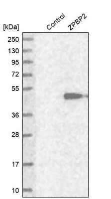
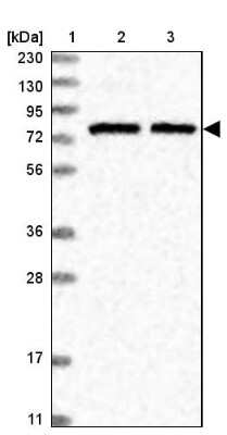
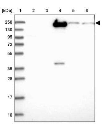
| SDS-PAGE | 11 kDa, reducing conditions |
|
| Publications |
|
| Storage | Use a manual defrost freezer and avoid repeated freeze-thaw cycles.
|
| Buffer | Lyophilized from a 0.2 μm filtered solution in PBS. |
| Purity | >95%, by SDS-PAGE under reducing conditions and visualized by silver stain |
| Reconstitution Instructions | Reconstitute at 300 μg/mL in sterile PBS. |
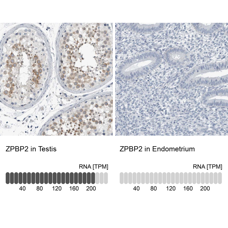
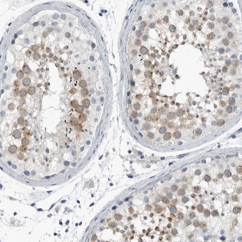
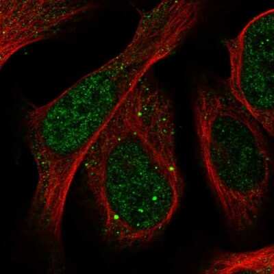
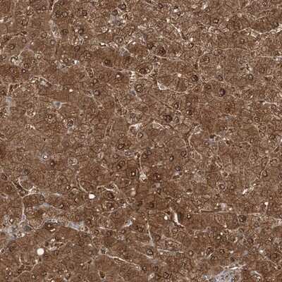
TAFA2 (also FAM19A2) is a secreted, 11 kDa member of the FAM19/TAFA family of chemokine-like proteins (1). It is synthesized as a 131 amino acid (aa) precursor that contains a 30 aa signal sequence and a 101 aa mature chain (SwissProt #: Q8N3H0). Like other members of the FAM19/TAFA family, with the exception of TAFA5, mature TAFA1 contains 10 regularly spaced cysteine residues that follow the pattern CX7CCX13CXCX14CX11CX4CX5CX10C, where C represents a conserved cysteine residue and X represents any noncysteine amino acid (1). Human TAFA2 is 97% aa identical to mouse TAFA2 (1). TAFA2 expression can be detected in the central nervous system (CNS), colon, heart, lung, spleen, kidney, and thymus, but its expression in the CNS is 50- to 1000-fold higher than in other tissues (1). Within the CNS, TAFA2 expression is highest in the occipital and frontal cortex (3- to 10-fold more abundantly expressed than in other cortical regions) and medulla (1). The biological functions of TAFA family members remain to be determined, but there are a few tentative hypotheses. First, TAFAs may modulate immune responses in the CNS by functioning as brain-specific chemokines, and may act with other chemokines to optimize the recruitment and activity of immune cells in the CNS (1). Second, TAFAs may represent a novel class of neurokines that act as regulators of immune nervous cells (1 - 2). Finally, TAFAs may control axonal sprouting following brain injury (1).


![ADAMTSL-1/Punctin Antibody [Unconjugated]](/sites/all/modules/enterprise-tech/et_datasheets/images/novus_guarantee.png "ADAMTSL-1/Punctin Antibody [Unconjugated]")
![Flow Cytometry NAALADase-like 2/NAALADL2 Antibody [Unconjugated]](https://images.novusbio.com/images/antibody/NAALADase-like_2_AF4665_Flow_Cytometry_8256.jpg)


The concentration calculator allows you to quickly calculate the volume, mass or concentration of your vial. Simply enter your mass, volume, or concentration values for your reagent and the calculator will determine the rest.


![Western Blot Ikaros/IKZF1 Antibody [Unconjugated]](https://images.novusbio.com/images/antibody/Ikaros_AF4984_Western_Blot_5594.jpg)
![Intracellular Staining by Flow Cytometry Ikaros/IKZF1 Antibody [Unconjugated]](https://images.novusbio.com/images/antibody/Ikaros_AF4984_Flow_Cytometry_8264.jpg)
![Simple Western Ikaros/IKZF1 Antibody [Unconjugated]](https://images.novusbio.com/images/antibody/Ikaros_AF4984_Simple_Western_16946.jpg)
![N/A BDNF [HRP]](https://images.novusbio.com/images/elisa/BDNF_DBD00_ELISA_36.jpg)
![N/A BDNF [HRP]](https://images.novusbio.com/images/elisa/DATA_BDNF_DBD00_ELISA_587.jpg)
![N/A BDNF [HRP]](https://images.novusbio.com/images/elisa/DATA_BDNF_DBD00_ELISA_588.jpg)
![SDS-Page IL-12 [Unconjugated]](https://images.novusbio.com/images/protein/IL-12_419-ML_403.jpg)
![Bioactivity IL-12 [Unconjugated]](https://images.novusbio.com/images/protein/IL-12_419-ML_405.jpg)
