| Reactivity | HuSpecies Glossary |
| Applications | Enzyme Activity |
| Format | Carrier-Free |
| Details of Functionality | Measured by its ability to cleave the fluorogenic peptide substrate Boc-QAR-AMC (Catalog # ES014). The specific activity is >300 pmol/min/µg, as measured under the described conditions. |
| Source | Mouse myeloma cell line, NS0-derived human Spinesin protein Tyr71-Leu457 (Phe369Leu), with an N-terminal 9-His tag Accession # NP_110397 |
| Accession # | |
| N-terminal Sequence | His |
| Structure / Form | Pro form |
| Protein/Peptide Type | Recombinant Enzymes |
| Gene | TMPRSS5 |
| Purity | >95%, by SDS-PAGE under reducing conditions and visualized by silver stain |
| Endotoxin Note | <1.0 EU per 1 μg of the protein by the LAL method. |
| Dilutions |
|
|
| Theoretical MW | 43 kDa. Disclaimer note: The observed molecular weight of the protein may vary from the listed predicted molecular weight due to post translational modifications, post translation cleavages, relative charges, and other experimental factors. |
|
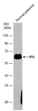
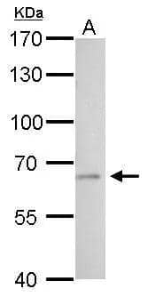
| SDS-PAGE | 63 kDa, reducing conditions |
|
| Publications |
|
| Storage | Use a manual defrost freezer and avoid repeated freeze-thaw cycles.
|
|||
| Buffer | Supplied as a 0.2 μm filtered solution in MES and NaCl. |
|||
| Purity | >95%, by SDS-PAGE under reducing conditions and visualized by silver stain |
|||
| Assay Procedure |
*Adjusted for Substrate Blank
|
Spinesin, encoded by the TMPRSS5 gene, is a member of type II transmembrane serine proteases (TTSPs) (1). Human Spinesin contains the following structural domains: a short N-terminal cytoplasmic tail (amino acid residues 1-49), a transmembrane domain (residues 50-70), a stem region and a serine protease domain (residues 71-457) (2). The domain structure of Spinesin is common to other TTSPs, many of which have additional domains. The stem region of Spinesin contains a scavenger receptor-like domain. The ectodomain of human Spinesin (residues 71-457) was expressed and purified as a single chain pro-enzyme. The deduced amino acid sequence contains a Leu instead of a Phe residue at position 369; the former is identical to the mouse protein (3, 4). The pro-enzyme can be activated and the resulting enzyme activity can be measured as described in the Activity Assay Protocol.


The concentration calculator allows you to quickly calculate the volume, mass or concentration of your vial. Simply enter your mass, volume, or concentration values for your reagent and the calculator will determine the rest.


![Hepsin [Unconjugated]](/sites/all/modules/enterprise-tech/et_datasheets/images/novus_guarantee.png "Hepsin [Unconjugated]")
![N/A Corin [Biotin]](https://images.novusbio.com/images/elisa/DATA_Corin_DY2209_ELISA_1787.jpg)


![Intracellular Staining by Flow Cytometry Matriptase/ST14 Antibody [Unconjugated]](https://images.novusbio.com/images/antibody/Matriptase_AF3946_Flow_Cytometry_8218.jpg)
![Simple Western Matriptase/ST14 Antibody [Unconjugated]](https://images.novusbio.com/images/af3946_human-matriptase-st14-affinity-purified-polyclonal-ab-4120241239322.jpg)
![Simple Western Matriptase/ST14 Antibody [Unconjugated]](https://images.novusbio.com/images/af3946_human-matriptase-st14-affinity-purified-polyclonal-ab-41202412401222.jpg)