HAF017). Specific bands were detected for TNF-a at approximately 17 and 26 kDa (as indicated). This experiment was conducted under reducing conditions and using
Immunoblot Buffer Group 1." class="big_lightbox">
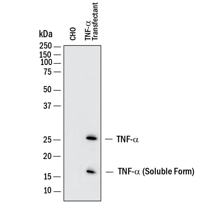
HAF017). Specific bands were detected for TNF-a at approximately 17 and 26 kDa (as indicated). This experiment was conducted under reducing conditions and using
Immunoblot Buffer Group 1." alt="Western blot shows lysates of CHO Chinese hamster ovary cell line either mock transfected or transfected with human TNF- alpha. PVDF membrane was probed with 1 µg/mL of Goat Anti-Human/Mouse TNF-a Antigen Affinity-purified Polyclonal Antibody (Catalog # AF-410-NA) followed by HRP-conjugated Anti-Goat IgG Secondary Antibody (
HAF017). Specific bands were detected for TNF-a at approximately 17 and 26 kDa (as indicated). This experiment was conducted under reducing conditions and using
Immunoblot Buffer Group 1." class="big_thumb" />
HAF017). Specific bands were detected for TNF-a at approximately 16 and 25 kDa (as indicated). This experiment was conducted under reducing conditions and using
Immunoblot Buffer Group 1." class="big_lightbox">

HAF017). Specific bands were detected for TNF-a at approximately 16 and 25 kDa (as indicated). This experiment was conducted under reducing conditions and using
Immunoblot Buffer Group 1." class="big_thumb" />
VC004). Tissue was stained using DAB (brown) and counterstained with hematoxylin (blue). Specific staining was localized to lymphocytes. Staining was performed using our IHC Staining with VisUCyte HRP Polymer Detection Reagents." class="big_lightbox">
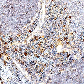
VC004). Tissue was stained using DAB (brown) and counterstained with hematoxylin (blue). Specific staining was localized to lymphocytes. Staining was performed using our IHC Staining with VisUCyte HRP Polymer Detection Reagents." class="big_thumb" />
410-MT) induces cytotoxicity in the the L-929 mouse fibroblast cell line in a dose-dependent manner (orange line). Cytotoxicity elicited by Recombinant Mouse TNF-a (0.1 ng/mL) is neutralized (green line) by increasing concentrations of Human/Mouse TNF-a Antigen Affinity-purified Polyclonal Antibody (Catalog # AF-410-NA). The ND
50 is typically 1.5-10 ng/mL in the presence of the metabolic inhibitor actinomycin D." class="big_lightbox">
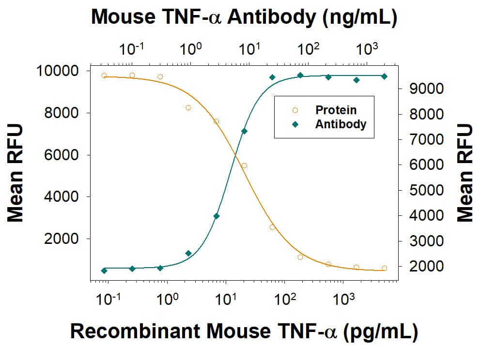
410-MT) induces cytotoxicity in the the L-929 mouse fibroblast cell line in a dose-dependent manner (orange line). Cytotoxicity elicited by Recombinant Mouse TNF-a (0.1 ng/mL) is neutralized (green line) by increasing concentrations of Human/Mouse TNF-a Antigen Affinity-purified Polyclonal Antibody (Catalog # AF-410-NA). The ND
50 is typically 1.5-10 ng/mL in the presence of the metabolic inhibitor actinomycin D." class="big_thumb" />
VC004). Before incubation with the primary antibody, tissue was subjected to heat-induced epitope retrieval using VisUCyte Antigen Retrieval Reagent-Basic (Catalog #
VCTS021). Tissue was stained using DAB (brown) and counterstained with hematoxylin (blue). Specific staining was localized to cytoplasm in lymphocytes. View our protocol for
IHC Staining with VisUCyte HRP Polymer Detection Reagents." class="big_lightbox">

VC004). Before incubation with the primary antibody, tissue was subjected to heat-induced epitope retrieval using VisUCyte Antigen Retrieval Reagent-Basic (Catalog #
VCTS021). Tissue was stained using DAB (brown) and counterstained with hematoxylin (blue). Specific staining was localized to cytoplasm in lymphocytes. View our protocol for
IHC Staining with VisUCyte HRP Polymer Detection Reagents." class="big_thumb" />
![WNT-5A induces a proinflammatory transformation in mouse microglia. (A, B) iNOS, COX-2 and TNF alpha were detected by immunoblotting in lysates from mouse primary microglia after WNT-5A stimulation (ctrl, 300 ng/ml, 6 hours). (B’) shows TNF alpha levels in the supernatant of primary microglia under ctrl conditions and upon WNT-5A stimulation (ctrl, 300 ng/ml, 24 hours; n = 4). At least three experiments are summarized in the bar graphs. Data are normalized to ctrl. Error bars give s.e.m. (C) Microglial proliferation was assessed by an MTT assay monitoring mitochondrial activity, which is proportional to cell number [see Additional file 1: Figure S5]. Stimulation with WNT-5A (300 ng/ml, 24 hours) increased MTT, which was blocked by PTX (100 ng/ml, overnight) or the MEK1/2 (10 μM) inhibitor, SL327. (D). Experiments were done in triplicate and data from three independent experiments are shown. *, P < 0.01; ***, P < 0.001: Error bars show s.e.m.. (E) Cell tracker (red)-stained primary microglia were seeded on top of a collagen matrix in 35 mm glass bottom dishes. One day after ctrl or 300 ng/ml WNT-5A stimulation, invasion was observed by confocal microscopy and Z-stacking using a Zeiss LSM710 microscope and subsequent analysis with the Bitplane Imaris software. The size of the collagen cube shown is 1,000 (Z) x 1,400 (Y) x 1,400 (X) μm. Three invasion experiments in the absence and presence of the MEK1/2 inhibitor SL327 were quantified. Data are presented in a bar graph (F). *, P < 0.05; error bars show s.e.m.. (G) cDNA of ctrl stimulated (−) and WNT-5A stimulated (+) primary microglia was analyzed by QPCR for expression of inflammatory genes. At least three independent experiments are summarized. Gene expression is normalized to the housekeeping gene GAPDH and expressed as arbitrary units (2-delta ct). *, P < 0.05; **, P < 0.01. COX-2, cyclooxygenase 2; iNOS, inducible nitric oxide synthase; MTT, 3-(4,5-dimethylthiazol-2-yl)-2,5-diphenyltetrazolium bromide; n, number; s.e.m., standa](//images.novusbio.com/images/af-410-na_mouse-tnf-alpha-affinity-purified-polyclonal-ab-41202410331929.jpg)
AB-108-C, open histogram) followed by Allophycocyanin-conjugated Anti-Goat IgG Secondary Antibody (Catalog #
F0108). To facilitate intracellular staining, cells were fixed with Flow Cytometry Fixation Buffer (Catalog #
FC004) and permeabilized with saponin. View our protocol for
Staining Intracellular Molecules." class="big_lightbox">
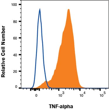
AB-108-C, open histogram) followed by Allophycocyanin-conjugated Anti-Goat IgG Secondary Antibody (Catalog #
F0108). To facilitate intracellular staining, cells were fixed with Flow Cytometry Fixation Buffer (Catalog #
FC004) and permeabilized with saponin. View our protocol for
Staining Intracellular Molecules." class="big_thumb" />
410-MT) was serially diluted and captured by Goat Anti-Human/Mouse TNF‑ alpha Antigen Affinity-purified Polyclonal Antibody (Catalog # AF-410-NA) coated on a Clear Polystyrene Microplate (Catalog #
DY990). Goat Anti-Human/Mouse TNF‑ alpha Antigen Affinity-purified Polyclonal Antibody (Catalog #
AF-410-NA) was biotinylated and incubated with the protein captured on the plate. Detection of the standard curve was achieved by incubating Streptavidin-HRP (Catalog #
DY998)" class="big_lightbox">

410-MT) was serially diluted and captured by Goat Anti-Human/Mouse TNF‑ alpha Antigen Affinity-purified Polyclonal Antibody (Catalog # AF-410-NA) coated on a Clear Polystyrene Microplate (Catalog #
DY990). Goat Anti-Human/Mouse TNF‑ alpha Antigen Affinity-purified Polyclonal Antibody (Catalog #
AF-410-NA) was biotinylated and incubated with the protein captured on the plate. Detection of the standard curve was achieved by incubating Streptavidin-HRP (Catalog #
DY998)" class="big_thumb" />
410-MT) was serially diluted and captured by Goat Anti-Human/Mouse TNF‑ alpha Antigen Affinity-purified Polyclonal Antibody (Catalog #
AF-410-NA) coated on a Clear Polystyrene Microplate (Catalog #
DY990). Goat Anti-Human/Mouse TNF‑ alpha Antigen Affinity-purified Polyclonal Antibody (Catalog # AF-410-NA) was biotinylated and incubated with the protein captured on the plate. Detection of the standard curve was achieved by incubating Streptavidin-HRP (Catalog #
DY998)" class="big_lightbox">

410-MT) was serially diluted and captured by Goat Anti-Human/Mouse TNF‑ alpha Antigen Affinity-purified Polyclonal Antibody (Catalog #
AF-410-NA) coated on a Clear Polystyrene Microplate (Catalog #
DY990). Goat Anti-Human/Mouse TNF‑ alpha Antigen Affinity-purified Polyclonal Antibody (Catalog # AF-410-NA) was biotinylated and incubated with the protein captured on the plate. Detection of the standard curve was achieved by incubating Streptavidin-HRP (Catalog #
DY998)" class="big_thumb" />
 HAF017). Specific bands were detected for TNF-a at approximately 17 and 26 kDa (as indicated). This experiment was conducted under reducing conditions and using Immunoblot Buffer Group 1." alt="Western blot shows lysates of CHO Chinese hamster ovary cell line either mock transfected or transfected with human TNF- alpha. PVDF membrane was probed with 1 µg/mL of Goat Anti-Human/Mouse TNF-a Antigen Affinity-purified Polyclonal Antibody (Catalog # AF-410-NA) followed by HRP-conjugated Anti-Goat IgG Secondary Antibody (HAF017). Specific bands were detected for TNF-a at approximately 17 and 26 kDa (as indicated). This experiment was conducted under reducing conditions and using Immunoblot Buffer Group 1." class="big_thumb" />
 210-TA), Recombinant Mouse TNF-a aa 80-235 (Catalog # 410-MT), Recombinant Rat TNF-a (Catalog # 510-RT), and Recombinant Human APRIL/TNFSF13 (Catalog # 5860-AP). PVDF Membrane was probed with 1 µg/mL of Mouse Anti-Human TNF-a Monoclonal Antibody (Catalog # MAB610) followed by HRP-conjugated Anti-Mouse IgG Secondary Antibody (Catalog # HAF007). A specific band was detected for TNF-a at approximately 15 kDa (as indicated). This experiment was conducted under reducing conditions and using Immunoblot Buffer Group 3." alt="Western blot shows 25 ng of Recombinant Human TNF-a (Catalog # 210-TA), Recombinant Mouse TNF-a aa 80-235 (Catalog # 410-MT), Recombinant Rat TNF-a (Catalog # 510-RT), and Recombinant Human APRIL/TNFSF13 (Catalog # 5860-AP). PVDF Membrane was probed with 1 µg/mL of Mouse Anti-Human TNF-a Monoclonal Antibody (Catalog # MAB610) followed by HRP-conjugated Anti-Mouse IgG Secondary Antibody (Catalog # HAF007). A specific band was detected for TNF-a at approximately 15 kDa (as indicated). This experiment was conducted under reducing conditions and using Immunoblot Buffer Group 3." class="big_thumb" />
 510-RT) induces cytotoxicity in the the L-929 mouse fibroblast cell line in a dose-dependent manner (orange line), as measured by Resazurin. Cytotoxicity elicited by Recombinant Rat TNF-a (0.25 ng/mL) is neutralized (green line) by increasing concentrations of Goat Anti-Rat TNF-a Antigen Affinity-purified Polyclonal Antibody (Catalog # AF-510-NA). The ND50 is typically 0.15-0.75 µg/mL in the presence of the metabolic inhibitor actinomycin D (1 µg/mL)." alt="Recombinant Rat TNF-a (Catalog # 510-RT) induces cytotoxicity in the the L-929 mouse fibroblast cell line in a dose-dependent manner (orange line), as measured by Resazurin. Cytotoxicity elicited by Recombinant Rat TNF-a (0.25 ng/mL) is neutralized (green line) by increasing concentrations of Goat Anti-Rat TNF-a Antigen Affinity-purified Polyclonal Antibody (Catalog # AF-510-NA). The ND50 is typically 0.15-0.75 µg/mL in the presence of the metabolic inhibitor actinomycin D (1 µg/mL)." class="big_thumb" />




![N/A TNF-alpha [Biotin]](http://images.novusbio.com/fullsize/elisa/DATA_TNFalpha_DY410_ELISA_2323.jpg "N/A TNF-alpha [Biotin]")
![N/A TNF-alpha [Biotin]](http://images.novusbio.com/fullsize/elisa/DATA_TNFalpha_DY210_ELISA_1766.jpg "N/A TNF-alpha [Biotin]")

 410-MT) induces cytotoxicity in the L-929 mouse fibroblast cell line in the presence of the metabolic inhibitor actinomycin D. The ED50 for this effect is 8-50 pg/mL." class="big_thumb" />
![N/A TNF-alpha [HRP]](http://images.novusbio.com/fullsize/elisa/DATA_TNFalpha_RTA00_ELISA_1099.jpg "N/A TNF-alpha [HRP]")
![N/A TNF-alpha [HRP]](http://images.novusbio.com/fullsize/elisa/TNF-alpha_PRTA00_ELISA_365.jpg)
 HAF017). Specific bands were detected for TNF-a at approximately 16 and 25 kDa (as indicated). This experiment was conducted under reducing conditions and using
  NL001) and counterstained with DAPI (blue). View our protocol for
  Fluorescent ICC Staining of Non-adherent Cells." class="big_thumb" />
 VC004). Tissue was stained using DAB (brown) and counterstained with hematoxylin (blue). Specific staining was localized to lymphocytes. Staining was performed using our IHC Staining with VisUCyte HRP Polymer Detection Reagents." class="big_thumb" />
 410-MT) induces cytotoxicity in the the L-929 mouse fibroblast cell line in a dose-dependent manner (orange line). Cytotoxicity elicited by Recombinant Mouse TNF-a (0.1 ng/mL) is neutralized (green line) by increasing concentrations of Human/Mouse TNF-a Antigen Affinity-purified Polyclonal Antibody (Catalog # AF-410-NA). The ND50 is typically 1.5-10 ng/mL in the presence of the metabolic inhibitor actinomycin D." class="big_thumb" />
 VC004). Before incubation with the primary antibody, tissue was subjected to heat-induced epitope retrieval using VisUCyte Antigen Retrieval Reagent-Basic (Catalog #
 ![WNT-5A induces a proinflammatory transformation in mouse microglia. (A, B) iNOS, COX-2 and TNF alpha were detected by immunoblotting in lysates from mouse primary microglia after WNT-5A stimulation (ctrl, 300 ng/ml, 6 hours). (B’) shows TNF alpha levels in the supernatant of primary microglia under ctrl conditions and upon WNT-5A stimulation (ctrl, 300 ng/ml, 24 hours; n = 4). At least three experiments are summarized in the bar graphs. Data are normalized to ctrl. Error bars give s.e.m. (C) Microglial proliferation was assessed by an MTT assay monitoring mitochondrial activity, which is proportional to cell number [see Additional file 1: Figure S5]. Stimulation with WNT-5A (300 ng/ml, 24 hours) increased MTT, which was blocked by PTX (100 ng/ml, overnight) or the MEK1/2 (10 μM) inhibitor, SL327. (D). Experiments were done in triplicate and data from three independent experiments are shown. *, P < 0.01; ***, P < 0.001: Error bars show s.e.m.. (E) Cell tracker (red)-stained primary microglia were seeded on top of a collagen matrix in 35 mm glass bottom dishes. One day after ctrl or 300 ng/ml WNT-5A stimulation, invasion was observed by confocal microscopy and Z-stacking using a Zeiss LSM710 microscope and subsequent analysis with the Bitplane Imaris software. The size of the collagen cube shown is 1,000 (Z) x 1,400 (Y) x 1,400 (X) μm. Three invasion experiments in the absence and presence of the MEK1/2 inhibitor SL327 were quantified. Data are presented in a bar graph (F). *, P < 0.05; error bars show s.e.m.. (G) cDNA of ctrl stimulated (−) and WNT-5A stimulated (+) primary microglia was analyzed by QPCR for expression of inflammatory genes. At least three independent experiments are summarized. Gene expression is normalized to the housekeeping gene GAPDH and expressed as arbitrary units (2-delta ct). *, P < 0.05; **, P < 0.01. COX-2, cyclooxygenase 2; iNOS, inducible nitric oxide synthase; MTT, 3-(4,5-dimethylthiazol-2-yl)-2,5-diphenyltetrazolium bromide; n, number; s.e.m., standa](http://images.novusbio.com/fullsize/af-410-na_mouse-tnf-alpha-affinity-purified-polyclonal-ab-41202410331929.jpg)
 AB-108-C, open histogram) followed by Allophycocyanin-conjugated Anti-Goat IgG Secondary Antibody (Catalog #
  410-MT) was serially diluted and captured by Goat Anti-Human/Mouse TNF‑ alpha Antigen Affinity-purified Polyclonal Antibody (Catalog # AF-410-NA) coated on a Clear Polystyrene Microplate (Catalog #
  410-MT) was serially diluted and captured by Goat Anti-Human/Mouse TNF‑ alpha Antigen Affinity-purified Polyclonal Antibody (Catalog #
 ![Western Blot: TNF-alpha Antibody [NBP1-19532] - Recombinant human TNF alpha (10 ng) was separated on a 12% gel by SDS-PAGE, transferred to 0.2 um PVDF membrane and blocked in 5% non-fat milk in TBST. The membrane was probed with 2.0 ug/ml anti-TNF alpha in 5% block buffer and detected with an anti-rabbit HRP secondary antibody using chemiluminescence.](http://images.novusbio.com/fullsize/TNF-alpha-Antibody-Western-Blot-NBP1-19532-img0007.jpg "Western Blot: TNF-alpha Antibody [NBP1-19532] - Recombinant human TNF alpha (10 ng) was separated on a 12% gel by SDS-PAGE, transferred to 0.2 um PVDF membrane and blocked in 5% non-fat milk in TBST. The membrane was probed with 2.0 ug/ml anti-TNF alpha in 5% block buffer and detected with an anti-rabbit HRP secondary antibody using chemiluminescence.")

![Immunohistochemistry-Paraffin: TNF-alpha Antibody [NBP1-19532] - Analysis of a FFPE tissue section of mouse intestine using TNF-alpha antibody (NBP1-19532) at 1:300 dilution. The binding of this primary antibody to TNF-alpha protein in the section was detected using HRP-labeled secondary antibody and DAB reagent, and nuclei of cells were counterstained using hematoxylin. This TNF-alpha antibody generated an expected diffused immunostaining of this protein in the tested tissue. Staining was primarily observed in the epithelial cells and some cells showed membrane positivity also.](http://images.novusbio.com/fullsize/TNF-alpha-Antibody-Immunohistochemistry-Paraffin-NBP1-19532-img0005.jpg)
![Immunohistochemistry-Paraffin: TNF-alpha Antibody [NBP1-19532] - Analysis of a FFPE tissue section of mouse intestine using TNF-alpha antibody (NBP1-19532) at 1:300 dilution. The binding of this primary antibody to TNF-alpha protein in the section was detected using HRP-labeled secondary antibody and DAB reagent, and nuclei of cells were counterstained using hematoxylin. This TNF-alpha antibody generated an expected immunopositivity of this protein in the tested tissue. Staining was primarily observed in the epithelial cells while some staining was present in mucosa muscularis also.](http://images.novusbio.com/fullsize/TNF-alpha-Antibody-Immunohistochemistry-Paraffin-NBP1-19532-img0006.jpg)

![Immunocytochemistry/ Immunofluorescence: TNF-alpha Antibody - BSA Free [NBP1-19532] - Effects of femoral artery occlusion on TNF-alpha signal in sensory nerves. (A) Seventy-two hours of femoral artery occlusion increased the levels of TNF-alpha in the DRG tissues as compared with them in DRG of control limbs (n = 6 in each group); & PTX given into the hindlimb muscles attenuated amplification of TNF-alpha in the DRG tissues of limbs with femoral artery occlusion (n = 8). ∗P < 0.05, occlusion group vs. control & occlusion with prior PTX. (B) Immunofluorescence was used to examine double-labeling for TNF-alpha & peripherin/NF-200 (n = 3). Peripherin was used to label DRG neurons that project thin C-fibers. NF200 was used to identify A-fibers of DRG neurons. Representative photomicrographs show co-existence of TNF-alpha & peripherin staining in DRG neurons (top panel), whereas few TNF-alpha & NF-200 staining were observed in DRG neurons (bottom panel). Arrows indicate representative positive cells for both TNF-alpha & peripherin after they were merged. Scale bar = 50 μm. (C) Representative bands (left panel) & averaged data (right panel), demonstrating that femoral artery occlusion upregulated protein expression TNF-alpha receptor subtype TNFR1, but not TNFR2. A significant difference in TNFR1 was seen between control & occluded groups. ∗P < 0.05 vs. control. n = 6 in each group. Image collected & cropped by CiteAb from the following publication (//pubmed.ncbi.nlm.nih.gov/30374312), licensed under a CC-BY license. Not internally tested by Novus Biologicals.](http://images.novusbio.com/fullsize/nbp1-19532_rabbit-polyclonal-tnf-alpha-antibody-31020241618927.jpg)
![Immunocytochemistry/ Immunofluorescence: TNF-alpha Antibody - BSA Free [NBP1-19532] - Immunoflorescence staining results of TNF-alpha (96 h). A). Lamella of gill tissue (arrow). Control group. IF. 20 μm. B) Positive reaction in lamellar cells of gill tissue (arrows). Low dose toxic group. IF. 20 μm. C) Positivity in lamellar cells of gill tissue (arrow). High dose toxic group. IF. 20 μm. D) Liver tissue, vena centralis (arrow). Control group. IF. 20 μm. E) Positive reactions in hepatocytes (arrows). Low dose toxic group. IF. 20 μm. F) Positive reactions in hepatocytes (arrows). High dose toxic group. IF. 20 μm. G) Weak positive reactions in neurons (arrows). Control group. IF. 20 μm. H) Weak positivity in neurons (arrows). Low dose toxic group. IF. 20 μm. I) Positivity in neurons (arrows). High dose toxic group. IF. 20 μm. Image collected & cropped by CiteAb from the following publication (//pubmed.ncbi.nlm.nih.gov/29321977), licensed under a CC-BY license. Not internally tested by Novus Biologicals.](http://images.novusbio.com/fullsize/nbp1-19532_rabbit-polyclonal-tnf-alpha-antibody-310202416171446.jpg)


![N/A TNF-alpha [Biotin]](http://images.novusbio.com/fullsize/elisa/DATA_TNFalpha_DY510_ELISA_2079.jpg "N/A TNF-alpha [Biotin]")
![N/A TNF-alpha [HRP]](http://images.novusbio.com/fullsize/elisa/DATA_TNFalpha_PTA00_ELISA_1061.jpg "N/A TNF-alpha [HRP]")
![N/A TNF-alpha [HRP]](http://images.novusbio.com/fullsize/elisa/DATA_TNFalpha_PTA00_ELISA_1062.jpg)
![N/A TNF-alpha [HRP]](http://images.novusbio.com/fullsize/elisa/TNF-alpha_PTA00_ELISA_343.jpg)
 NL007) and counterstained with DAPI (blue). View our protocol for
  210-TA) induces cytotoxicity in the the L-929 mouse fibroblast cell line in a dose-dependent manner (orange line). Cytotoxicity elicited by Recombinant Human TNF-a (0.25 ng/mL) is neutral-ized (green line) by increasing concentrations of Mouse Anti-Human TNF-a Monoclonal Antibody (Catalog # MAB610). The ND50 is typically 0.01-0.04 µg/mL in the presence of the metabolic inhibitor actinomycin D." class="big_thumb" />
 DY990). Goat Anti-Human TNF‑ alpha Antigen Affinity-purified Polyclonal Antibody (Catalog #
  NL001) and counterstained with DAPI (blue). Specific staining was localized to cytoplasmic. View our protocol for

 510-RT) was serially diluted and captured by Mouse Anti-Rat TNF‑ alpha Monoclonal Antibody (Catalog #


