2929-TM), Recombinant Human TIM-1/KIM-1/HAVCR (Catalog #
1750-TM), Recombinant Human TIM-3 Fc Chimera (Catalog #
2365-TM), Recombinant Mouse TIM-1/KIM-1/HAVCR (Catalog #
1817-TM), Recombinant Mouse TIM-2 (Catalog #
1885-TM), Recombinant Mouse TIM-3 Fc Chimera (Catalog #
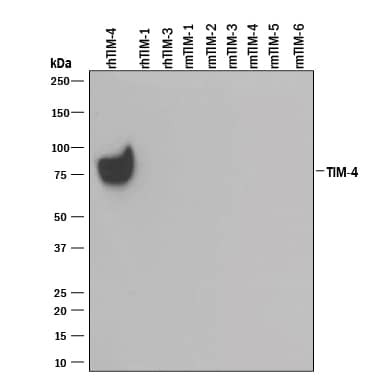
1529-TM), Recombinant Mouse TIM-4 (Catalog #
2826-TI), Recombinant Mouse TIM-5, and Recombinant Mouse TIM-6. PVDF Membrane was probed with 0.1 µg/mL of Goat Anti-Human TIM-4 Antigen Affinity-purified Polyclonal Antibody (Catalog # AF2929) followed by HRP-conjugated Anti-Goat IgG Secondary Antibody (Catalog #
HAF109). A specific band was detected for TIM-4 at approximately 75 kDa (as indicated). This experiment was conducted under reducing conditions and using
Immunoblot Buffer Group 3." class="big_lightbox">
2929-TM), Recombinant Human TIM-1/KIM-1/HAVCR (Catalog #
1750-TM), Recombinant Human TIM-3 Fc Chimera (Catalog #
2365-TM), Recombinant Mouse TIM-1/KIM-1/HAVCR (Catalog #
1817-TM), Recombinant Mouse TIM-2 (Catalog #
1885-TM), Recombinant Mouse TIM-3 Fc Chimera (Catalog #
1529-TM), Recombinant Mouse TIM-4 (Catalog #
2826-TI), Recombinant Mouse TIM-5, and Recombinant Mouse TIM-6. PVDF Membrane was probed with 0.1 µg/mL of Goat Anti-Human TIM-4 Antigen Affinity-purified Polyclonal Antibody (Catalog # AF2929) followed by HRP-conjugated Anti-Goat IgG Secondary Antibody (Catalog #
HAF109). A specific band was detected for TIM-4 at approximately 75 kDa (as indicated). This experiment was conducted under reducing conditions and using
Immunoblot Buffer Group 3." alt="Western blot shows 25 ng of Recombinant Human TIM-4 His-tag (Catalog #
2929-TM), Recombinant Human TIM-1/KIM-1/HAVCR (Catalog #
1750-TM), Recombinant Human TIM-3 Fc Chimera (Catalog #
2365-TM), Recombinant Mouse TIM-1/KIM-1/HAVCR (Catalog #
1817-TM), Recombinant Mouse TIM-2 (Catalog #
1885-TM), Recombinant Mouse TIM-3 Fc Chimera (Catalog #
1529-TM), Recombinant Mouse TIM-4 (Catalog #
2826-TI), Recombinant Mouse TIM-5, and Recombinant Mouse TIM-6. PVDF Membrane was probed with 0.1 µg/mL of Goat Anti-Human TIM-4 Antigen Affinity-purified Polyclonal Antibody (Catalog # AF2929) followed by HRP-conjugated Anti-Goat IgG Secondary Antibody (Catalog #
HAF109). A specific band was detected for TIM-4 at approximately 75 kDa (as indicated). This experiment was conducted under reducing conditions and using
Immunoblot Buffer Group 3." class="big_thumb" />
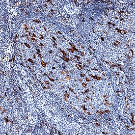
VC004). Before incubation with the primary antibody, tissue was subjected to heat-induced epitope retrieval using Antigen Retrieval Reagent-Basic (Catalog #
CTS013). Tissue was stained using DAB (brown) and counterstained with hematoxylin (blue). Specific staining was localized to cell membranes of macrophages. View our protocol for
IHC Staining with VisUCyte HRP Polymer Detection Reagents." class="big_lightbox">
VC004). Before incubation with the primary antibody, tissue was subjected to heat-induced epitope retrieval using Antigen Retrieval Reagent-Basic (Catalog #
CTS013). Tissue was stained using DAB (brown) and counterstained with hematoxylin (blue). Specific staining was localized to cell membranes of macrophages. View our protocol for
IHC Staining with VisUCyte HRP Polymer Detection Reagents." class="big_thumb" />
 2929-TM), Recombinant Human TIM-1/KIM-1/HAVCR (Catalog # 1750-TM), Recombinant Human TIM-3 Fc Chimera (Catalog # 2365-TM), Recombinant Mouse TIM-1/KIM-1/HAVCR (Catalog # 1817-TM), Recombinant Mouse TIM-2 (Catalog # 1885-TM), Recombinant Mouse TIM-3 Fc Chimera (Catalog # 1529-TM), Recombinant Mouse TIM-4 (Catalog # 2826-TI), Recombinant Mouse TIM-5, and Recombinant Mouse TIM-6. PVDF Membrane was probed with 0.1 µg/mL of Goat Anti-Human TIM-4 Antigen Affinity-purified Polyclonal Antibody (Catalog # AF2929) followed by HRP-conjugated Anti-Goat IgG Secondary Antibody (Catalog # HAF109). A specific band was detected for TIM-4 at approximately 75 kDa (as indicated). This experiment was conducted under reducing conditions and using Immunoblot Buffer Group 3." alt="Western blot shows 25 ng of Recombinant Human TIM-4 His-tag (Catalog # 2929-TM), Recombinant Human TIM-1/KIM-1/HAVCR (Catalog # 1750-TM), Recombinant Human TIM-3 Fc Chimera (Catalog # 2365-TM), Recombinant Mouse TIM-1/KIM-1/HAVCR (Catalog # 1817-TM), Recombinant Mouse TIM-2 (Catalog # 1885-TM), Recombinant Mouse TIM-3 Fc Chimera (Catalog # 1529-TM), Recombinant Mouse TIM-4 (Catalog # 2826-TI), Recombinant Mouse TIM-5, and Recombinant Mouse TIM-6. PVDF Membrane was probed with 0.1 µg/mL of Goat Anti-Human TIM-4 Antigen Affinity-purified Polyclonal Antibody (Catalog # AF2929) followed by HRP-conjugated Anti-Goat IgG Secondary Antibody (Catalog # HAF109). A specific band was detected for TIM-4 at approximately 75 kDa (as indicated). This experiment was conducted under reducing conditions and using Immunoblot Buffer Group 3." class="big_thumb" />
 MAB002) followed by Phycoerythrin-conjugated Anti-Mouse IgG Secondary Antibody (Catalog # F0102B). View our protocol for Staining Membrane-associated Proteins." alt="HEK293 human embryonic kidney cell line transfected with human TIM-4 and eGFP was stained with and either (A) Mouse Anti-Human TIM-4 Monoclonal Antibody (Catalog # MAB2929) or (B) Mouse IgG1Isotype Control (Catalog # MAB002) followed by Phycoerythrin-conjugated Anti-Mouse IgG Secondary Antibody (Catalog # F0102B). View our protocol for Staining Membrane-associated Proteins." class="big_thumb" />


 VC004). Before incubation with the primary antibody, tissue was subjected to heat-induced epitope retrieval using Antigen Retrieval Reagent-Basic (Catalog #
 ![Western Blot: TIM-4 Antibody [NBP1-76702] - Analysis of TIM-4 in RAW264.7 cell lysate with TIM-4 antibody at 2 ug/mL in (A) the absence and (B) the presence of blocking peptide.](http://images.novusbio.com/fullsize/TIM-4-Antibody-Western-Blot-NBP1-76702-img0008.jpg "Western Blot: TIM-4 Antibody [NBP1-76702] - Analysis of TIM-4 in RAW264.7 cell lysate with TIM-4 antibody at 2 ug/mL in (A) the absence and (B) the presence of blocking peptide.")
![Immunocytochemistry/ Immunofluorescence: TIM-4 Antibody - BSA Free [NBP1-76702] - Immunofluorescence of TIM-4 in Jurkat cells with TIM-4 antibody at 20 ug/mL.](http://images.novusbio.com/fullsize/nbp1-76702_rabbit-polyclonal-tim-4-antibody-271120242074681.jpg)
![Immunocytochemistry/ Immunofluorescence: TIM-4 Antibody - BSA Free [NBP1-76702] - Immunocytochemistry of TIM-4 in Jurkat cells with TIM-4 antibody at 10 ug/mL.](http://images.novusbio.com/fullsize/nbp1-76702_rabbit-polyclonal-tim-4-antibody-2711202420392754.jpg)
![Immunocytochemistry/ Immunofluorescence: TIM-4 Antibody - BSA Free [NBP1-76702] - Immunofluorescence of TIM-4 in human spleen tissue with TIM-4 antibody at 20 ug/ml.Green: TIM-4 Antibody Blue: DAPI staining](http://images.novusbio.com/fullsize/nbp1-76702_rabbit-polyclonal-tim-4-antibody-271120241928879.gif)
![Immunohistochemistry: TIM-4 Antibody - BSA Free [NBP1-76702] - Immunohistochemistry of TIM-4 in human spleen tissue with TIM-4 antibody at 2.5 ug/ml.](http://images.novusbio.com/fullsize/nbp1-76702_rabbit-polyclonal-tim-4-antibody-271120241926183.gif)
![Immunohistochemistry: TIM-4 Antibody - BSA Free [NBP1-76702] - Immunohistochemistry of TIM-4 in mouse liver tissue with TIM-4 antibody at 2 ug/ml.](http://images.novusbio.com/fullsize/nbp1-76702_rabbit-polyclonal-tim-4-antibody-2711202419531672.gif)

![Western Blot: TIM-4 Antibody - BSA Free [NB100-80837] - Jurkat lysate with TIM-4 antibody at (A) 1 and (B) 2 ug/ml.](http://images.novusbio.com/fullsize/TIM-4-Antibody---BSA-Free-Western-Blot-NB100-80837-img0007.jpg "Western Blot: TIM-4 Antibody - BSA Free [NB100-80837] - Jurkat lysate with TIM-4 antibody at (A) 1 and (B) 2 ug/ml.")
![Immunohistochemistry-Paraffin: TIM-4 Antibody - BSA Free [NB100-80837] - Staining of human thymus. Immunohistochemistry of formalin-fixed, paraffin-embedded tissue after heat-induced antigen retrieval. Antibody concentration 5 ug/ml.](http://images.novusbio.com/fullsize/TIM-4-Antibody---BSA-Free-Immunohistochemistry-Paraffin-NB100-80837-img0004.jpg)
![Immunocytochemistry/Immunofluorescence: TIM-4 Antibody - BSA Free [NB100-80837] - Jurkat cells with TIM-4 antibody at 10 ug/ml.](http://images.novusbio.com/fullsize/TIM-4-Antibody---BSA-Free-Immunocytochemistry-Immunofluorescence-NB100-80837-img0005.jpg)
![Immunocytochemistry/Immunofluorescence: TIM-4 Antibody - BSA Free [NB100-80837] - Jurkat cells with TIM-4 antibody at 20 ug/ml.](http://images.novusbio.com/fullsize/TIM-4-Antibody---BSA-Free-Immunocytochemistry-Immunofluorescence-NB100-80837-img0006.jpg)
![Immunohistochemistry-Paraffin: TIM-4 Antibody - BSA Free [NB100-80837] - Staining of mouse spleen. Immunohistochemistry of formalin-fixed, paraffin-embedded tissue after heat-induced antigen retrieval. Antibody concentration 5 ug/ml.](http://images.novusbio.com/fullsize/TIM-4-Antibody---BSA-Free-Immunohistochemistry-Paraffin-NB100-80837-img0003.jpg)
![Immunocytochemistry/Immunofluorescence: TIM-4 Antibody (5G3) - Chimeric [NBP2-81121] - Immunofluorescence staining of fixed mouse splenocytes with anti-Tim-4 antibody (5G3) [NBP2-81121]. Immunofluorescence analysis of paraformaldehyde fixed mouse splenocytes permeabilized with 0.15% Triton and stained with the chimeric mouse IgG1 version of 5G3 [NBP2-81121] at 10 ug/ml for 1h followed by Alexa Fluor 488 secondary antibody (2 ug/ml), showing membrane staining. The nuclear stain is DAPI (blue). Panels show from left-right, top-bottom NBP2-81121, DAPI, merged channels and an isotype control. The isotype control was stained with an anti-unknown specificity antibody followed by Alexa Fluor 488 secondary antibody.](http://images.novusbio.com/fullsize/TIM-4-Antibody-5G3-Chimeric-Immunocytochemistry-Immunofluorescence-NBP2-81121-img0001.jpg "Immunocytochemistry/Immunofluorescence: TIM-4 Antibody (5G3) - Chimeric [NBP2-81121] - Immunofluorescence staining of fixed mouse splenocytes with anti-Tim-4 antibody (5G3) [NBP2-81121]. Immunofluorescence analysis of paraformaldehyde fixed mouse splenocytes permeabilized with 0.15% Triton and stained with the chimeric mouse IgG1 version of 5G3 [NBP2-81121] at 10 ug/ml for 1h followed by Alexa Fluor 488 secondary antibody (2 ug/ml), showing membrane staining. The nuclear stain is DAPI (blue). Panels show from left-right, top-bottom NBP2-81121, DAPI, merged channels and an isotype control. The isotype control was stained with an anti-unknown specificity antibody followed by Alexa Fluor 488 secondary antibody.")

![Western Blot: TIM-4 Antibody [NBP1-87569] - Analysis in control (vector only transfected HEK293T lysate) and TIMD4 over-expression lysate (Co-expressed with a C-terminal myc-DDK tag (3.1 kDa) in mammalian HEK293T cells).](http://images.novusbio.com/fullsize/TIM-4-Antibody-Western-Blot-NBP1-87569-img0009.jpg "Western Blot: TIM-4 Antibody [NBP1-87569] - Analysis in control (vector only transfected HEK293T lysate) and TIMD4 over-expression lysate (Co-expressed with a C-terminal myc-DDK tag (3.1 kDa) in mammalian HEK293T cells).")
![Immunohistochemistry-Paraffin: TIM-4 Antibody [NBP1-87569] - Staining of human lymph node shows distinct positivity in macrophages.](http://images.novusbio.com/fullsize/TIM-4-Antibody-Immunohistochemistry-Paraffin-NBP1-87569-img0008.jpg)
![Immunocytochemistry/Immunofluorescence: TIM-4 Antibody (3A1) - Chimeric [NBP3-11990] - Immunofluorescence analysis of paraformaldehyde fixed RAW 264.7 cells stained with the chimeric rabbit IgG version of 3A1 (NBP3-11990) at 10 ug/ml followed by Alexa Fluor(R) 488 secondary antibody (2 ug/ml), showing cytoplasmic staining. The nuclear stain is DAPI (blue). Panels show from left-right, top-bottom NBP3-11990, DAPI, merged channels and an isotype control. The isotype control was stained with anti-Fluorescein antibody (NBP2-52638) followed by Alexa Fluor(R) 488 secondary antibody.](http://images.novusbio.com/fullsize/TIM-4-Antibody-3A1-Chimeric-Immunocytochemistry-Immunofluorescence-NBP3-11990-img0001.jpg "Immunocytochemistry/Immunofluorescence: TIM-4 Antibody (3A1) - Chimeric [NBP3-11990] - Immunofluorescence analysis of paraformaldehyde fixed RAW 264.7 cells stained with the chimeric rabbit IgG version of 3A1 (NBP3-11990) at 10 ug/ml followed by Alexa Fluor(R) 488 secondary antibody (2 ug/ml), showing cytoplasmic staining. The nuclear stain is DAPI (blue). Panels show from left-right, top-bottom NBP3-11990, DAPI, merged channels and an isotype control. The isotype control was stained with anti-Fluorescein antibody (NBP2-52638) followed by Alexa Fluor(R) 488 secondary antibody.")
![ELISA: Human TIM-4 ELISA Kit (Colorimetric) [NBP3-40186] - Standard Curve Reference](http://images.novusbio.com/fullsize/nbp3-40186_human-tim-4-elisa-kit-colorimetric-2110202415292726.png "ELISA: Human TIM-4 ELISA Kit (Colorimetric) [NBP3-40186] - Standard Curve Reference")
![ELISA: Human TIM-4 - Ready-To-Use ELISA Kit (Colorimetric) [NBP3-40185] - Standard Curve Reference](http://images.novusbio.com/fullsize/nbp3-40185_human-tim-4-ready-to-use-elisa-kit-colorimetric-2110202415362388.png "ELISA: Human TIM-4 - Ready-To-Use ELISA Kit (Colorimetric) [NBP3-40185] - Standard Curve Reference")


