HAF019). A specific band was detected for Cathepsin B at approximately 25-30 kDa (as indicated). This experiment was conducted under reducing conditions and using
Immunoblot Buffer Group 1." class="big_lightbox">
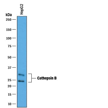
HAF019). A specific band was detected for Cathepsin B at approximately 25-30 kDa (as indicated). This experiment was conducted under reducing conditions and using
Immunoblot Buffer Group 1." alt="Western blot shows lysates of HepG2 human hepatocellular carcinoma cell line. PVDF membrane was probed with 0.25 µg/mL of Goat Anti-Human Cathepsin B Antigen Affinity-purified Polyclonal Antibody (Catalog # AF953) followed by HRP-conjugated Anti-Goat IgG Secondary Antibody (Catalog #
HAF019). A specific band was detected for Cathepsin B at approximately 25-30 kDa (as indicated). This experiment was conducted under reducing conditions and using
Immunoblot Buffer Group 1." class="big_thumb" />
CTS008) and counterstained with hematoxylin (blue). Lower panel shows a lack of labeling if primary antibodies are omitted and tissue is stained only with secondary antibody followed by incubation with detection reagents. View our protocol for
Chromogenic IHC Staining of Paraffin-embedded Tissue Sections." class="big_lightbox">
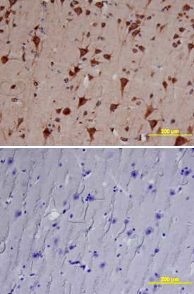
CTS008) and counterstained with hematoxylin (blue). Lower panel shows a lack of labeling if primary antibodies are omitted and tissue is stained only with secondary antibody followed by incubation with detection reagents. View our protocol for
Chromogenic IHC Staining of Paraffin-embedded Tissue Sections." class="big_thumb" />
HAF109). This experiment was conducted under reducing conditions and using the 12-230 kDa separation system." class="big_lightbox">
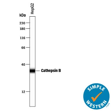
HAF109). This experiment was conducted under reducing conditions and using the 12-230 kDa separation system." class="big_thumb" />
HAF017). Specific bands were detected for Cathepsin B at approximately 25-45 kDa (as indicated) in the parental HeLa cell line, but is not detectable in knockout HeLa cell line. GAPDH (Catalog #
AF5718) is shown as a loading control. This experiment was conducted under reducing conditions and using
Immunoblot Buffer Group 1. New adjunct appears with knockout cell line." class="big_lightbox">
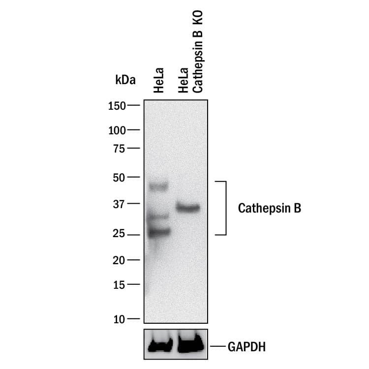
HAF017). Specific bands were detected for Cathepsin B at approximately 25-45 kDa (as indicated) in the parental HeLa cell line, but is not detectable in knockout HeLa cell line. GAPDH (Catalog #
AF5718) is shown as a loading control. This experiment was conducted under reducing conditions and using
Immunoblot Buffer Group 1. New adjunct appears with knockout cell line." class="big_thumb" />


953-CY) was serially diluted and captured by Mouse Anti-Human Pro Cathepsin B Monoclonal Antibody (Catalog #
MAB2176) coated on a Clear Polystyrene Microplate (Catalog #
DY990). Goat Anti-Human Cathepsin B Antigen Affinity-purified Polyclonal Antibody (Catalog # AF953) was biotinylated and incubated with the protein captured on the plate. Detection of the standard curve was achieved by incubating Streptavidin-HRP (Catalog #
DY998)" class="big_lightbox">

953-CY) was serially diluted and captured by Mouse Anti-Human Pro Cathepsin B Monoclonal Antibody (Catalog #
MAB2176) coated on a Clear Polystyrene Microplate (Catalog #
DY990). Goat Anti-Human Cathepsin B Antigen Affinity-purified Polyclonal Antibody (Catalog # AF953) was biotinylated and incubated with the protein captured on the plate. Detection of the standard curve was achieved by incubating Streptavidin-HRP (Catalog #
DY998)" class="big_thumb" />
 HAF019). A specific band was detected for Cathepsin B at approximately 25-30 kDa (as indicated). This experiment was conducted under reducing conditions and using Immunoblot Buffer Group 1." alt="Western blot shows lysates of HepG2 human hepatocellular carcinoma cell line. PVDF membrane was probed with 0.25 µg/mL of Goat Anti-Human Cathepsin B Antigen Affinity-purified Polyclonal Antibody (Catalog # AF953) followed by HRP-conjugated Anti-Goat IgG Secondary Antibody (Catalog # HAF019). A specific band was detected for Cathepsin B at approximately 25-30 kDa (as indicated). This experiment was conducted under reducing conditions and using Immunoblot Buffer Group 1." class="big_thumb" />


 CTS008) and counterstained with hematoxylin (blue). Lower panel shows a lack of labeling if primary antibodies are omitted and tissue is stained only with secondary antibody followed by incubation with detection reagents. View our protocol for
  HAF109). This experiment was conducted under reducing conditions and using the 12-230 kDa separation system." class="big_thumb" />
 HAF017). Specific bands were detected for Cathepsin B at approximately 25-45 kDa (as indicated) in the parental HeLa cell line, but is not detectable in knockout HeLa cell line. GAPDH (Catalog #
  NL001) and counterstained with DAPI (blue). Specific staining was localized to cell surface. View our protocol for


 953-CY) was serially diluted and captured by Mouse Anti-Human Pro Cathepsin B Monoclonal Antibody (Catalog #
![N/A Cathepsin B [HRP]](http://images.novusbio.com/fullsize/elisa/DATA_Cathepsin_B_DCATB0_ELISA_608.jpg "N/A Cathepsin B [HRP]")
![N/A Cathepsin B [HRP]](http://images.novusbio.com/fullsize/elisa/DATA_Cathepsin_B_DCATB0_ELISA_609.jpg)
![N/A Cathepsin B [HRP]](http://images.novusbio.com/fullsize/elisa/Cathepsin_B_DCATB0_ELISA_50.jpg)
![N/A Cathepsin B [Biotin]](http://images.novusbio.com/fullsize/elisa/DATA_Cathepsin_B_DY2176_ELISA_1778.jpg "N/A Cathepsin B [Biotin]")

 953-CY) was serially diluted and captured by Mouse Anti-Human Pro Cathepsin B Monoclonal Antibody (Catalog # MAB2176) coated on a Clear Polystyrene Microplate (Catalog #
 ![Immunohistochemistry-Paraffin: Cathepsin B Antibody [NBP1-86048] - Staining in human thyroid gland and skeletal muscle tissues . Corresponding CTSB RNA-seq data are presented for the same tissues.](http://images.novusbio.com/fullsize/Cathepsin-B-Antibody-Immunohistochemistry-Paraffin-NBP1-86048-img0019.jpg "<b>Orthogonal Strategies Validation. </b>Immunohistochemistry-Paraffin: Cathepsin B Antibody [NBP1-86048] - Staining in human thyroid gland and skeletal muscle tissues . Corresponding CTSB RNA-seq data are presented for the same tissues.")
![<b>Orthogonal Strategies Validation. </b>Immunocytochemistry/Immunofluorescence: Cathepsin B Antibody [NBP1-86048] - Staining of human cell line A-431 shows localization to nucleoli & vesicles. Antibody staining is shown in green.](http://images.novusbio.com/fullsize/Cathepsin-B-Antibody-Immunocytochemistry-Immunofluorescence-NBP1-86048-img0010.jpg)
![Immunohistochemistry-Paraffin: Cathepsin B Antibody [NBP1-86048] - Staining of human thyroid gland shows moderate granular cytoplasmic positivity in glandular cells.](http://images.novusbio.com/fullsize/Cathepsin-B-Antibody-Immunohistochemistry-Paraffin-NBP1-86048-img0018.jpg)
![Immunohistochemistry-Paraffin: Cathepsin B Antibody [NBP1-86048] - Staining of human cerebral cortex shows granular cytoplasmic positivity in neuronal cells.](http://images.novusbio.com/fullsize/Cathepsin-B-Antibody-Immunohistochemistry-Paraffin-NBP1-86048-img0015.jpg)
![Immunohistochemistry-Paraffin: Cathepsin B Antibody [NBP1-86048] - Staining of human lung shows cytoplasmic positivity in macrophages.](http://images.novusbio.com/fullsize/Cathepsin-B-Antibody-Immunohistochemistry-Paraffin-NBP1-86048-img0016.jpg)
![Immunohistochemistry-Paraffin: Cathepsin B Antibody [NBP1-86048] - Staining of human skeletal muscle shows no positivity in myocytes as expected.](http://images.novusbio.com/fullsize/Cathepsin-B-Antibody-Immunohistochemistry-Paraffin-NBP1-86048-img0017.jpg)
![Western Blot: Cathepsin B Antibody (6I5D7) [NBP3-15614] - Western blot analysis of extracts of HepG2 cells, using Cathepsin B antibody (NBP3-15614) at 1:1000 dilution. Secondary antibody: HRP Goat Anti-Rabbit IgG (H+L) at 1:10000 dilution. Lysates/proteins: 25ug per lane. Blocking buffer: 3% nonfat dry milk in TBST. Detection: ECL Basic Kit. Exposure time: 1min.](http://images.novusbio.com/fullsize/Cathepsin-B-Antibody-6I5D7-Western-Blot-NBP3-15614-img0002.jpg "Western Blot: Cathepsin B Antibody (6I5D7) [NBP3-15614] - Western blot analysis of extracts of HepG2 cells, using Cathepsin B antibody (NBP3-15614) at 1:1000 dilution. Secondary antibody: HRP Goat Anti-Rabbit IgG (H+L) at 1:10000 dilution. Lysates/proteins: 25ug per lane. Blocking buffer: 3% nonfat dry milk in TBST. Detection: ECL Basic Kit. Exposure time: 1min.")
![Immunohistochemistry-Paraffin: Cathepsin B Antibody (6I5D7) [NBP3-15614] - Immunohistochemistry of paraffin-embedded human lung cancer using Cathepsin B Rabbit mAb (NBP3-15614) at dilution of 1:100 (40x lens).Perform microwave antigen retrieval with 10 mM PBS buffer pH 7.2 before commencing with IHC staining protocol.](http://images.novusbio.com/fullsize/Cathepsin-B-Antibody-6I5D7-Immunohistochemistry-Paraffin-NBP3-15614-img0001.jpg)
![Immunohistochemistry-Paraffin: Cathepsin B Antibody (010) [NBP2-90402] - Staining of mouse CTSB in mouse kidney with rabbit monoclonal antibody (1:200).](http://images.novusbio.com/fullsize/Cathepsin-B-Antibody-010-Immunohistochemistry-Paraffin-NBP2-90402-img0002.jpg "Immunohistochemistry-Paraffin: Cathepsin B Antibody (010) [NBP2-90402] - Staining of mouse CTSB in mouse kidney with rabbit monoclonal antibody (1:200).")
![Immunohistochemistry-Paraffin: Cathepsin B Antibody (010) [NBP2-90402] - Staining of mouse CTSB in mouse liver with rabbit monoclonal antibody (1:200).](http://images.novusbio.com/fullsize/Cathepsin-B-Antibody-010-Immunohistochemistry-Paraffin-NBP2-90402-img0001.jpg)
![ELISA: Mouse Cathepsin B ELISA Kit (Colorimetric) [NBP2-67254] - Samples were spiked with high concentrations of Mouse Cathepsin B and diluted with Reference Standard & Sample Diluent to produce samples with values within the range of the assay.](http://images.novusbio.com/fullsize/nbp2-67254_mouse-cathepsin-b-elisa-kit-colorimetric-132202416143031.jpg "ELISA: Mouse Cathepsin B ELISA Kit (Colorimetric) [NBP2-67254] - Samples were spiked with high concentrations of Mouse Cathepsin B and diluted with Reference Standard & Sample Diluent to produce samples with values within the range of the assay.")
![ELISA: Mouse Cathepsin B ELISA Kit (Colorimetric) [NBP2-67254] - Standard Curve Reference](http://images.novusbio.com/fullsize/nbp2-67254_mouse-cathepsin-b-elisa-kit-colorimetric-43202495020.jpg)


