HAF109). For additional reference, recombinant human beta -catenin (1 ng) was included. A specific band for beta-Catenin was detected at approximately 95 kDa (as indicated). This experiment was conducted under reducing conditions and using
Immunoblot Buffer Group 1." class="big_lightbox">

HAF109). For additional reference, recombinant human beta -catenin (1 ng) was included. A specific band for beta-Catenin was detected at approximately 95 kDa (as indicated). This experiment was conducted under reducing conditions and using
Immunoblot Buffer Group 1." alt="Western blot shows lysates of HeLa human cervical epithelial carcinoma cell line, C6 rat glioma cell line, and NIH-3T3 mouse embryonic fibroblast cell line. PVDF membrane was probed with 1 µg/mL Goat Anti-Human/Mouse/Rat beta -Catenin Antigen Affinity-purified Polyclonal Antibody (Catalog # AF1329) followed by HRP-conjugated Anti-Goat IgG Secondary Antibody (Catalog #
HAF109). For additional reference, recombinant human beta -catenin (1 ng) was included. A specific band for beta-Catenin was detected at approximately 95 kDa (as indicated). This experiment was conducted under reducing conditions and using
Immunoblot Buffer Group 1." class="big_thumb" />
AB-108-C) for 15 minutes in an ultrasonic bath, followed by Biotinylated Anti-Goat IgG Secondary Antibody (Catalog #
BAF109). Immuno-complexes were captured using 50 µL of MagCellect Streptavidin Ferrofluid (Catalog #
MAG999) and DNA was purified using chelating resin solution. The
SU(Z)12promoter was detected by standard PCR." class="big_lightbox">
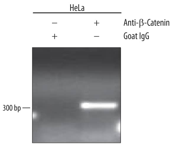
AB-108-C) for 15 minutes in an ultrasonic bath, followed by Biotinylated Anti-Goat IgG Secondary Antibody (Catalog #
BAF109). Immuno-complexes were captured using 50 µL of MagCellect Streptavidin Ferrofluid (Catalog #
MAG999) and DNA was purified using chelating resin solution. The
SU(Z)12promoter was detected by standard PCR." class="big_thumb" />
CTS008) and counterstained with hematoxylin (blue). Lower panel shows a lack of labeling if primary antibodies are omitted and tissue is stained only with secondary antibody followed by incubation with detection reagents. View our protocol for
Chromogenic IHC Staining of Paraffin-embedded Tissue Sections." class="big_lightbox">
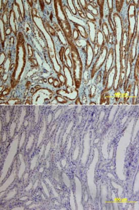
CTS008) and counterstained with hematoxylin (blue). Lower panel shows a lack of labeling if primary antibodies are omitted and tissue is stained only with secondary antibody followed by incubation with detection reagents. View our protocol for
Chromogenic IHC Staining of Paraffin-embedded Tissue Sections." class="big_thumb" />
AB-108-C, open histogram), followed by Allophycocyanin-conjugated Anti-Goat IgG Secondary Antibody (Catalog #
F0108). To facilitate intracellular staining, cells were fixed with paraformaldehyde and permeabilized with saponin." class="big_lightbox">

AB-108-C, open histogram), followed by Allophycocyanin-conjugated Anti-Goat IgG Secondary Antibody (Catalog #
F0108). To facilitate intracellular staining, cells were fixed with paraformaldehyde and permeabilized with saponin." class="big_thumb" />
HAF109). This experiment was conducted under reducing conditions and using the 12-230 kDa separation system. Non-specific interaction with the 230 kDa Simple Western standard may be seen with this antibody." class="big_lightbox">
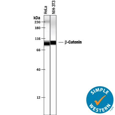
HAF109). This experiment was conducted under reducing conditions and using the 12-230 kDa separation system. Non-specific interaction with the 230 kDa Simple Western standard may be seen with this antibody." class="big_thumb" />
 HAF109). For additional reference, recombinant human beta -catenin (1 ng) was included. A specific band for beta-Catenin was detected at approximately 95 kDa (as indicated). This experiment was conducted under reducing conditions and using Immunoblot Buffer Group 1." alt="Western blot shows lysates of HeLa human cervical epithelial carcinoma cell line, C6 rat glioma cell line, and NIH-3T3 mouse embryonic fibroblast cell line. PVDF membrane was probed with 1 µg/mL Goat Anti-Human/Mouse/Rat beta -Catenin Antigen Affinity-purified Polyclonal Antibody (Catalog # AF1329) followed by HRP-conjugated Anti-Goat IgG Secondary Antibody (Catalog # HAF109). For additional reference, recombinant human beta -catenin (1 ng) was included. A specific band for beta-Catenin was detected at approximately 95 kDa (as indicated). This experiment was conducted under reducing conditions and using Immunoblot Buffer Group 1." class="big_thumb" />
 NL007) and counter-stained with DAPI (blue). View our protocol for Fluorescent ICC Staining of Cells on Coverslips." alt=" beta -Catenin was detected in immersion fixed SW480 human colorectal adenocarcinoma cell line using 10 µg/mL Mouse Anti-Human beta -Catenin Monoclonal Antibody (Catalog # MAB13291) for 3 hours at room temperature. Cells were stained with the NorthernLights™ 557-conjugated Anti-Mouse IgG Secondary Antibody (red; Catalog # NL007) and counter-stained with DAPI (blue). View our protocol for Fluorescent ICC Staining of Cells on Coverslips." class="big_thumb" />
 MAB1329) at 15 µg/mL at 37° Celsius for 2 minutes. Before incubation with the primary antibody, tissue underwent an all-in-one dewaxing and antigen retrieval preprocessing using PreTreatment Module (PT Module) and Dewax and HIER Buffer H (pH 9). Tissue was stained using the Alexa Fluor™ 647 Goat anti-Mouse IgG Secondary Antibody at 1:200 at 37 ° Celsius for 2 minutes. (Yellow; Lunaphore Catalog # DR647MS) and counterstained with DAPI (blue; Lunaphore Catalog # DR100). Specific staining was localized to the cytoplasm and cell membrane. Protocol available in COMET™ Panel Builder." alt="beta-Catenin was detected in immersion fixed paraffin-embedded sections of human colon cancer using Mouse Anti-Human beta-Catenin Monoclonal Antibody (Catalog # MAB1329) at 15 µg/mL at 37° Celsius for 2 minutes. Before incubation with the primary antibody, tissue underwent an all-in-one dewaxing and antigen retrieval preprocessing using PreTreatment Module (PT Module) and Dewax and HIER Buffer H (pH 9). Tissue was stained using the Alexa Fluor™ 647 Goat anti-Mouse IgG Secondary Antibody at 1:200 at 37 ° Celsius for 2 minutes. (Yellow; Lunaphore Catalog # DR647MS) and counterstained with DAPI (blue; Lunaphore Catalog # DR100). Specific staining was localized to the cytoplasm and cell membrane. Protocol available in COMET™ Panel Builder." class="big_thumb" />


 AB-108-C) for 15 minutes in an ultrasonic bath, followed by Biotinylated Anti-Goat IgG Secondary Antibody (Catalog #
  NL001) and counterstained with DAPI (blue). Specific staining was localized to cytoplasm and nuclei. View our protocol for
  CTS008) and counterstained with hematoxylin (blue). Lower panel shows a lack of labeling if primary antibodies are omitted and tissue is stained only with secondary antibody followed by incubation with detection reagents. View our protocol for
  CTS008) and counterstained with hematoxylin (blue). Specific labeling was localized to epithelial cells in collecting tubules in the medulla. View our protocol for
  AB-108-C, open histogram), followed by Allophycocyanin-conjugated Anti-Goat IgG Secondary Antibody (Catalog #
  HAF109). This experiment was conducted under reducing conditions and using the 12-230 kDa separation system. Non-specific interaction with the 230 kDa Simple Western standard may be seen with this antibody." class="big_thumb" />


![Western Blot: beta-Catenin Antibody (12F7) [NBP1-54467] - Total protein from human HepG2 and HeLa cells, rat PC12 cells and mouse 3T3 cells was separated on a 7.5% gel by SDS-PAGE, transferred to PVDF membrane and blocked in 5% non-fat milk in TBST. The membrane was probed with 1.0 ug/ml anti-Beta catenin in 1% non-fat milk in TBST and detected with an anti-mouse HRP secondary antibody using chemiluminescence.](http://images.novusbio.com/fullsize/beta-Catenin-Antibody-12F7-Western-Blot-NBP1-54467-img0008.jpg "<b>Biological Strategies Validation. </b>Western Blot: beta-Catenin Antibody (12F7) [NBP1-54467] - Total protein from human HepG2 and HeLa cells, rat PC12 cells and mouse 3T3 cells was separated on a 7.5% gel by SDS-PAGE, transferred to PVDF membrane and blocked in 5% non-fat milk in TBST. The membrane was probed with 1.0 ug/ml anti-Beta catenin in 1% non-fat milk in TBST and detected with an anti-mouse HRP secondary antibody using chemiluminescence.")

![Immunocytochemistry/Immunofluorescence: beta-Catenin Antibody (12F7) [NBP1-54467] - The beta- Catenin antibody was tested in MCF-7 cells against Dylight 488 (Green). Actin and nuclei were counterstained against Phalloidin 550 (Red) and DAPI (Blue).](http://images.novusbio.com/fullsize/beta-Catenin-Antibody-12F7-Immunocytochemistry-Immunofluorescence-NBP1-54467-img0004.jpg)
![Immunohistochemistry: beta-Catenin Antibody (12F7) [NBP1-54467] - Beta-catening (green) was detected in human skin (psoriasis) using beta-catenin-FITC antibody (1:40) in PBS for 1 hour. Nuclei were stained with Dapi (blue). Image from verified customer review. Image using the FITC format of this antibody.](http://images.novusbio.com/fullsize/beta-Catenin-Antibody-12F7-Immunohistochemistry-NBP1-54467-img0013.jpg)
![Flow Cytometry: beta-Catenin Antibody (12F7) [NBP1-54467] - An intracellular stain was performed on SK-MEL-28 cells with beta-Catenin Antibody [12F7] NBP1-54467B (blue) and a matched isotype control (orange). Both antibodies were conjugated to Biotin. Cells were fixed with 4% PFA and then permeabilized with 0.1% saponin. Cells were incubated in an antibody dilution of 2.5 ug/mL for 30 minutes at room temperature, followed by Streptavidin - R-Phycoerythrin Protein (2012-1000, Novus Biologicals).](http://images.novusbio.com/fullsize/beta-Catenin-Antibody-12F7-Flow-Cytometry-NBP1-54467-img0012.jpg)
![Western Blot: beta-Catenin Antibody (12F7) [NBP1-54467] - Analysis of beta Catenin expression in 1) HepG2, 2) MCF7, and 3) Cos7 whole cell lysates using NBP1-54467.](http://images.novusbio.com/fullsize/beta-Catenin-Antibody-12F7-Western-Blot-NBP1-54467-img0001.jpg)
![Western Blot: beta-Catenin Antibody (12F7) [NBP1-54467] - Analysis of beta- Catenin in embryonic lung (lane 1) and embryonic limb (lane 2) lysates using anti-beta- Catenin antibody. Each lane was loaded with 5ug of protein sample. Image from verified customer review.](http://images.novusbio.com/fullsize/beta-Catenin-Antibody-12F7-Western-Blot-NBP1-54467-img0005.jpg)
![Immunohistochemistry-Paraffin: beta-Catenin Antibody (12F7) [NBP1-54467] - Analysis of beta Catenin in mouse intestine using DAB with hematoxylin counterstain.](http://images.novusbio.com/fullsize/beta-Catenin-Antibody-12F7-Immunohistochemistry-Paraffin-NBP1-54467-img0002.jpg)
![Immunohistochemistry-Paraffin: beta-Catenin Antibody (12F7) [NBP1-54467] - Analysis of beta- Catenin in mouse intestine using DAB with hematoxylin counterstain.](http://images.novusbio.com/fullsize/beta-Catenin-Antibody-12F7-Immunohistochemistry-Paraffin-NBP1-54467-img0003.jpg)
![Flow (Intracellular): beta-Catenin Antibody (12F7) [NBP1-54467] - An intracellular stain was performed on HeLa cells with beta-Catenin Antibody (12F7) NBP1-54467AF647 (blue) and a matched isotype control (orange). Cells were fixed with 4% PFA and then permeabilized with 0.1% saponin. Cells were incubated in an antibody dilution of 5 ug/mL for 30 minutes at room temperature. Both antibodies were conjugated to Alexa Fluor 647.](http://images.novusbio.com/fullsize/beta-Catenin-Antibody-12F7-Flow-Intracellular-NBP1-54467-img0009.jpg)
![Simple Western: beta-Catenin Antibody (12F7) [NBP1-54467] - Simple Western lane view shows a specific band for Beta- Catenin in 0.5 mg/ml of HepG2 lysate. This experiment was performed under reducing conditions using the 12-230 kDa separation system.](http://images.novusbio.com/fullsize/beta-Catenin-Antibody-12F7-Simple-Western-NBP1-54467-img0006.jpg)


![Western Blot: beta-Catenin Antibody [NBP1-32239] - Various whole cell extracts (30 ug) were separated by 7.5% SDS-PAGE, and the membrane was blotted with beta Catenin antibody [N1N2-2], N-term diluted at 1:3000.](http://images.novusbio.com/fullsize/beta-Catenin-Antibody-Western-Blot-NBP1-32239-img0033.jpg "Western Blot: beta-Catenin Antibody [NBP1-32239] - Various whole cell extracts (30 ug) were separated by 7.5% SDS-PAGE, and the membrane was blotted with beta Catenin antibody [N1N2-2], N-term diluted at 1:3000.")
![Immunocytochemistry/Immunofluorescence: beta-Catenin Antibody [NBP1-32239] - Paraformaldehyde-fixed A431, using beta- Catenin antibody (Green) at 1:200 dilution. Alpha-tubulin filaments were labeled with an alpha Tubulin antibody (Red) at 1:2000.](http://images.novusbio.com/fullsize/beta-Catenin-Antibody-Immunocytochemistry-Immunofluorescence-NBP1-32239-img0017.jpg)

![Chromatin Immunoprecipitation (ChIP): beta-Catenin Antibody [NBP1-32239] - Cross-linked ChIP was performed with HCT116 chromatin extract and 5 ug of either control rabbit IgG or anti-beta Catenin antibody. The precipitated DNA was detected by PCR with primer set targeting to c-Myc promoter.](http://images.novusbio.com/fullsize/beta-Catenin-Antibody-Chromatin-Immunoprecipitation-ChIP-NBP1-32239-img0024.jpg)
![<b>Biological Strategies Validation. </b>Western Blot: beta-Catenin Antibody [NBP1-32239] - Western blot for beta-catenin in HEK 293 cells treated with LiCl (10mM) for 1 hr. Image from verified customer review.](http://images.novusbio.com/fullsize/beta-Catenin-Antibody-Western-Blot-NBP1-32239-img0008.jpg)
![Western Blot: beta-Catenin Antibody [NBP1-32239] - Sample (50 ug of whole cell lysate) A: mouse brain 7.5% SDS PAGE diluted at 1:1000](http://images.novusbio.com/fullsize/beta-Catenin-Antibody-Western-Blot-NBP1-32239-img0013.jpg)
![Western Blot: beta-Catenin Antibody [NBP1-32239] - A. 30 ug PC-12 whole cell lysate/extract 7.5 % SDS-PAGEbeta Catenin antibody [N1N2-2], N-term dilution: 1:1000](http://images.novusbio.com/fullsize/beta-Catenin-Antibody-Western-Blot-NBP1-32239-img0014.jpg)
![<b>Genetic Strategies Validation. </b>Western Blot: beta-Catenin Antibody [NBP1-32239] - Non-transfected (-) and transfected (+) HeLa whole cell extracts (30 ug) were separated by 7.5% SDS-PAGE, and the membrane was blotted with beta Catenin antibody [N1N2-2], N-term.](http://images.novusbio.com/fullsize/beta-Catenin-Antibody-Western-Blot-NBP1-32239-img0026.jpg)
![Immunocytochemistry/Immunofluorescence: beta-Catenin Antibody [NBP1-32239] - HeLa cells were fixed in 4% paraformaldehyde at RT for 15 min. Green: beta Catenin protein stained by beta Catenin antibody [N1N2-2], N-term diluted at 1:500. Blue: Hoechst 33342 staining.](http://images.novusbio.com/fullsize/beta-Catenin-Antibody-Immunocytochemistry-Immunofluorescence-NBP1-32239-img0032.jpg)
![Immunohistochemistry-Paraffin: beta-Catenin Antibody [NBP1-32239] - Paraffin-embedded mouse urinary bladder diluted at 1:500.](http://images.novusbio.com/fullsize/beta-Catenin-Antibody-Immunohistochemistry-Paraffin-NBP1-32239-img0019.jpg)
![Immunohistochemistry-Paraffin: beta-Catenin Antibody [NBP1-32239] - Paraffin-embedded mouse skin dilution: 1:500.](http://images.novusbio.com/fullsize/beta-Catenin-Antibody-Immunohistochemistry-Paraffin-NBP1-32239-img0020.jpg)
![Immunohistochemistry-Paraffin: beta-Catenin Antibody [NBP1-32239] - Paraffin-embedded mouse colon dilution: 1:500.](http://images.novusbio.com/fullsize/beta-Catenin-Antibody-Immunohistochemistry-Paraffin-NBP1-32239-img0021.jpg)
![Immunohistochemistry-Paraffin: beta-Catenin Antibody [NBP1-32239] - Paraffin-embedded mouse duodenum. beta Catenin antibody [N1N2-2], N-term diluted at 1:500.](http://images.novusbio.com/fullsize/beta-Catenin-Antibody-Immunohistochemistry-Paraffin-NBP1-32239-img0036.jpg)
![Immunohistochemistry-Paraffin: beta-Catenin Antibody [NBP1-32239] - Paraffin-embedded human esophagus. beta Catenin antibody [N1N2-2], N-term diluted at 1:500.](http://images.novusbio.com/fullsize/beta-Catenin-Antibody-Immunohistochemistry-Paraffin-NBP1-32239-img0037.jpg)
![Immunohistochemistry-Paraffin: beta-Catenin Antibody [NBP1-32239] - Paraffin-embedded human cervix. beta Catenin antibody [N1N2-2], N-term diluted at 1:500.](http://images.novusbio.com/fullsize/beta-Catenin-Antibody-Immunohistochemistry-Paraffin-NBP1-32239-img0038.jpg)
![Immunohistochemistry-Paraffin: beta-Catenin Antibody [NBP1-32239] - Paraffin-embedded mouse duodenum. beta Catenin antibody [N1N2-2], N-term diluted at 1:500.](http://images.novusbio.com/fullsize/beta-Catenin-Antibody-Immunohistochemistry-Paraffin-NBP1-32239-img0039.jpg)
![Immunohistochemistry-Paraffin: beta-Catenin Antibody [NBP1-32239] - Paraffin-embedded rat colon. beta Catenin antibody [N1N2-2], N-term diluted at 1:500.](http://images.novusbio.com/fullsize/beta-Catenin-Antibody-Immunohistochemistry-Paraffin-NBP1-32239-img0040.jpg)
![Immunohistochemistry-Paraffin: beta-Catenin Antibody [NBP1-32239] - Paraffin-embedded rat duodenum. beta Catenin antibody [N1N2-2], N-term diluted at 1:500.](http://images.novusbio.com/fullsize/beta-Catenin-Antibody-Immunohistochemistry-Paraffin-NBP1-32239-img0041.jpg)
![Immunohistochemistry-Paraffin: beta-Catenin Antibody [NBP1-32239] - Paraffin-embedded mouse intestine. beta Catenin antibody [N1N2-2], N-term diluted at 1:500.](http://images.novusbio.com/fullsize/beta-Catenin-Antibody-Immunohistochemistry-Paraffin-NBP1-32239-img0042.jpg)
![Immunocytochemistry/ Immunofluorescence: beta-Catenin Antibody [NBP1-32239] - P-cadherin is co-localized with other junctional proteins at the RPE cell border in mice.Immunofluorescence of mouse RPE flat-mounts. Double staining: P-cadherin (red; A, E, I) & either ZO-1 (green; B), beta -catenin (green; F), or F-actin (green; J), with nuclear stain by DAPI (blue; C, G, K). Merged images (D, H, L) show the co-localization of P-cadherin with ZO-1 (tight junction), beta -catenin (adherens junction), & F-actin (adherens junction) at the cell-cell border. Image collected & cropped by CiteAb from the following publication (//pubmed.ncbi.nlm.nih.gov/29338041), licensed under a CC-BY license. Not internally tested by Novus Biologicals.](http://images.novusbio.com/fullsize/nbp1-32239_rabbit-polyclonal-beta-catenin-antibody-310202415171344.jpg)
![Immunocytochemistry/ Immunofluorescence: beta-Catenin Antibody [NBP1-32239] - WNT10A/ beta -catenin signalling is required for region-specific differentiation.(a–d) Filiform papillae are present in Wnt10a−/− & inducible beta -catenin mutant dorsal tongue (yellow arrows), but horny structures & expression of hard keratins (in situ hybridization, purple signals) are decreased (red arrows). (e–l) Epithelial deletion of Wnt10a (e–f″,i,j) or beta -catenin (g–h″,k,l) induced from P25, P110 or P15 as indicated causes decreased expression of nuclear beta -catenin, LEF1 & HOXC13 (white arrows, LEF1+ proliferating cells; yellow arrows, HOXC13+ differentiating cells). (m) qPCR shows significantly decreased Hoxc13 levels in Wnt10a & beta -catenin mutant tongue epithelium. (n–r) IF & qPCR reveal reduced levels of KRT9 protein (n–q) & mRNA (r) in Wnt10a−/− & inducible beta -catenin mutant footpad epidermis. (s–v″) Co-IF for KRT9 & KRT10 in plantar epidermis from patients homozygous for WNT10A c.756+1G>A (s–t″) or WNT10A c.391G>A (u–v″) compared with similarly aged sex-matched controls. For qPCR, RNA levels were quantified in six control & six mutant (P40) or four control & four mutant (P20-100) samples with three technical replicates for each, & normalized to beta -actin mRNA. Significance was calculated with two-tailed Student's t-test. Error bars indicate s.e.m. Scale bar, 25 μm (e–l) or 50 μm (a–d,n–q,s–v″). Image collected & cropped by CiteAb from the following publication (//www.nature.com/articles/ncomms15397), licensed under a CC-BY license. Not internally tested by Novus Biologicals.](http://images.novusbio.com/fullsize/nbp1-32239_rabbit-polyclonal-beta-catenin-antibody-31020241617146.jpg)
![Immunocytochemistry/ Immunofluorescence: beta-Catenin Antibody [NBP1-32239] - WNT10A/ beta -catenin signalling is required for region-specific differentiation.(a–d) Filiform papillae are present in Wnt10a−/− & inducible beta -catenin mutant dorsal tongue (yellow arrows), but horny structures & expression of hard keratins (in situ hybridization, purple signals) are decreased (red arrows). (e–l) Epithelial deletion of Wnt10a (e–f″,i,j) or beta -catenin (g–h″,k,l) induced from P25, P110 or P15 as indicated causes decreased expression of nuclear beta -catenin, LEF1 & HOXC13 (white arrows, LEF1+ proliferating cells; yellow arrows, HOXC13+ differentiating cells). (m) qPCR shows significantly decreased Hoxc13 levels in Wnt10a & beta -catenin mutant tongue epithelium. (n–r) IF & qPCR reveal reduced levels of KRT9 protein (n–q) & mRNA (r) in Wnt10a−/− & inducible beta -catenin mutant footpad epidermis. (s–v″) Co-IF for KRT9 & KRT10 in plantar epidermis from patients homozygous for WNT10A c.756+1G>A (s–t″) or WNT10A c.391G>A (u–v″) compared with similarly aged sex-matched controls. For qPCR, RNA levels were quantified in six control & six mutant (P40) or four control & four mutant (P20-100) samples with three technical replicates for each, & normalized to beta -actin mRNA. Significance was calculated with two-tailed Student's t-test. Error bars indicate s.e.m. Scale bar, 25 μm (e–l) or 50 μm (a–d,n–q,s–v″). Image collected & cropped by CiteAb from the following publication (//www.nature.com/articles/ncomms15397), licensed under a CC-BY license. Not internally tested by Novus Biologicals.](http://images.novusbio.com/fullsize/nbp1-32239_rabbit-polyclonal-beta-catenin-antibody-310202416171420.jpg)
![Immunocytochemistry/ Immunofluorescence: beta-Catenin Antibody [NBP1-32239] - WNT10A/ beta -catenin signalling is required for region-specific differentiation.(a–d) Filiform papillae are present in Wnt10a−/− & inducible beta -catenin mutant dorsal tongue (yellow arrows), but horny structures & expression of hard keratins (in situ hybridization, purple signals) are decreased (red arrows). (e–l) Epithelial deletion of Wnt10a (e–f″,i,j) or beta -catenin (g–h″,k,l) induced from P25, P110 or P15 as indicated causes decreased expression of nuclear beta -catenin, LEF1 & HOXC13 (white arrows, LEF1+ proliferating cells; yellow arrows, HOXC13+ differentiating cells). (m) qPCR shows significantly decreased Hoxc13 levels in Wnt10a & beta -catenin mutant tongue epithelium. (n–r) IF & qPCR reveal reduced levels of KRT9 protein (n–q) & mRNA (r) in Wnt10a−/− & inducible beta -catenin mutant footpad epidermis. (s–v″) Co-IF for KRT9 & KRT10 in plantar epidermis from patients homozygous for WNT10A c.756+1G>A (s–t″) or WNT10A c.391G>A (u–v″) compared with similarly aged sex-matched controls. For qPCR, RNA levels were quantified in six control & six mutant (P40) or four control & four mutant (P20-100) samples with three technical replicates for each, & normalized to beta -actin mRNA. Significance was calculated with two-tailed Student's t-test. Error bars indicate s.e.m. Scale bar, 25 μm (e–l) or 50 μm (a–d,n–q,s–v″). Image collected & cropped by CiteAb from the following publication (//www.nature.com/articles/ncomms15397), licensed under a CC-BY license. Not internally tested by Novus Biologicals.](http://images.novusbio.com/fullsize/nbp1-32239_rabbit-polyclonal-beta-catenin-antibody-31020241618265.jpg)
![Immunocytochemistry/ Immunofluorescence: beta-Catenin Antibody [NBP1-32239] - WNT10A/ beta -catenin signalling is required for region-specific differentiation.(a–d) Filiform papillae are present in Wnt10a−/− & inducible beta -catenin mutant dorsal tongue (yellow arrows), but horny structures & expression of hard keratins (in situ hybridization, purple signals) are decreased (red arrows). (e–l) Epithelial deletion of Wnt10a (e–f″,i,j) or beta -catenin (g–h″,k,l) induced from P25, P110 or P15 as indicated causes decreased expression of nuclear beta -catenin, LEF1 & HOXC13 (white arrows, LEF1+ proliferating cells; yellow arrows, HOXC13+ differentiating cells). (m) qPCR shows significantly decreased Hoxc13 levels in Wnt10a & beta -catenin mutant tongue epithelium. (n–r) IF & qPCR reveal reduced levels of KRT9 protein (n–q) & mRNA (r) in Wnt10a−/− & inducible beta -catenin mutant footpad epidermis. (s–v″) Co-IF for KRT9 & KRT10 in plantar epidermis from patients homozygous for WNT10A c.756+1G>A (s–t″) or WNT10A c.391G>A (u–v″) compared with similarly aged sex-matched controls. For qPCR, RNA levels were quantified in six control & six mutant (P40) or four control & four mutant (P20-100) samples with three technical replicates for each, & normalized to beta -actin mRNA. Significance was calculated with two-tailed Student's t-test. Error bars indicate s.e.m. Scale bar, 25 μm (e–l) or 50 μm (a–d,n–q,s–v″). Image collected & cropped by CiteAb from the following publication (//www.nature.com/articles/ncomms15397), licensed under a CC-BY license. Not internally tested by Novus Biologicals.](http://images.novusbio.com/fullsize/nbp1-32239_rabbit-polyclonal-beta-catenin-antibody-31020241618934.jpg)
![Immunocytochemistry/ Immunofluorescence: beta-Catenin Antibody [NBP1-32239] - Oxidative stress-induced dissociation of adherens junctions results in nuclear translocation of beta -catenin & an increase of EMT-related factors in mouse RPE.(A) Immunofluorescence of mouse RPE flat-mounts. Mice were injected with NaIO3 (15 mg/kg body weight) on Day 0, & the localization of beta -catenin (green) & P-cadherin (red) was analyzed along with nuclear stain by DAPI (blue) on Days 0 (a-c), 1 (d-f), 3 (g-i) & 7 (j-l). Double staining: beta -catenin (a, d, g, j), P-cadherin (b, e, h, k), & merged images with DAPI (c, f, i, l). The localization of beta -catenin & P-cadherin at the cell-cell border was significantly disrupted, & instead prominently detected on/in the nucleus on Day 3. (B) Immunofluorescence of mouse retinal sections with a focus on the RPE nuclei. Mice were injected with NaIO3 (15 mg/kg body weight) on Day 0, & the localization of beta -catenin (green) & P-cadherin (red) was analyzed along with nuclear stain by DAPI (blue) on Days 0 (m-o) & 3 (two representative nuclei; p-r & s-u). Double staining: beta -catenin (m, p, s), P-cadherin (n, q, t), & merged images with DAPI (o, r, u). On Day 3, beta -catenin was detected in the nuclei of mouse RPE. (C) Western blot analyses of mouse RPE proteins. Mice were injected with NaIO3 (15 mg/kg body weight) on Day 0, & RPE protein lysates were prepared on Days 0, 1, 3, & 7. The protein levels were analyzed using Western blotting with antibodies against P-cadherin, beta -catenin, SNAI1 (Snail), vimentin, & control beta -actin. The protein levels of beta -catenin & SNAI1 increased similarly on Day 1 following oxidative stress. Image collected & cropped by CiteAb from the following publication (//pubmed.ncbi.nlm.nih.gov/29338041), licensed under a CC-BY license. Not internally tested by Novus Biologicals.](http://images.novusbio.com/fullsize/nbp1-32239_rabbit-polyclonal-beta-catenin-antibody-310202416165641.jpg)
![Western Blot: beta-Catenin Antibody [NBP1-32239] - Whole cell extract (30 ug) was separated by 7.5% SDS-PAGE, and the membrane was blotted with beta-Catenin antibody (NBP1-32239) diluted at 1:1000. The HRP-conjugated anti-rabbit IgG antibody was used to detect the primary antibody, and the signal was developed with Trident ECL plus-Enhanced.](http://images.novusbio.com/fullsize/nbp1-32239_rabbit-polyclonal-beta-catenin-antibody-91020242011415.jpg)
![Immunohistochemistry-Paraffin: beta-Catenin Antibody [NBP1-32239] - beta-Catenin antibody detects beta-Catenin protein at cell membrane by immunohistochemical analysis.Sample: Paraffin-embedded cat liver.beta-Catenin stained by beta-Catenin antibody (NBP1-32239) diluted at 1:500.Antigen Retrieval: Citrate buffer, pH 6.0, 15 min](http://images.novusbio.com/fullsize/nbp1-32239_rabbit-polyclonal-beta-catenin-antibody-91020242075974.jpg)
![Immunohistochemistry-Paraffin: beta-Catenin Antibody [NBP1-32239] - beta-Catenin antibody detects beta-Catenin protein at cell membrane by immunohistochemical analysis.Sample: Paraffin-embedded cat colon.beta-Catenin stained by beta-Catenin antibody (NBP1-32239) diluted at 1:500.Antigen Retrieval: Citrate buffer, pH 6.0, 15 min](http://images.novusbio.com/fullsize/nbp1-32239_rabbit-polyclonal-beta-catenin-antibody-910202420122025.jpg)
![Immunohistochemistry: beta-Catenin Antibody [NBP1-32239] - beta-Catenin antibody [N1N2-2], N-term detects Ctnnb1 protein on zebrafish by whole mount immunohistochemical analysis. Sample: 2 days-post-fertilization zebrafish embryo. beta-Catenin antibody [N1N2-2], N-term (NBP1-32239) dilution: 1:100.](http://images.novusbio.com/fullsize/nbp1-32239_rabbit-polyclonal-beta-catenin-antibody-910202420134025.jpg)
![Western Blot: beta-Catenin Antibody [NBP1-32239] - Various tissue extracts (30 ug) were separated by 7.5% SDS-PAGE, and the membrane was blotted with beta-Catenin antibody (NBP1-32239) diluted at 1:500. The HRP-conjugated anti-rabbit IgG antibody was used to detect the primary antibody.](http://images.novusbio.com/fullsize/nbp1-32239_rabbit-polyclonal-beta-catenin-antibody-910202420104461.jpg)
![Immunohistochemistry: beta-Catenin Antibody [NBP1-32239] - Immunohistochemical analysis of agarose-embedded zebrafish embryo, using beta-Catenin antibody [N1N2-2], N-term NBP1-32239) at 1:100. dilution. (This image was provided courtesy of the Schilling Lab at UC, Irvine.)](http://images.novusbio.com/fullsize/nbp1-32239_rabbit-polyclonal-beta-catenin-antibody-910202420115056.jpg)
![Immunohistochemistry: beta-Catenin Antibody [NBP1-32239] - beta-Catenin antibody [N1N2-2], N-term detects Ctnnb1 protein on zebrafish by whole mount immunohistochemical analysis. Sample: 1 day-post-fertilization zebrafish embryo. beta-Catenin antibody [N1N2-2], N-term (NBP1-32239) dilution: 1:100.](http://images.novusbio.com/fullsize/nbp1-32239_rabbit-polyclonal-beta-catenin-antibody-910202420564.jpg)
![Immunohistochemistry-Paraffin: beta-Catenin Antibody [NBP1-32239] - beta-Catenin antibody [N1N2-2], N-term detects beta-Catenin protein by immunohistochemical analysis.Sample: Paraffin-embedded rat tissues.beta-Catenin stained by beta-Catenin antibody [N1N2-2], N-term (NBP1-32239) diluted at 1:500.Antigen Retrieval: Citrate buffer, pH 6.0, 15 min](http://images.novusbio.com/fullsize/nbp1-32239_rabbit-polyclonal-beta-catenin-antibody-9102024202461.jpg)
![Immunohistochemistry-Paraffin: beta-Catenin Antibody [NBP1-32239] - beta-Catenin antibody [N1N2-2] detects beta-Catenin protein at cell membrane in mouse colon by immunohistochemical analysis. Sample: Paraffin-embedded mouse colon. Green: beta-Catenin antibody [N1N2-2] (NBP1-32239) diluted at 1:500.Red: alpha Tubulin antibody [GT114] diluted at 1:500. Blue: Hoechst 33342 staining.Antigen Retrieval: Citrate buffer, pH 6.0, 15 min](http://images.novusbio.com/fullsize/nbp1-32239_rabbit-polyclonal-beta-catenin-antibody-91020242001070.jpg)
![Immunohistochemistry: beta-Catenin Antibody [NBP1-32239] - beta-Catenin antibody [N1N2-2], N-term detects Ctnnb1 protein on zebrafish by whole mount immunohistochemical analysis. Sample: 2 days-post-fertilization zebrafish embryo. beta-Catenin antibody [N1N2-2], N-term (NBP1-32239) dilution: 1:100.](http://images.novusbio.com/fullsize/nbp1-32239_rabbit-polyclonal-beta-catenin-antibody-9102024202198.jpg)
![Immunocytochemistry/ Immunofluorescence: beta-Catenin Antibody [NBP1-32239] - beta-Catenin antibody detects beta-Catenin protein at cell membrane by immunofluorescent analysis.Sample: MDCK cells were fixed in 4% paraformaldehyde at RT for 15 min.Green: beta-Catenin stained by beta-Catenin antibody (NBP1-32239) diluted at 1:1000.](http://images.novusbio.com/fullsize/nbp1-32239_rabbit-polyclonal-beta-catenin-antibody-91020242010443.jpg)
![Immunohistochemistry: beta-Catenin Antibody [NBP1-32239] - Immunohistochemical analysis of paraffin-embedded zebrafish tissue, using beta-Catenin antibody [N1N2-2], N-term (NBP1-32239) at 1:300 dilution.](http://images.novusbio.com/fullsize/nbp1-32239_rabbit-polyclonal-beta-catenin-antibody-91020242075983.jpg)
![Immunohistochemistry-Paraffin: beta-Catenin Antibody [NBP1-32239] - beta-Catenin antibody [N1N2-2], N-term detects beta-Catenin protein by immunohistochemical analysis.Sample: Paraffin-embedded mouse tissues.beta-Catenin stained by beta-Catenin antibody [N1N2-2], N-term (NBP1-32239) diluted at 1:500.Antigen Retrieval: Citrate buffer, pH 6.0, 15 min](http://images.novusbio.com/fullsize/nbp1-32239_rabbit-polyclonal-beta-catenin-antibody-91020242075987.jpg)
![Western Blot: beta-Catenin Antibody [NBP1-32239] - Whole cell extract (30 ug) was separated by 7.5% SDS-PAGE, and the membrane was blotted with beta-Catenin antibody (NBP1-32239) diluted at 1:1000. The HRP-conjugated anti-rabbit IgG antibody was used to detect the primary antibody.](http://images.novusbio.com/fullsize/nbp1-32239_rabbit-polyclonal-beta-catenin-antibody-910202420131247.jpg)
![<b>Genetic Strategies Validation. </b>Western Blot: beta-Catenin Antibody [NBP1-32239] - Wild-type (WT) and beta Catenin knockout (KO) 293T cell extracts (9 ug) were separated by 7.5% SDS-PAGE, and the membrane was blotted with beta Catenin antibody [N1N2-2], N-term diluted at 1:1000. The HRP-conjugated anti-rabbit IgG antibody was used to detect the primary antibody.](http://images.novusbio.com/fullsize/nbp1-32239_rabbit-polyclonal-beta-catenin-antibody-1610202419405728.jpg)
![beta-Catenin antibody [N1N2-2] detects beta-Catenin protein at cell membrane in mouse colon by immunohistochemical analysis. Sample: Paraffin-embedded mouse colon. Green: beta-Catenin antibody [N1N2-2] (NBP1-32239) diluted at 1:500.Red: alpha Tubulin antibody [GT114] diluted at 1:500. Blue: Hoechst 33342 staining.Antigen Retrieval: Citrate buffer, pH 6.0, 15 min](http://images.novusbio.com/fullsize/nbp1-32239_rabbit-polyclonal-beta-catenin-antibody-immunohistochemistry-paraffin-1411202518321620.jpg)
![beta-Catenin antibody [N1N2-2], N-term (NBP1-32239) detects CTNNB1 protein by flow cytometry analysis. Sample: HeLa cell. Black: Unlabelled sample was used as a control. Red: beta-Catenin antibody [N1N2-2], N-term (NBP1-32239) dilution: 1:50. Acquisition of 20,000 events were collected for FACS analysis.](http://images.novusbio.com/fullsize/nbp1-32239_rabbit-polyclonal-beta-catenin-antibody-flow-cytometry-1411202518333124.jpg)
![beta-Catenin antibody [N1N2-2], N-term detects beta-Catenin protein at cell membrane by immunofluorescent analysis.Sample: HCT 116 cells were fixed in 4% paraformaldehyde at RT for 15 min.Green: beta-Catenin protein stained by beta-Catenin antibody [N1N2-2], N-term (NBP1-32239) diluted at 1:500.Blue: Hoechst 33342 staining.](http://images.novusbio.com/fullsize/nbp1-32239_rabbit-polyclonal-beta-catenin-antibody-immunocytochemistry-immunofluorescence-1411202518324119.jpg)

![Western Blot: beta-Catenin Antibody (9L6X0) [NBP3-15842] - Western blot analysis of extracts of Mouse brain, using beta-Catenin antibody (NBP3-15842) at 1:1000 dilution. Secondary antibody: HRP Goat Anti-Rabbit IgG (H+L) at 1:10000 dilution. Lysates/proteins: 25ug per lane. Blocking buffer: 3% nonfat dry milk in TBST. Detection: ECL Basic Kit. Exposure time: 10s.](http://images.novusbio.com/fullsize/beta-Catenin-Antibody-9L6X0-Western-Blot-NBP3-15842-img0008.jpg "Western Blot: beta-Catenin Antibody (9L6X0) [NBP3-15842] - Western blot analysis of extracts of Mouse brain, using beta-Catenin antibody (NBP3-15842) at 1:1000 dilution. Secondary antibody: HRP Goat Anti-Rabbit IgG (H+L) at 1:10000 dilution. Lysates/proteins: 25ug per lane. Blocking buffer: 3% nonfat dry milk in TBST. Detection: ECL Basic Kit. Exposure time: 10s.")
![Western Blot: beta-Catenin Antibody (9L6X0) [NBP3-15842] - Western blot analysis of extracts of HeLa cells, using beta-Catenin antibody (NBP3-15842) at 1:1000 dilution. Secondary antibody: HRP Goat Anti-Rabbit IgG (H+L) at 1:10000 dilution. Lysates/proteins: 25ug per lane. Blocking buffer: 3% nonfat dry milk in TBST. Detection: ECL Basic Kit. Exposure time: 10s.](http://images.novusbio.com/fullsize/beta-Catenin-Antibody-9L6X0-Western-Blot-NBP3-15842-img0007.jpg)
![<b>Genetic Strategies Validation. </b>Western Blot: beta-Catenin Antibody (9L6X0) [NBP3-15842] - Western blot analysis of extracts from normal (control) and beta-Catenin knockout (KO) 293T cells, using beta-Catenin antibody (NBP3-15842) at 1:1000 dilution. Secondary antibody: HRP Goat Anti-Rabbit IgG (H+L) at 1:10000 dilution. Lysates/proteins: 25ug per lane. Blocking buffer: 3% nonfat dry milk in TBST. Detection: ECL Basic Kit. Exposure time: 10s.](http://images.novusbio.com/fullsize/beta-Catenin-Antibody-9L6X0-Knockout-Validated-NBP3-15842-img0001.jpg)
![Immunoprecipitation: beta-Catenin Antibody (9L6X0) [NBP3-15842] - Immunoprecipitation analysis of 600ug extracts of Mouse brain using 3ug beta-Catenin antibody (NBP3-15842). Western blot was performed from the immunoprecipitate using beta-Catenin (NBP3-15842) at a dilition of 1:1000.](http://images.novusbio.com/fullsize/beta-Catenin-Antibody-9L6X0-Immunoprecipitation-NBP3-15842-img0009.jpg)
![Immunocytochemistry/ Immunofluorescence: beta-Catenin Antibody (6A3P2) [beta-Catenin] - Confocal imaging of paraffin-embedded Human colon tissue using [KO Validated] beta-Catenin Rabbit mAb followed by a further incubation with Cy3 Goat Anti-Rabbit IgG (H+L) . DAPI was used for nuclear staining (Blue). High pressure antigen retrieval performed with 0.01M Citrate Buffer (pH 6.0) prior to IF staining. Objective: 40x.](http://images.novusbio.com/fullsize/nbp3-15842_rabbit-beta-catenin-mab-6a3p2-52202517284937.jpg)
![Immunohistochemistry: beta-Catenin Antibody (6A3P2) [beta-Catenin] - Immunohistochemistry analysis of paraffin-embedded Human liver using [KO Validated] beta-Catenin Rabbit mAb at dilution of 1:100 (40x lens). High pressure antigen retrieval performed with 0.01M Citrate Bufferr (pH 6.0) prior to IHC staining.](http://images.novusbio.com/fullsize/nbp3-15842_rabbit-beta-catenin-mab-6a3p2-52202517184515.jpg)
![Immunohistochemistry: beta-Catenin Antibody (6A3P2) [beta-Catenin] - Immunohistochemistry analysis of paraffin-embedded Human liver cancer using [KO Validated] beta-Catenin Rabbit mAb at dilution of 1:100 (40x lens). High pressure antigen retrieval performed with 0.01M Citrate Bufferr (pH 6.0) prior to IHC staining.](http://images.novusbio.com/fullsize/nbp3-15842_rabbit-beta-catenin-mab-6a3p2-52202517162378.jpg)
![Western Blot: beta-Catenin Antibody (6A3P2) [beta-Catenin] - Western blot analysis of lysates from NIH/3T3 cells, using [KO Validated] beta-Catenin Rabbit mAb at 1:1000 dilution.<br>Lysates/proteins: 25ug per lane.<br>Blocking buffer: 3% nonfat dry milk in TBST.<br>Detection: ECL Basic Kit .<br>Exposure time: 30s.](http://images.novusbio.com/fullsize/nbp3-15842_rabbit-beta-catenin-mab-6a3p2-52202517223995.jpg)
![<b>Genetic Strategies Validation. </b>Western Blot: beta-Catenin Antibody (6A3P2) [beta-Catenin] - Western blot analysis of lysates from wild type(WT) and beta-Catenin knockout (KO) 293T(KO) cells, using [KO Validated] beta-Catenin Rabbit mAb at 1:1000 dilution.<br>Secondary antibody: HRP-conjugated Goat anti-Rabbit IgG (H+L) at 1:10000 dilution.<br>Lysates/proteins: 25ug per lane.<br>Blocking buffer: 3% nonfat dry milk in TBST.<br>Detection: ECL Basic Kit .<br>Exposure time: 30s.](http://images.novusbio.com/fullsize/nbp3-15842_rabbit-beta-catenin-mab-6a3p2-52202517202968.jpg)
![Immunohistochemistry: beta-Catenin Antibody (6A3P2) [beta-Catenin] - Immunohistochemistry analysis of paraffin-embedded Human kidney using [KO Validated] beta-Catenin Rabbit mAb at dilution of 1:100 (40x lens). High pressure antigen retrieval performed with 0.01M Citrate Bufferr (pH 6.0) prior to IHC staining.](http://images.novusbio.com/fullsize/nbp3-15842_rabbit-beta-catenin-mab-6a3p2-522025171893.jpg)
![Immunocytochemistry/ Immunofluorescence: beta-Catenin Antibody (6A3P2) [beta-Catenin] - Confocal imaging of paraffin-embedded Mouse colon tissue using [KO Validated] beta-Catenin Rabbit mAb followed by a further incubation with Cy3 Goat Anti-Rabbit IgG (H+L) . DAPI was used for nuclear staining (Blue). High pressure antigen retrieval performed with 0.01M Citrate Buffer (pH 6.0) prior to IF staining. Objective: 40x.](http://images.novusbio.com/fullsize/nbp3-15842_rabbit-beta-catenin-mab-6a3p2-52202517223986.jpg)
![Immunohistochemistry: beta-Catenin Antibody (6A3P2) [beta-Catenin] - Immunohistochemistry analysis of paraffin-embedded Human solitary fibrous tumor tissue using [KO Validated] beta-Catenin Rabbit mAb at a dilution of 1:200 (40x lens). High pressure antigen retrieval was performed with 0.01 M Tris-EDTA buffer (pH 9.0) prior to IHC staining.](http://images.novusbio.com/fullsize/nbp3-15842_rabbit-beta-catenin-mab-6a3p2-52202517172874.jpg)
![Immunocytochemistry/ Immunofluorescence: beta-Catenin Antibody (6A3P2) [beta-Catenin] - Confocal imaging of paraffin-embedded Human prostate cancer tissue using [KO Validated] beta-Catenin Rabbit mAb followed by a further incubation with Cy3 Goat Anti-Rabbit IgG (H+L) . DAPI was used for nuclear staining (Blue). High pressure antigen retrieval performed with 0.01M Citrate Buffer (pH 6.0) prior to IF staining. Objective: 40x.](http://images.novusbio.com/fullsize/nbp3-15842_rabbit-beta-catenin-mab-6a3p2-5220251718979.jpg)
![Immunohistochemistry: beta-Catenin Antibody (6A3P2) [beta-Catenin] - Immunohistochemistry analysis of paraffin-embedded Human thyroid cancer using [KO Validated] beta-Catenin Rabbit mAb at dilution of 1:100 (40x lens). High pressure antigen retrieval performed with 0.01M Citrate Bufferr (pH 6.0) prior to IHC staining.](http://images.novusbio.com/fullsize/nbp3-15842_rabbit-beta-catenin-mab-6a3p2-52202517273553.jpg)
![Immunocytochemistry/ Immunofluorescence: beta-Catenin Antibody (6A3P2) [beta-Catenin] - Confocal imaging of paraffin-embedded Rat colon tissue using [KO Validated] beta-Catenin Rabbit mAb followed by a further incubation with Cy3 Goat Anti-Rabbit IgG (H+L) . DAPI was used for nuclear staining (Blue). High pressure antigen retrieval performed with 0.01M Citrate Buffer (pH 6.0) prior to IF staining. Objective: 40x.](http://images.novusbio.com/fullsize/nbp3-15842_rabbit-beta-catenin-mab-6a3p2-52202517284993.jpg)
 CTS002) and counterstained with hematoxylin (blue). Specific staining was localized to plasma membranes. View our protocol for
 HAF007). A specific band was detected for beta -Catenin at approximately 95 kDa (as indicated). This experiment was conducted under reducing conditions and using
  CTS002) and counter-stained with hematoxylin (blue). Specific staining was localized to plasma membranes. View our protocol for
 ![Immunocytochemistry/Immunofluorescence: beta-Catenin Antibody [NBP1-89989] - Staining of human cell line U-2 OS shows localization to plasma membrane. Antibody staining is shown in green.](http://images.novusbio.com/fullsize/beta-Catenin-Antibody-Immunocytochemistry-Immunofluorescence-NBP1-89989-img0011.jpg "Immunocytochemistry/Immunofluorescence: beta-Catenin Antibody [NBP1-89989] - Staining of human cell line U-2 OS shows localization to plasma membrane. Antibody staining is shown in green.")
![<b>Orthogonal Strategies Validation. </b>Immunohistochemistry-Paraffin: beta-Catenin Antibody [NBP1-89989] - Staining in human cervix, uterine and skeletal muscle tissues . Corresponding CTNNB1 RNA-seq data are presented for the same tissues.](http://images.novusbio.com/fullsize/beta-Catenin-Antibody-Immunohistochemistry-Paraffin-NBP1-89989-img0025.jpg)
![Immunohistochemistry-Paraffin: beta-Catenin Antibody [NBP1-89989] - Staining of human cervix, uterine shows moderate membranous positivity in squamous epithelial cells.](http://images.novusbio.com/fullsize/beta-Catenin-Antibody-Immunohistochemistry-Paraffin-NBP1-89989-img0020.jpg)
![Immunohistochemistry-Paraffin: beta-Catenin Antibody [NBP1-89989] - Staining of human skeletal muscle shows weak positivity in myocytes.](http://images.novusbio.com/fullsize/beta-Catenin-Antibody-Immunohistochemistry-Paraffin-NBP1-89989-img0024.jpg)
![Immunohistochemistry-Paraffin: beta-Catenin Antibody [NBP1-89989] - Staining of human prostate shows moderate membranous positivity in glandular cells.](http://images.novusbio.com/fullsize/beta-Catenin-Antibody-Immunohistochemistry-Paraffin-NBP1-89989-img0023.jpg)
![Immunohistochemistry-Paraffin: beta-Catenin Antibody [NBP1-89989] - Staining of human duodenum shows moderate membranous positivity in glandular cells.](http://images.novusbio.com/fullsize/beta-Catenin-Antibody-Immunohistochemistry-Paraffin-NBP1-89989-img0021.jpg)
![Immunohistochemistry-Paraffin: beta-Catenin Antibody [NBP1-89989] - Staining of human pancreas shows moderate membranous positivity in exocrine glandular cells.](http://images.novusbio.com/fullsize/beta-Catenin-Antibody-Immunohistochemistry-Paraffin-NBP1-89989-img0022.jpg)


![ELISA beta-Catenin [Biotin]](http://images.novusbio.com/fullsize/dyc1329-2_human-total-beta-catenin-duoset-ic-elisa-2-plate-elisa-1592020123438.jpg "ELISA beta-Catenin [Biotin]")
![Western Blot: beta-Catenin Antibody (CTNNB1/1509) [NBP2-53366] - Breast cancer cell lysates were probed with Mouse anti-Human Beta-Catenin antibody (#NBP2-53366-20ug) at 0.5 ug/mL, followed by 1:5,000 anti-Mouse secondary antibody. Image is courtesy of customer review.](http://images.novusbio.com/fullsize/beta-Catenin-Antibody-CTNNB1-1509-Western-Blot-NBP2-53366-img0005.jpg "Western Blot: beta-Catenin Antibody (CTNNB1/1509) [NBP2-53366] - Breast cancer cell lysates were probed with Mouse anti-Human Beta-Catenin antibody (#NBP2-53366-20ug) at 0.5 ug/mL, followed by 1:5,000 anti-Mouse secondary antibody. Image is courtesy of customer review.")
![Immunohistochemistry-Paraffin: beta-Catenin Antibody (CTNNB1/1509) [NBP2-53366] - Formalin-fixed, paraffin-embedded Rat Colon stained with Beta-Catenin Monoclonal Antibody (CTNNB1/1509).](http://images.novusbio.com/fullsize/beta-Catenin-Antibody-CTNNB1-1509-Immunohistochemistry-Paraffin-NBP2-53366-img0004.jpg)
![Immunohistochemistry-Paraffin: beta-Catenin Antibody (CTNNB1/1509) [NBP2-53366] - Formalin-fixed, paraffin-embedded human Pancreas stained with Beta-Catenin Monoclonal Antibody (CTNNB1/1509).](http://images.novusbio.com/fullsize/beta-Catenin-Antibody-CTNNB1-1509-Immunohistochemistry-Paraffin-NBP2-53366-img0001.jpg)
![Immunohistochemistry-Paraffin: beta-Catenin Antibody (CTNNB1/1509) [NBP2-53366] - Formalin-fixed, paraffin-embedded human Tonsil stained with Beta-Catenin Monoclonal Antibody (CTNNB1/1509).](http://images.novusbio.com/fullsize/beta-Catenin-Antibody-CTNNB1-1509-Immunohistochemistry-Paraffin-NBP2-53366-img0002.jpg)
![Immunohistochemistry-Paraffin: beta-Catenin Antibody (CTNNB1/1509) [NBP2-53366] - Formalin-fixed, paraffin-embedded Mouse Colon stained with Beta-Catenin Monoclonal Antibody (CTNNB1/1509).](http://images.novusbio.com/fullsize/beta-Catenin-Antibody-CTNNB1-1509-Immunohistochemistry-Paraffin-NBP2-53366-img0003.jpg)
![Western Blot: beta-Catenin Antibody (12H7) [NBP2-03615] - Analysis of extracts (35ug) from 9 different cell lines by using anti-beta Catenin monoclonal antibody.](http://images.novusbio.com/fullsize/beta-Catenin-Antibody-12H7-Western-Blot-NBP2-03615-img0024.jpg "Western Blot: beta-Catenin Antibody (12H7) [NBP2-03615] - Analysis of extracts (35ug) from 9 different cell lines by using anti-beta Catenin monoclonal antibody.")
![Immunocytochemistry/Immunofluorescence: beta-Catenin Antibody (12H7) [NBP2-03615] - Staining of COS7 cells transiently transfected by pCMV6-ENTRY beta Catenin.](http://images.novusbio.com/fullsize/beta-Catenin-Antibody-12H7-Immunocytochemistry-Immunofluorescence-NBP2-03615-img0015.jpg)
![Immunohistochemistry-Paraffin: beta-Catenin Antibody (12H7) [NBP2-03615] - Staining of paraffin-embedded Human prostate tissue using anti-beta Catenin mouse monoclonal antibody.](http://images.novusbio.com/fullsize/beta-Catenin-Antibody-12H7-Immunohistochemistry-Paraffin-NBP2-03615-img0022.jpg)
![Flow Cytometry: beta-Catenin Antibody (12H7) [NBP2-03615] - HEK293T cells transfected with either overexpression plasmid (Red) or empty vector control plasmid (Blue) were immunostained by anti-beta Catenin antibody, and then analyzed by flow cytometry.](http://images.novusbio.com/fullsize/beta-Catenin-Antibody-12H7-Flow-Cytometry-NBP2-03615-img0014.jpg)
![Western Blot: beta-Catenin Antibody (12H7) [NBP2-03615] - HEK293T cells were transfected with the pCMV6-ENTRY control (Left lane) or pCMV6-ENTRY beta Catenin (Right lane) cDNA for 48 hrs and lysed. Equivalent amounts of cell lysates (5 ug per lane) were separated by SDS-PAGE and immunoblotted with anti-beta Catenin.](http://images.novusbio.com/fullsize/beta-Catenin-Antibody-12H7-Western-Blot-NBP2-03615-img0023.jpg)
![Immunohistochemistry-Paraffin: beta-Catenin Antibody (12H7) [NBP2-03615] - Staining of paraffin-embedded Adenocarcinoma of Human endometrium tissue using anti-beta Catenin mouse monoclonal antibody.](http://images.novusbio.com/fullsize/beta-Catenin-Antibody-12H7-Immunohistochemistry-Paraffin-NBP2-03615-img0016.jpg)
![Immunohistochemistry-Paraffin: beta-Catenin Antibody (12H7) [NBP2-03615] - Staining of paraffin-embedded Carcinoma of Human pancreas tissue using anti-beta Catenin mouse monoclonal antibody.](http://images.novusbio.com/fullsize/beta-Catenin-Antibody-12H7-Immunohistochemistry-Paraffin-NBP2-03615-img0017.jpg)
![Immunohistochemistry-Paraffin: beta-Catenin Antibody (12H7) [NBP2-03615] - Staining of paraffin-embedded Carcinoma of Human prostate tissue using anti-beta Catenin mouse monoclonal antibody.](http://images.novusbio.com/fullsize/beta-Catenin-Antibody-12H7-Immunohistochemistry-Paraffin-NBP2-03615-img0018.jpg)
![Immunohistochemistry-Paraffin: beta-Catenin Antibody (12H7) [NBP2-03615] - Staining of paraffin-embedded Carcinoma of Human thyroid tissue using anti-beta Catenin mouse monoclonal antibody.](http://images.novusbio.com/fullsize/beta-Catenin-Antibody-12H7-Immunohistochemistry-Paraffin-NBP2-03615-img0019.jpg)
![Immunohistochemistry-Paraffin: beta-Catenin Antibody (12H7) [NBP2-03615] - Staining of paraffin-embedded Human endometrium tissue using anti-beta Catenin mouse monoclonal antibody.](http://images.novusbio.com/fullsize/beta-Catenin-Antibody-12H7-Immunohistochemistry-Paraffin-NBP2-03615-img0020.jpg)
![Immunohistochemistry-Paraffin: beta-Catenin Antibody (12H7) [NBP2-03615] - Staining of paraffin-embedded Human pancreas tissue using anti-beta Catenin mouse monoclonal antibody.](http://images.novusbio.com/fullsize/beta-Catenin-Antibody-12H7-Immunohistochemistry-Paraffin-NBP2-03615-img0021.jpg)
![Flow Cytometry: beta-Catenin Antibody (12H7) [NBP2-03615] - Analysis of Hela cells, using anti-beta Catenin antibody, (Red), compared to a nonspecific negative control antibody (Blue).](http://images.novusbio.com/fullsize/beta-Catenin-Antibody-12H7-Flow-Cytometry-NBP2-03615-img0013.jpg)
![Western Blot: beta-Catenin Antibody (15B8) [NBP2-53358] - Western Blot Analysis of HeLa cell lysate using beta-Catenin Antibody (15B8).](http://images.novusbio.com/fullsize/beta-Catenin-Antibody-15B8-Western-Blot-NBP2-53358-img0004.jpg "Western Blot: beta-Catenin Antibody (15B8) [NBP2-53358] - Western Blot Analysis of HeLa cell lysate using beta-Catenin Antibody (15B8).")
![Immunocytochemistry/Immunofluorescence: beta-Catenin Antibody (15B8) [NBP2-53358] - Confocal immunofluorescence image of HeLa cells using beta-Catenin Antibody (15B8) labeled is Green (CF488) and Red Dot is used to label the nuclei Red.](http://images.novusbio.com/fullsize/beta-Catenin-Antibody-15B8-Immunocytochemistry-Immunofluorescence-NBP2-53358-img0003.jpg)
![Immunohistochemistry-Paraffin: beta-Catenin Antibody (15B8) [NBP2-53358] - Formalin-fixed, paraffin-embedded human Tonsil stained with Beta-Catenin (p120) Monoclonal Antibody (15B8).](http://images.novusbio.com/fullsize/beta-Catenin-Antibody-15B8-Immunohistochemistry-Paraffin-NBP2-53358-img0001.jpg)
![Western Blot: beta-Catenin Antibody (OTI9C1) [NBP2-46400] - Analysis of extracts (35ug) from 9 different cell lines (HepG2: human; HeLa: human; SVT2: mouse; A549: human; COS7: monkey; Jurkat: human; MDCK: canine; PC12: rat; MCF7: human).](http://images.novusbio.com/fullsize/beta-Catenin-Antibody-OTI9C1-Western-Blot-NBP2-46400-img0008.jpg "Western Blot: beta-Catenin Antibody (OTI9C1) [NBP2-46400] - Analysis of extracts (35ug) from 9 different cell lines (HepG2: human; HeLa: human; SVT2: mouse; A549: human; COS7: monkey; Jurkat: human; MDCK: canine; PC12: rat; MCF7: human).")
![Immunohistochemistry: beta-Catenin Antibody (OTI9C1) [NBP2-46400] - Analysis of Adenocarcinoma of Human ovary tissue. (Heat-induced epitope retrieval by 10mM citric buffer, pH6.0, 120C for 3min)](http://images.novusbio.com/fullsize/beta-Catenin-Antibody-OTI9C1-Immunohistochemistry-NBP2-46400-img0007.jpg)
![Western Blot: beta-Catenin Antibody (OTI9C1) [NBP2-46400] - Analysis of HEK293T cells were transfected with the pCMV6-ENTRY control (Left lane) or pCMV6-ENTRY beta-Catenin.](http://images.novusbio.com/fullsize/beta-Catenin-Antibody-OTI9C1-Western-Blot-NBP2-46400-img0009.jpg)
![Immunohistochemistry: beta-Catenin Antibody (OTI9C1) [NBP2-46400] - Analysis of Adenocarcinoma of Human endometrium tissue. (Heat-induced epitope retrieval by 10mM citric buffer, pH6.0, 120C for 3min)](http://images.novusbio.com/fullsize/beta-Catenin-Antibody-OTI9C1-Immunohistochemistry-NBP2-46400-img0003.jpg)
![Immunohistochemistry: beta-Catenin Antibody (OTI9C1) [NBP2-46400] - Analysis of Human pancreas tissue. (Heat-induced epitope retrieval by 10mM citric buffer, pH6.0, 120C for 3min)](http://images.novusbio.com/fullsize/beta-Catenin-Antibody-OTI9C1-Immunohistochemistry-NBP2-46400-img0004.jpg)
![Immunohistochemistry: beta-Catenin Antibody (OTI9C1) [NBP2-46400] - Analysis of Human liver tissue. (Heat-induced epitope retrieval by 10mM citric buffer, pH6.0, 120C for 3min)](http://images.novusbio.com/fullsize/beta-Catenin-Antibody-OTI9C1-Immunohistochemistry-NBP2-46400-img0005.jpg)
![Immunohistochemistry: beta-Catenin Antibody (OTI9C1) [NBP2-46400] - Analysis of Human prostate tissue. (Heat-induced epitope retrieval by 10mM citric buffer, pH6.0, 120C for 3min)](http://images.novusbio.com/fullsize/beta-Catenin-Antibody-OTI9C1-Immunohistochemistry-NBP2-46400-img0006.jpg)
![Immunofluorescence: beta-Catenin Antibody (OTI9C1) [NBP2-46400] - Analysis of COS7 cells transiently transfected by pCMV6-ENTRY beta-Catenin.](http://images.novusbio.com/fullsize/beta-Catenin-Antibody-OTI9C1-Immunofluorescence-NBP2-46400-img0001.jpg)
![Immunofluorescence: beta-Catenin Antibody (OTI9C1) [NBP2-46400] - Analysis of HeLa cells.](http://images.novusbio.com/fullsize/beta-Catenin-Antibody-OTI9C1-Immunofluorescence-NBP2-46400-img0002.jpg)


