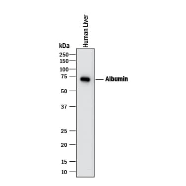
HAF018). A specific band was detected for Albumin at approximately 65-70 kDa (as indicated). This experiment was conducted under reducing conditions and using
Immunoblot Buffer Group 1." class="big_lightbox">
HAF018). A specific band was detected for Albumin at approximately 65-70 kDa (as indicated). This experiment was conducted under reducing conditions and using
Immunoblot Buffer Group 1." alt="Western blot shows lysate of human liver tissue. PVDF membrane was probed with 1 µg/mL of Mouse Anti-Human Serum Albumin Monoclonal Antibody (Catalog # MAB1455) followed by HRP-conjugated Anti-Mouse IgG Secondary Antibody (Catalog #
HAF018). A specific band was detected for Albumin at approximately 65-70 kDa (as indicated). This experiment was conducted under reducing conditions and using
Immunoblot Buffer Group 1." class="big_thumb" />
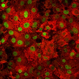
NL007) and counterstained with DAPI (blue). Specific staining was localized to cytoplasm. Cells were co-stained using Sheep Anti-Human CEBP alpha (Catalog #
AF7094) and NorthernLights™ 493-conjugated Anti-Sheep IgG Secondary Antibody (green, Catalog #
NL012). View our protocol for
Fluorescent ICC Staining of Cells on Coverslips." class="big_lightbox">
NL007) and counterstained with DAPI (blue). Specific staining was localized to cytoplasm. Cells were co-stained using Sheep Anti-Human CEBP alpha (Catalog #
AF7094) and NorthernLights™ 493-conjugated Anti-Sheep IgG Secondary Antibody (green, Catalog #
NL012). View our protocol for
Fluorescent ICC Staining of Cells on Coverslips." class="big_thumb" />
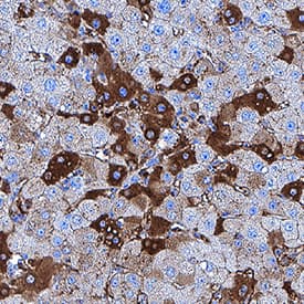
VC001). Before incubation with the primary antibody, tissue was subjected to heat-induced epitope retrieval using Antigen Retrieval Reagent-Basic (Catalog #
CTS013). Tissue was stained using DAB (brown) and counterstained with hematoxylin (blue). Specific staining was localized to cytoplasm and plasma membrane. View our protocol for
IHC Staining with VisUCyte HRP Polymer Detection Reagents." class="big_lightbox">
VC001). Before incubation with the primary antibody, tissue was subjected to heat-induced epitope retrieval using Antigen Retrieval Reagent-Basic (Catalog #
CTS013). Tissue was stained using DAB (brown) and counterstained with hematoxylin (blue). Specific staining was localized to cytoplasm and plasma membrane. View our protocol for
IHC Staining with VisUCyte HRP Polymer Detection Reagents." class="big_thumb" />
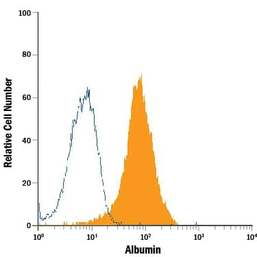
MAB003, open histogram), followed by Allophycocyanin-conjugated Anti-Mouse IgG Secondary Antibody (Catalog #
F0101B). To facilitate intracellular staining, cells were fixed with paraformaldehyde and permeabilized with saponin." class="big_lightbox">
MAB003, open histogram), followed by Allophycocyanin-conjugated Anti-Mouse IgG Secondary Antibody (Catalog #
F0101B). To facilitate intracellular staining, cells were fixed with paraformaldehyde and permeabilized with saponin." class="big_thumb" />
VC001) and DAB chromogen (lower image, yellow-brown). Tissues were counterstained with hematoxylin (blue)." class="big_lightbox">
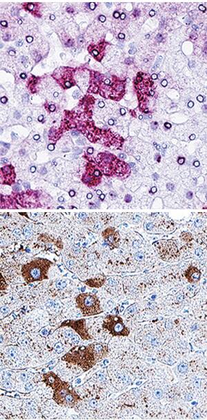
VC001) and DAB chromogen (lower image, yellow-brown). Tissues were counterstained with hematoxylin (blue)." class="big_thumb" />
human liver organoid culture protocol. Differentiated human liver organoids were stained using a (A) Mouse Anti-Human Serum Albumin Monoclonal Antibody (Catalog # MAB1455; red) and a (B) Goat Anti-Human HNF-3beta Antigen Affinity-purified Polyclonal Antibody (Catalog # AF2400; red) and counterstained with DAPI (Catalog # 5748; blue)." class="big_lightbox">

human liver organoid culture protocol. Differentiated human liver organoids were stained using a (A) Mouse Anti-Human Serum Albumin Monoclonal Antibody (Catalog # MAB1455; red) and a (B) Goat Anti-Human HNF-3beta Antigen Affinity-purified Polyclonal Antibody (Catalog # AF2400; red) and counterstained with DAPI (Catalog # 5748; blue)." class="big_thumb" />
 HAF018). A specific band was detected for Albumin at approximately 65-70 kDa (as indicated). This experiment was conducted under reducing conditions and using Immunoblot Buffer Group 1." alt="Western blot shows lysate of human liver tissue. PVDF membrane was probed with 1 µg/mL of Mouse Anti-Human Serum Albumin Monoclonal Antibody (Catalog # MAB1455) followed by HRP-conjugated Anti-Mouse IgG Secondary Antibody (Catalog # HAF018). A specific band was detected for Albumin at approximately 65-70 kDa (as indicated). This experiment was conducted under reducing conditions and using Immunoblot Buffer Group 1." class="big_thumb" />
 HAF017). A specific band was detected for Serum Albumin at approximately 65-70 kDa (as indicated). This experiment was conducted under reducing conditions and using Immunoblot Buffer Group 1." alt="Western blot shows lysates of human liver tissue and mouse liver tissue. PVDF membrane was probed with 0.1 µg/mL of Goat Anti-Human/Mouse Serum Albumin Antigen Affinity-purified Polyclonal Antibody (Catalog # AF3329) followed by HRP-conjugated Anti-Goat IgG Secondary Antibody (Catalog # HAF017). A specific band was detected for Serum Albumin at approximately 65-70 kDa (as indicated). This experiment was conducted under reducing conditions and using Immunoblot Buffer Group 1." class="big_thumb" />


 NL007) and counterstained with DAPI (blue). Specific staining was localized to cytoplasm. Cells were co-stained using Sheep Anti-Human CEBP alpha (Catalog #
  VC001). Before incubation with the primary antibody, tissue was subjected to heat-induced epitope retrieval using Antigen Retrieval Reagent-Basic (Catalog #
  MAB003, open histogram), followed by Allophycocyanin-conjugated Anti-Mouse IgG Secondary Antibody (Catalog #

 VC001) and DAB chromogen (lower image, yellow-brown). Tissues were counterstained with hematoxylin (blue)." class="big_thumb" />


 human liver organoid culture protocol. Differentiated human liver organoids were stained using a (A) Mouse Anti-Human Serum Albumin Monoclonal Antibody (Catalog # MAB1455; red) and a (B) Goat Anti-Human HNF-3beta Antigen Affinity-purified Polyclonal Antibody (Catalog # AF2400; red) and counterstained with DAPI (Catalog # 5748; blue)." class="big_thumb" />
![Western Blot: Albumin Antibody [NB600-41532] - Analysis using the HRP conjugate of NB600-41532. Detection of Albumin in whole mouse liver extracts. WB image submitted by a verified customer review.](http://images.novusbio.com/fullsize/Albumin-Antibody-Western-Blot-NB600-41532-img0002.jpg "Western Blot: Albumin Antibody [NB600-41532] - Analysis using the HRP conjugate of NB600-41532. Detection of Albumin in whole mouse liver extracts. WB image submitted by a verified customer review.")

![Western Blot: Albumin Antibody - BSA Free [NB600-41532] - Primary cell characterization.As per the materials & methods, tumors were isolated from mice & cultured. A, Primary murine hepatocytes were used as controls (Mu). Western blot revealed the primary cultures of APN KO A51, A48, A50 & A52 & WT C53, C54 & C55 cells to express p-mTOR, mTOR, p-Src, Src, PCNA, albumin & AFP. B, Immunofluorescence for K8 & pan-cytokeratin (PK) confirmed that A52 cells were epithelial (Scale bar 20 μM). C, Table showing in vitro & in vivo growth characteristics of WT & APN KO primary cultures. Scale of growth (+ to ++++). Image collected & cropped by CiteAb from the following publication (//pubmed.ncbi.nlm.nih.gov/30794695), licensed under a CC-BY license. Not internally tested by Novus Biologicals.](http://images.novusbio.com/fullsize/nb600-41532_goat-polyclonal-albumin-antibody-31020241537196.jpg)

![Immunocytochemistry/Immunofluorescence: Albumin Antibody [NBP1-32458] - A431 cells were fixed in ice-cold MeOH for 5 min. Green: Albumin protein stained by Albumin antibody diluted at 1:500. Blue: Hoechst 33342 staining. Scale bar = 10 um.](http://images.novusbio.com/fullsize/Albumin-Antibody-Immunocytochemistry-Immunofluorescence-NBP1-32458-img0021.jpg "Immunocytochemistry/Immunofluorescence: Albumin Antibody [NBP1-32458] - A431 cells were fixed in ice-cold MeOH for 5 min. Green: Albumin protein stained by Albumin antibody diluted at 1:500. Blue: Hoechst 33342 staining. Scale bar = 10 um.")
![Immunohistochemistry-Paraffin: Albumin Antibody [NBP1-32458] - Mouse liver. Albumin stained by Albumin antibody diluted at 1:1000.Antigen Retrieval: Citrate buffer, pH 6.0, 15 min.](http://images.novusbio.com/fullsize/Albumin-Antibody-Immunohistochemistry-Paraffin-NBP1-32458-img0025.jpg)
![Immunohistochemistry-Paraffin: Albumin Antibody [NBP1-32458] - Human hepatocellular carcinoma. Albumin antibody diluted at 1:500. Antigen Retrieval: Citrate buffer, pH 6.0, 15 min.](http://images.novusbio.com/fullsize/Albumin-Antibody-Immunohistochemistry-Paraffin-NBP1-32458-img0016.jpg)
![Immunohistochemistry-Frozen: Albumin Antibody [NBP1-32458] - Albumin antibody detects Albumin protein at cytoplasm in rat liver by immunohistochemical analysis. Sample: Frozen section of rat liver. Albumin antibody diluted at 1:200.](http://images.novusbio.com/fullsize/Albumin-Antibody-Immunohistochemistry-Frozen-NBP1-32458-img0017.jpg)
![Immunohistochemistry-Paraffin: Albumin Antibody [NBP1-32458] - Rat liver. Albumin stained by Albumin antibody diluted at 1:1000.Antigen Retrieval: Citrate buffer, pH 6.0, 15 min.](http://images.novusbio.com/fullsize/Albumin-Antibody-Immunohistochemistry-Paraffin-NBP1-32458-img0024.jpg)
![Immunohistochemistry-Paraffin: Albumin Antibody [NBP1-32458] - Dog liver.Albumin stained by Albumin antibody diluted at 1:500.Antigen Retrieval: Citrate buffer, pH 6.0, 15 min](http://images.novusbio.com/fullsize/antibody/nbp1-32458_rabbit-polyclonal-albumin-antibody-immunohistochemistry-paraffin-1482023124527..jpg)
![Immunohistochemistry-Paraffin: Albumin Antibody [NBP1-32458] - Dog kidney.Albumin stained by Albumin antibody diluted at 1:500.Antigen Retrieval: Citrate buffer, pH 6.0, 15 min](http://images.novusbio.com/fullsize/antibody/nbp1-32458_rabbit-polyclonal-albumin-antibody-immunohistochemistry-paraffin-1482023124716..jpg)
![Immunohistochemistry-Paraffin: Albumin Antibody [NBP1-32458] - Cat liver.Albumin stained by Albumin antibody diluted at 1:500.Antigen Retrieval: Citrate buffer, pH 6.0, 15 min](http://images.novusbio.com/fullsize/antibody/nbp1-32458_rabbit-polyclonal-albumin-antibody-immunohistochemistry-paraffin-1482023124837..jpg)
![Immunocytochemistry/ Immunofluorescence: Albumin Antibody [NBP1-32458] - Immunofluorescence localization of TNF-alpha , IL-1 beta & IL-6 in the liver tissue of animals treated with ConA or ConA + hVNS. a–c Representative immunofluorescence images of TNF-alpha , IL-1 beta , & IL-6 that were merged with CD11b. Signal intensities of TNF-alpha , IL-1 beta , & IL-6 from 4 to 6 images were averaged per each animal & compared among experimental groups (n = 3 independent experiments). One-way ANOVA with Tukey post hoc is shown. *p < 0.05, **p < 0.01, ***p < 0.001. d Enlarged views of TNF-alpha , IL-1 beta , & IL-6 signals merged with CD11b & Hoechst nuclear staining images in the liver sections from ConA + hVNS animal. e Immunofluorescence staining of liver tissue with anti-CD11b & anti-albumin antibodies. Representative immunofluorescence images using normal mouse IgG antibody instead of primary antibodies followed by reaction with fluorescein-goat anti-mouse IgG antibody are shown in the last column. Scale bars in a–c, d, & e are 100 μm, 10 μm, & 50 μm respectively Image collected & cropped by CiteAb from the following publication (//pubmed.ncbi.nlm.nih.gov/33272194), licensed under a CC-BY license. Not internally tested by Novus Biologicals.](http://images.novusbio.com/fullsize/nbp1-32458_rabbit-polyclonal-albumin-antibody-310202416175245.jpg)
![Western Blot: Albumin Antibody [NBP1-32458] - PG-4 whole cell extract (30 ug) was separated by 7.5% SDS-PAGE, and the membrane was blotted with Albumin antibody (NBP1-32458) diluted at 1:1000. The HRP-conjugated anti-rabbit IgG antibody was used to detect the primary antibody.](http://images.novusbio.com/fullsize/nbp1-32458_rabbit-polyclonal-albumin-antibody-910202419593558.jpg)
![Western Blot: Albumin Antibody [NBP1-32458] - Whole cell extract (30 ug) was separated by 7.5% SDS-PAGE, and the membrane was blotted with Albumin antibody (NBP1-32458) diluted at 1:1000. The HRP-conjugated anti-rabbit IgG antibody was used to detect the primary antibody, and the signal was developed with Trident ECL plus-Enhanced.](http://images.novusbio.com/fullsize/nbp1-32458_rabbit-polyclonal-albumin-antibody-910202420104429.jpg)
![Immunocytochemistry/ Immunofluorescence: Albumin Antibody [NBP1-32458] - Albumin antibody detects Albumin protein at cytoplasm by immunofluorescent analysis.</br>Sample: PG-4 cells were fixed in 4% paraformaldehyde at RT for 15 min.</br>Green: Albumin stained by Albumin antibody (NBP1-32458) diluted at 1:500.</br>Blue: Hoechst 33342 staining.](http://images.novusbio.com/fullsize/nbp1-32458_rabbit-polyclonal-albumin-antibody-91020241957221.jpg)
![Immunocytochemistry/ Immunofluorescence: Albumin Antibody [NBP1-32458] - Albumin antibody detects Albumin protein at cytoplasm by immunofluorescent analysis.</br>Sample: MDCK cells were fixed in 4% paraformaldehyde at RT for 15 min.</br>Green: Albumin stained by Albumin antibody (NBP1-32458) diluted at 1:500.</br>Blue: Hoechst 33342 staining.](http://images.novusbio.com/fullsize/nbp1-32458_rabbit-polyclonal-albumin-antibody-91020242063392.jpg)
![Western Blot: Albumin Antibody [NBP1-32458] - Various tissue extracts (50 ug) were separated by 7.5% SDS-PAGE, and the membrane was blotted with Albumin antibody (NBP1-32458) diluted at 1:1000. The HRP-conjugated anti-rabbit IgG antibody was used to detect the primary antibody.](http://images.novusbio.com/fullsize/nbp1-32458_rabbit-polyclonal-albumin-antibody-91020242041286.jpg)
![<b>Orthogonal Strategies Validation. </b>Western Blot: Albumin Antibody [NBP1-32458] - Various whole cell extracts (30 ug) were separated by 7.5% SDS-PAGE, and the membrane was blotted with Albumin antibody diluted at 1:1000. The HRP-conjugated anti-rabbit IgG antibody was used to detect the primary antibody. Corresponding RNA expression data for the same cell lines are based on Human Protein Atlas program.](http://images.novusbio.com/fullsize/nbp1-32458_rabbit-polyclonal-albumin-antibody-1610202419405735.jpg)


![Immunocytochemistry/ Immunofluorescence: Albumin Antibody - BSA Free [NBP1-77027] - Validation of Albumin in Mouse Liver. Immunofluorescent analysis of 4% paraformaldehyde-fixed mouse liver tissue labeling albumin with at 20mL, followed by goat anti-chicken IgY secondary antibody at 1/500 dilution (red) and DAPI staining (blue).](http://images.novusbio.com/fullsize/nbp1-77027_chicken-polyclonal-albumin-antibody-2711202420312949.gif "Immunocytochemistry/ Immunofluorescence: Albumin Antibody - BSA Free [NBP1-77027] - Validation of Albumin in Mouse Liver. Immunofluorescent analysis of 4% paraformaldehyde-fixed mouse liver tissue labeling albumin with at 20mL, followed by goat anti-chicken IgY secondary antibody at 1/500 dilution (red) and DAPI staining (blue).")
![Immunohistochemistry: Albumin Antibody - BSA Free [NBP1-77027] - Validation of Albumin in Human Liver.Immunohistochemical analysis of paraffin-embedded human liver tissue using anti-albumin antibody at 2 u/ml. Tissue was fixed with formaldehyde and blocked with 10% serum for 1 h at RT; antigen retrieval was by heat mediation with a citrate buffer (pH6). Samples were incubated with primary antibody overnight at 4C. A goat anti- chicken IgY (HRP) at 1/250 was used as secondary. Counter stained with Hematoxylin.](http://images.novusbio.com/fullsize/nbp1-77027_chicken-polyclonal-albumin-antibody-2711202419241330.jpg)
![Immunohistochemistry: Albumin Antibody - BSA Free [NBP1-77027] - Validation of Albumin in Mouse Liver.Immunohistochemical analysis of paraffin-embedded mouse liver tissue using anti-albumin antibody at 10 u/ml. Tissue was fixed with formaldehyde and blocked with 10% serum for 1 h at RT; antigen retrieval was by heat mediation with a citrate buffer (pH6). Samples were incubated with primary antibody overnight at 4C. A goat anti- chicken IgY (HRP) at 1/250 was used as secondary. Counter stained with Hematoxylin.](http://images.novusbio.com/fullsize/nbp1-77027_chicken-polyclonal-albumin-antibody-2711202419392785.jpg)
![Immunohistochemistry: Albumin Antibody - BSA Free [NBP1-77027] - Validation of Albumin in Rat Liver.Immunohistochemical analysis of paraffin-embedded rat liver tissue using anti-albumin antibody at 2 u/ml. Tissue was fixed with formaldehyde and blocked with 10% serum for 1 h at RT; antigen retrieval was by heat mediation with a citrate buffer (pH6). Samples were incubated with primary antibody overnight at 4C. A goat anti-chicken IgY (HRP) at 1/250 was used as secondary. Counter stained with Hematoxylin.](http://images.novusbio.com/fullsize/nbp1-77027_chicken-polyclonal-albumin-antibody-271120242045617.jpg)
![Western Blot: Albumin Antibody - BSA Free [NBP1-77027] - Validation in Rat Liver. Loading: 10 ug of lysates per lane.Antibodies: Albumin, 1 u/mL, 1h incubation at RT in 5% NFDM/TBST.Secondary: Goat anti- chicken IgY HRP conjugate at 1:10000 dilution.](http://images.novusbio.com/fullsize/nbp1-77027_chicken-polyclonal-albumin-antibody-2711202420285845.gif)
![Western Blot: Albumin Antibody - BSA Free [NBP1-77027] - Validation in Human Liver.Loading: 10 ug of lysates per lane.Antibodies: Albumin, , 1 u/mL, 1h incubation at RT in 5% NFDM/TBST.Secondary: Goat anti- chicken IgY HRP conjugate at 1:10000 dilution.](http://images.novusbio.com/fullsize/nbp1-77027_chicken-polyclonal-albumin-antibody-2711202419392736.gif)
![Western Blot: Albumin Antibody - BSA Free [NBP1-77027] - Loading: 10 µg of lysates per lane.Antibodies: Albumin, 1 u/mL, 1h incubation at RT in 5% NFDM/TBST.Secondary: Goat anti- chicken IgY HRP conjugate at 1:10000 dilution.](http://images.novusbio.com/fullsize/nbp1-77027_chicken-polyclonal-albumin-antibody-2711202419293848.gif)
![N/A Albumin [Biotin]](http://images.novusbio.com/fullsize/elisa/DATA_Albumin_DY1455_ELISA_1653.jpg "N/A Albumin [Biotin]")
 NL001) and counterstained with DAPI (blue). Specific staining was localized to cytoplasm. View our protocol for
  HAF109). This experiment was conducted under reducing conditions and using the 12-230 kDa separation system." class="big_thumb" />
 HAF109). This experiment was conducted under reducing conditions and using the 12-230 kDa separation system." class="big_thumb" />
 HAF109). This experiment was conducted under reducing conditions and using the 12-230 kDa separation system." class="big_thumb" />
![Western Blot: Albumin Antibody (AL-01) [NB500-417] - Analysis (reducing conditions) of human serum albumin using anti-human Albumin (AL-01). Lane 1: Human blood plasma Lane 2: Human blood serum.](http://images.novusbio.com/fullsize/Albumin-Antibody-AL-01-Western-Blot-NB500-417-img0001.jpg "Western Blot: Albumin Antibody (AL-01) [NB500-417] - Analysis (reducing conditions) of human serum albumin using anti-human Albumin (AL-01). Lane 1: Human blood plasma Lane 2: Human blood serum.")
![Western Blot: Human Serum Albumin Antibody (JF32-10) [NBP2-66894] - Western blot analysis of Human Serum Albumin on different lysates. Proteins were transferred to a PVDF membrane and blocked with 5% BSA in PBS for 1 hour at room temperature. The primary antibody (1/500) was used in 5% BSA at room temperature for 2 hours. Goat Anti-Rabbit IgG - HRP Secondary Antibody (HA1001) at 1:5,000 dilution was used for 1 hour at room temperature. Positive control: Lane 1: Hela cell lysate Lane 2: HepG2 cell lysate](http://images.novusbio.com/fullsize/Human-Serum-Albumin-Antibody-JF32-10-Western-Blot-NBP2-66894-img0008.jpg "Western Blot: Human Serum Albumin Antibody (JF32-10) [NBP2-66894] - Western blot analysis of Human Serum Albumin on different lysates. Proteins were transferred to a PVDF membrane and blocked with 5% BSA in PBS for 1 hour at room temperature. The primary antibody (1/500) was used in 5% BSA at room temperature for 2 hours. Goat Anti-Rabbit IgG - HRP Secondary Antibody (HA1001) at 1:5,000 dilution was used for 1 hour at room temperature. Positive control: Lane 1: Hela cell lysate Lane 2: HepG2 cell lysate")
![Immunohistochemistry-Paraffin: Human Serum Albumin Antibody (JF32-10) [NBP2-66894] - Analysis of paraffin-embedded mouse lung tissue using anti-Albumin antibody. Counter stained with hematoxylin.](http://images.novusbio.com/fullsize/Human-Serum-Albumin-Antibody-JF32-10-Immunohistochemistry-Paraffin-NBP2-66894-img0006.jpg)
![Immunohistochemistry-Paraffin: Human Serum Albumin Antibody (JF32-10) [NBP2-66894] - Analysis of paraffin-embedded human kidney tissue using anti-Albumin antibody. Counter stained with hematoxylin.](http://images.novusbio.com/fullsize/Human-Serum-Albumin-Antibody-JF32-10-Immunohistochemistry-Paraffin-NBP2-66894-img0001.jpg)
![Immunohistochemistry-Paraffin: Human Serum Albumin Antibody (JF32-10) [NBP2-66894] - Analysis of paraffin-embedded human liver tissue using anti-Albumin antibody. Counter stained with hematoxylin.](http://images.novusbio.com/fullsize/Human-Serum-Albumin-Antibody-JF32-10-Immunohistochemistry-Paraffin-NBP2-66894-img0002.jpg)
![Immunohistochemistry-Paraffin: Human Serum Albumin Antibody (JF32-10) [NBP2-66894] - Analysis of paraffin-embedded human lung tissue using anti-Albumin antibody. Counter stained with hematoxylin.](http://images.novusbio.com/fullsize/Human-Serum-Albumin-Antibody-JF32-10-Immunohistochemistry-Paraffin-NBP2-66894-img0003.jpg)
![Immunohistochemistry-Paraffin: Human Serum Albumin Antibody (JF32-10) [NBP2-66894] - Analysis of paraffin-embedded human spleen tissue using anti-Albumin antibody. Counter stained with hematoxylin.](http://images.novusbio.com/fullsize/Human-Serum-Albumin-Antibody-JF32-10-Immunohistochemistry-Paraffin-NBP2-66894-img0004.jpg)
![Immunohistochemistry-Paraffin: Human Serum Albumin Antibody (JF32-10) [NBP2-66894] - Analysis of paraffin-embedded mouse liver tissue using anti-Albumin antibody. Counter stained with hematoxylin.](http://images.novusbio.com/fullsize/Human-Serum-Albumin-Antibody-JF32-10-Immunohistochemistry-Paraffin-NBP2-66894-img0005.jpg)
![Western blot analysis on different lysates with NBP2-66894 at 1/40,000 dilution. Lane 1: Mouse liver tissue lysate Lane 2: Mouse spleen tissue lysate Lane 3: Rat liver tissue lysate Lysates/proteins at 20 µg/Lane. Predicted band size: 69 kDa Observed band size: 69 kDa Exposure time: 10 seconds; 4-20% SDS-PAGE gel. Proteins were transferred to a PVDF membrane and blocked with 5% NFDM/TBST for 1 hour at room temperature. NBP2-66894 at 1/40,000 dilution was used in 5% NFDM/TBST at 4℃ overnight. Goat Anti-Rabbit IgG [HRP] Secondary Antibody at 1/50,000 dilution was used for 1 hour at room temperature.](http://images.novusbio.com/fullsize/nbp2-66894_-western-blot-639204597489887500.jpg)
![SDS-Page: Albumin Native Protein [NBP1-97049] - SDS PAGE of Human Albumin. Lane 1: Non-Reduced Human Albumin. Lane 2: 5 uL Opal Prestained Marker. Lane 3: Reduced Human Albumin. Load: 1 ug per lane. Predicted/Observed size: Non-Reduced at 63 kDa/Observed at 55 kDa; Reduced at 63 kDa. Non-reduced migrates farther on gel due to native albumin's capacity to bind ions, increasing its charge and, therefore, ability to migrate farther down gel.](http://images.novusbio.com/fullsize/Albumin-Native-Protein-SDS-Page-NBP1-97049-img0002.jpg "SDS-Page: Albumin Native Protein [NBP1-97049] - SDS PAGE of Human Albumin. Lane 1: Non-Reduced Human Albumin. Lane 2: 5 uL Opal Prestained Marker. Lane 3: Reduced Human Albumin. Load: 1 ug per lane. Predicted/Observed size: Non-Reduced at 63 kDa/Observed at 55 kDa; Reduced at 63 kDa. Non-reduced migrates farther on gel due to native albumin's capacity to bind ions, increasing its charge and, therefore, ability to migrate farther down gel.")
![Dot Blot: Albumin Native Protein [NBP1-97049] - Human Albumin Rhodamine Conjugated. Antigen: Rhodamine Conjugated Human Albumin. Load: 3-fold serial dilution starting at 200 ng. Primary antibody: None. Secondary antibody: None. Block for 1 HR at RT.. Using the Rhodamine format of this antibody.](http://images.novusbio.com/fullsize/Albumin-Native-Protein-Dot-Blot-NBP1-97049-img0004.jpg)
![Dot Blot: Albumin Native Protein [NBP1-97049] - Human Albumin Rhodamine Conjugated. Antigen: Rhodamine Conjugated Human Albumin. Load: 3-fold serial dilution starting at 200 ng. Primary antibody: None. Secondary antibody: None. Block for 1 HR at RT.. Using the Rhodamine format of this antibody.](http://images.novusbio.com/fullsize/Albumin-Native-Protein-Dot-Blot-NBP1-97049-img0005.jpg)
![SDS-Page: Albumin Native Protein [NBP1-97049] - SDS-PAGE results of Human Albumin. Lane 1: reduced human albumin. Lane 2: opal prestained molecular weight ladder. Lane 3: non-reduced human albumin. Load: 1ug. 4-20% Lonza SDS-PAGE; Coomassie Stained; ChemiDoc Imaged.](http://images.novusbio.com/fullsize/Albumin Native Protein-SDS-Page-NBP1-97049-img0006.jpg)
![ELISA: Bovine Albumin ELISA Kit [NBP3-12180] - Typical standard curve.](http://images.novusbio.com/fullsize/Bovine-Albumin-ELISA-Kit-ELISA-NBP3-12180-img0001.jpg "ELISA: Bovine Albumin ELISA Kit [NBP3-12180] - Typical standard curve.")
![Western Blot: Mouse Serum Albumin Antibody [NBP2-26510] - Western Blot of Mouse Serum Albumin antibody. Lane 1: molecular weight. Lane 2: mouse Mouse Serum Albumin. Load: 0.1 ug.Primary antibody: MSA antibody at 1:1000 in 1% BSA-TTBS for overnight at 4C.Secondary antibody: IRDye800(TM) rabbit secondary antibody at 1:20,000 for 45 min at RT.Block: Blocking Buffer for Fluorescent Western Blotting 30 min at 4C.Predicted/Observed size: 69 kDa, 72 kDa for MSA. Other band(s): none.](http://images.novusbio.com/fullsize/Mouse-Serum-Albumin-Antibody-Western-Blot-NBP2-26510-img0010.jpg "Western Blot: Mouse Serum Albumin Antibody [NBP2-26510] - Western Blot of Mouse Serum Albumin antibody. Lane 1: molecular weight. Lane 2: mouse Mouse Serum Albumin. Load: 0.1 ug.Primary antibody: MSA antibody at 1:1000 in 1% BSA-TTBS for overnight at 4C.Secondary antibody: IRDye800(TM) rabbit secondary antibody at 1:20,000 for 45 min at RT.Block: Blocking Buffer for Fluorescent Western Blotting 30 min at 4C.Predicted/Observed size: 69 kDa, 72 kDa for MSA. Other band(s): none.")
![Western Blot: Mouse Serum Albumin Antibody [NBP2-26510] - Western Blot: Albumin Antibody [Biotin] [NB600-929] - Lane 1: Human Serum Albumin. Load: 50 ng per lane. Primary antibody: Rabbit anti-Human Serum Albumin Antibody Biotin Conjugated at 1:1,000 overnight at 4C. Secondary antibody: HRP Streptavidin secondary antibody at 1:40,000 for 30 min at RT. Block: incubated with blocking buffer for 30 min at RT. Predicted/Observed size: 66 kDa, 60 kDa for Human Serum Albumin. Image using the Biotin form of this antibody.](http://images.novusbio.com/fullsize/Mouse-Serum-Albumin-Antibody-Western-Blot-NBP2-26510-img0005.jpg)
![SDS-Page: Mouse Serum Albumin Antibody [NBP2-26510]](http://images.novusbio.com/fullsize/Mouse-Serum-Albumin-Antibody-SDS-Page-NBP2-26510-img0007.jpg)
![SDS-Page: Mouse Serum Albumin Antibody [NBP2-26510] - SDS-PAGE of Mouse Mouse Serum Albumin antibody.Lane 1: mouse serum albumin reduced. Lane 2: molecular weight. Lane 3: mouse serum albumin non-reduced. Load: 1.0 ug.Predicted/Observed size: Analysis by SDS-PAGE shows a banding pattern consistent with purified Rabbit IgG.Other band(s): none.](http://images.novusbio.com/fullsize/Mouse-Serum-Albumin-Antibody-SDS-Page-NBP2-26510-img0011.jpg)
![ELISA: Mouse Serum Albumin Antibody [NBP2-26510] - ELISA results of purified Mouse Mouse Serum Albumin antibody tested against Mouse Mouse Serum Albumin. Each well was coated in duplicate with 1.0 ug of conjugate. The starting dilution of antibody was 5ug/ml and the X-axis represents the Log10 of a 3-fold dilution. This titration is a 4-parameter curve fit where the IC50 is defined as the titer of the antibody. Assay performed using 3% fish gel, Goat anti-Rabbit IgG Antibody Peroxidase Conjugated (Min X Bv Ch Gt GP Ham Hs Hu Ms Rt & Sh Serum Proteins) and TMB ELISA Peroxidase Substrate.](http://images.novusbio.com/fullsize/Mouse-Serum-Albumin-Antibody-ELISA-NBP2-26510-img0012.jpg)

![ELISA: Mouse Albumin ELISA Kit [NBP3-12188] - Typical standard curve.](http://images.novusbio.com/fullsize/Mouse-Albumin-ELISA-Kit-ELISA-NBP3-12188-img0001.jpg "ELISA: Mouse Albumin ELISA Kit [NBP3-12188] - Typical standard curve.")


