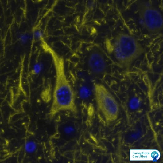
MAB8304) at 20ug/mL at 37 ° Celsius for 8 minutes. Before incubation with the primary antibody, tissue underwent an all-in-one dewaxing and antigen retrieval preprocessing using PreTreatment Module (PT Module) and Dewax and HIER Buffer H (pH 9; Epredia Catalog # TA-999-DHBH). Tissue was stained using the Alexa Fluor™ 647 Goat anti-Mouse IgG Secondary Antibody at 1:200 at 37 ° Celsius for 2 minutes. (Yellow; Lunaphore Catalog #
DR647MS) and counterstained with DAPI (blue; Lunaphore Catalog #
DR100). Specific staining was localized to the cytoplasm. Protocol available in
COMET™ Panel Builder." class="big_lightbox">
MAB8304) at 20ug/mL at 37 ° Celsius for 8 minutes. Before incubation with the primary antibody, tissue underwent an all-in-one dewaxing and antigen retrieval preprocessing using PreTreatment Module (PT Module) and Dewax and HIER Buffer H (pH 9; Epredia Catalog # TA-999-DHBH). Tissue was stained using the Alexa Fluor™ 647 Goat anti-Mouse IgG Secondary Antibody at 1:200 at 37 ° Celsius for 2 minutes. (Yellow; Lunaphore Catalog #
DR647MS) and counterstained with DAPI (blue; Lunaphore Catalog #
DR100). Specific staining was localized to the cytoplasm. Protocol available in
COMET™ Panel Builder." alt="MAP2 was detected in immersion fixed paraffin-embedded sections of human Brain Cortex using Mouse Anti-Human MAP2, Monoclonal Antibody (Catalog #
MAB8304) at 20ug/mL at 37 ° Celsius for 8 minutes. Before incubation with the primary antibody, tissue underwent an all-in-one dewaxing and antigen retrieval preprocessing using PreTreatment Module (PT Module) and Dewax and HIER Buffer H (pH 9; Epredia Catalog # TA-999-DHBH). Tissue was stained using the Alexa Fluor™ 647 Goat anti-Mouse IgG Secondary Antibody at 1:200 at 37 ° Celsius for 2 minutes. (Yellow; Lunaphore Catalog #
DR647MS) and counterstained with DAPI (blue; Lunaphore Catalog #
DR100). Specific staining was localized to the cytoplasm. Protocol available in
COMET™ Panel Builder." class="big_thumb" />
 DR647MS) and counterstained with DAPI (blue; Lunaphore Catalog # DR100). Specific staining was localized to the cytoplasm. Protocol available in COMET™ Panel Builder." alt="MAP2 was detected in immersion fixed paraffin-embedded sections of mouse Hippocampus using Mouse Anti-Human Mouse MAP2, Monoclonal Antibody (Catalog # NBP2-50032) at 1:12,000 dilution at 37 ° Celsius for 4 minutes. Before incubation with the primary antibody, tissue underwent an all-in-one dewaxing and antigen retrieval preprocessing using PreTreatment Module (PT Module) and Dewax and HIER Buffer H (pH 9; Epredia Catalog # TA-999-DHBH). Tissue was stained using the Alexa Fluor™ 647 Goat anti-Mouse IgG Secondary Antibody at 1:200 at 37 ° Celsius for 2 minutes. (Yellow; Lunaphore Catalog # DR647MS) and counterstained with DAPI (blue; Lunaphore Catalog # DR100). Specific staining was localized to the cytoplasm. Protocol available in COMET™ Panel Builder." class="big_thumb" />
 MAB8304) at 20ug/mL at 37 ° Celsius for 8 minutes. Before incubation with the primary antibody, tissue underwent an all-in-one dewaxing and antigen retrieval preprocessing using PreTreatment Module (PT Module) and Dewax and HIER Buffer H (pH 9; Epredia Catalog # TA-999-DHBH). Tissue was stained using the Alexa Fluor™ 647 Goat anti-Mouse IgG Secondary Antibody at 1:200 at 37 ° Celsius for 2 minutes. (Yellow; Lunaphore Catalog # DR647MS) and counterstained with DAPI (blue; Lunaphore Catalog # DR100). Specific staining was localized to the cytoplasm. Protocol available in COMET™ Panel Builder." alt="MAP2 was detected in immersion fixed paraffin-embedded sections of human Brain Cortex using Mouse Anti-Human MAP2, Monoclonal Antibody (Catalog # MAB8304) at 20ug/mL at 37 ° Celsius for 8 minutes. Before incubation with the primary antibody, tissue underwent an all-in-one dewaxing and antigen retrieval preprocessing using PreTreatment Module (PT Module) and Dewax and HIER Buffer H (pH 9; Epredia Catalog # TA-999-DHBH). Tissue was stained using the Alexa Fluor™ 647 Goat anti-Mouse IgG Secondary Antibody at 1:200 at 37 ° Celsius for 2 minutes. (Yellow; Lunaphore Catalog # DR647MS) and counterstained with DAPI (blue; Lunaphore Catalog # DR100). Specific staining was localized to the cytoplasm. Protocol available in COMET™ Panel Builder." class="big_thumb" />


![Western blot analysis of whole brain tissue lysates using chicken pAb to microtubule associated protein 2 (MAP2), NB300-213, dilution 1:50,000 in green: [1] protein standard (red), [2] adult rat brain, [3] embryonic E20 rat brain, [4] adult mouse brain. Strong band observed at ~280 kDa mark corresponds to two major isoforms of MAP2 protein referred to as MAP2A and MAP2B. Smaller fragments of these isoforms are also detected if the antibody is used at higher concentrations.](http://images.novusbio.com/fullsize/MAP2-Antibody-Western-Blot-NB300-213-img0005.jpg)


![Immunocytochemistry/ Immunofluorescence: MAP2 Antibody [NB300-213] - SUMO1 conjugates are not localized to amyloid plaques. (a) Sagittal brain sections of 24‐week‐old KI/AD & WT/AD animals were stained using anti‐HA antibodies (red), 6E10 antibodies (green) that label amyloid beta 1–42 among other amyloid beta variants (epitope lies within amino acids 3–8 of amyloid beta) & anti‐MAP2 antibodies (blue). Sections of the hippocampal subiculum are shown. Images are representatives of three independent experiments. Scale bar, 25 μm. (b) Anti‐HA signal intensity in amyloid plaques & in cell nuclei was quantified using ImageJ (N = 3, ***significance between WT/AD & KI/AD, p = .0007 in Student's t test) Image collected & cropped by CiteAb from the following publication (//pubmed.ncbi.nlm.nih.gov/29633471), licensed under a CC-BY license. Not internally tested by Novus Biologicals.](http://images.novusbio.com/fullsize/nb300-213_chicken-map2-pab-310202415392532.jpg)
![Immunocytochemistry/ Immunofluorescence: MAP2 Antibody [NB300-213] - Oxytocin receptors (Oxtr) are expressed in mouse hippocampus.(a & b) Coronal sections of the hippocampus (a) & striatum (b) from OxtrVn/+ & Oxtr+/+ mice stained for Venus & DAPI (ac, anterior commisure; cx, cortex; hip, hippocampus; str, striatum). (c) Confocal images of rostral Oxtr+/+ & OxtrVn/+ brain sections including hippocampus, double-labeled for Venus (green) & NeuN (red). (d) Confocal images of Oxtr-expressing neurons in the CA1 subfields of the medial & caudal hippocampus from OxtrVn/+ sections, triple stained with antibodies directed against NeuN (red), MAP2 (magenta), & Venus (green). (e) Representative images of hippocampal mass cultures obtained from OxtrVn/+ mice at P0 mice & processed for immunofluorescence staining at 5, 10, & 14 DIV using antibodies to Venus (green) & GAD67 (red), & DAPI (blue). (f) Quantitative analysis of Venus-positive neurons with (GAD67+) & without colabeling for GAD67 (GAD67-) in hippocampal mass cultures obtained from OxtrVn/+ mice at P0 mice & processed for immunofluorescence staining at 5, 10, & 14 DIV. Data are shown as mean ± SEM.DOI://dx.doi.org/10.7554/eLife.22466.015 Image collected & cropped by CiteAb from the following publication (//pubmed.ncbi.nlm.nih.gov/28231043), licensed under a CC-BY license. Not internally tested by Novus Biologicals.](http://images.novusbio.com/fullsize/nb300-213_chicken-map2-pab-310202416212347.jpg)
![Immunocytochemistry/ Immunofluorescence: MAP2 Antibody [NB300-213] - SUMO1 conjugates remain nuclear during increased amyloid burden. (a) Western blot analysis of subcellular fractions of 36‐week‐old KI/WT & KI/AD mouse brain using anti‐HA antibody (upper two panels) & antibodies to GluN1 (a marker of the postsynaptic compartment) & Synaptophysin (a marker of the presynaptic compartment) to validate the fractionation procedure (lower two panels). H, homogenate; P1, nuclear pellet; S1, supernatant after P1 sedimentation; P2, crude synaptosomal pellet; S2, supernatant after P2 sedimentation; LP1, lysed synaptosomal membranes; LS1, supernatant after LP1 sedimentation; LP2, pellet after LS1 sedimentation, SPM, synaptic plasma membranes. Bracket indicates the anti‐HA signal representing SUMOylation, arrow indicates RanGAP1, stars indicate nonspecific signal detected by the anti‐HA antibody. (b) Brain sagittal sections of aged (36 weeks old) KI/AD (left panels) & WT/AD (right panels) mice were immunostained using antibodies directed against HA (red, labels HA‐HA conjugates), MAP2 (green, labels neuronal somata & dendrites), & Synapsin1 (Syn1, magenta, labels synapses). The images show triple‐labeled neurons of hippocampal subiculum (b), cortical layer 5 (c), & proximal apical dendrite from hippocampal CA3 (d). The white line shows the orientation of the line‐scan used to generate the image stack shown in the bottom side view. Scale bar: 10 μm. Note that the anti‐HA immunosignal is mainly located in neuronal nuclei, only background staining is observed in WT/AD mice. Little anti‐HA signal is observed along MAP2‐positive structures & does not colocalize with Synapsin1 Image collected & cropped by CiteAb from the following publication (//pubmed.ncbi.nlm.nih.gov/29633471), licensed under a CC-BY license. Not internally tested by Novus Biologicals.](http://images.novusbio.com/fullsize/nb300-213_chicken-map2-pab-310202416212317.jpg)
![Immunocytochemistry/ Immunofluorescence: MAP2 Antibody [NB300-213] - Validation of the CRISPR/Cas9 Alsin-/- cells in iPSC, NPC & iPSC-sMN.(A) Electrophoresis result of the PCR reaction using primers flanking exon three & within exon 3. Homozygous deletion was confirmed by the absence of a 2.8 kb. (B) Protein lysates from WT KOLF & Alsin-/- iPSC were loaded onto SDS-PAGE & immunoblotted with Alsin antibody. A band detected at ~184 kDa, but absent in Alsin-/-, corresponds to full-length Alsin. (C) WT KOLF, Alsin-/- iPSC, & neuroprogenitor cells (NPC) were immunostained with pluripotency markers Oct4 & Lin28, & neuroprogenitor markers Sox2 & Pax6, respectively. (D–E) WT KOLF & Alsin-/- iPSC-sMN were immunostained with motor neuron markers such as ChAT, HB9, & ISL1, nuclear dye DAPI, & cytoskeletal marker MAP2. (F) WT KOLF & Alsin-/- iPSC-sMN were immunostained with antibodies against Rab5 & Alsin, along with DAPI (nuclear) & phalloidin (actin) stains. Scale bars, 10 μm. Image collected & cropped by CiteAb from the following publication (//pubmed.ncbi.nlm.nih.gov/29469808), licensed under a CC-BY license. Not internally tested by Novus Biologicals.](http://images.novusbio.com/fullsize/nb300-213_chicken-map2-pab-310202416203714.jpg)
![Immunocytochemistry/ Immunofluorescence: MAP2 Antibody [NB300-213] - PEA decreases astrocyte activation in organotypic cultures of rat hippocampi & rescues neuronal CA3 damage caused by A beta challenge. A beta -challenged (1 μg/ml) slices of rat hippocampi treated for 24 hours w/ PEA (0.1 μM) in presence of selective PPAR gamma antagonist (GW9662, 9 nM) or selective PPAR alpha antagonist (MK886, 3 μM). (A) Nissl staining showing effect of treatment on morphology of organotypic hippocampal slices. Scale bar: 80 μm. A beta caused a marked neuronal loss, mainly in CA3 region of hippocampus, as highlighted by arrows. PEA able to reverse this effect. (B) Representative photomicrographs of CA3 region showing Results of immunofluorescence experiments aimed at investigating effect of treatments on astrocyte activation & neuronal loss, as determined by immunostaining for GFAP (green) & MAP2 (red) alone or merged, respectively. Nuclei stained w/ Hoechst (blue). Scale bar: 10 μm. Arrows in photomicrographs indicate astrocyte infiltration events & apoptotic condensation in nuclei of adjacent neurons. (C) Relative quantification of GFAP-positive cell number as a count of astrocyte proliferation. (D) Apoptotic events detected on MAP2-expressing cells as an indication of neuronal death. For (C) & (D), average value determined by counting cells in at least five microscopic fields for each treatment. Results are presented as means ± SEM of four separate experiments. At least four slices from each experimental group observed for each experiment. Statistical analysis performed using parametric one-way analysis of variance, & multiple comparisons performed using Bonferroni test. ***P < 0.001 & *P < 0.05 vs. control; ∘∘P < 0.01 & ∘P < 0.05 vs. A beta -challenge slices. Image collected & cropped by CiteAb from following publication (//jneuroinflammation.biomedcentral.com/articles/10.1186/1742-2094-9-49), licensed under a CC-BY license. Not internally tested by Novus Biologicals.](http://images.novusbio.com/fullsize/nb300-213_chicken-map2-pab-310202416203712.jpg)
![Immunocytochemistry/ Immunofluorescence: MAP2 Antibody [NB300-213] - PEA inhibits astroglial proliferation & reduces neuronal loss in mixed neuroglia co-cultures exposed to A beta . A beta -challenged (1 μg/ml) astrocyte/neuron mixed cultures were treated with PEA (0.1 μM) in the presence of the selective PPAR gamma antagonist (GW9662, 9 nM) or the selective PPAR alpha antagonist (MK886, 3 μM). After 24 hours of treatment, cells were processed for analyses. (A) Immunofluorescence photomicrographs showing the effect of the treatments on astrocyte proliferation & neuronal loss, as determined by immunostaining for GFAP (green) & MAP2 (red), respectively. Arrows indicate chromatin condensation in nuclei stained with Hoechst dye (blue) as markers of apoptotic events. Scale bar: 10 μm. (B) Relative quantification of GFAP-positive cell number as a count of astrocyte proliferation. (C) Apoptotic events detected on MAP2-expressing cells as an indication of neuronal death. For (B) & (C), the average value was determined by counting cells in at least nine microscopic fields for each treatment. Results are presented as means ± SEM of three separate experiments. Statistical analysis was performed using parametric one-way analysis of variance, & multiple comparisons were performed using the Bonferroni test. ***P < 0.001 vs. unstimulated cells, ∘∘P < 0.01 & ∘P < 0.05 vs. A beta -stimulated cultures. Image collected & cropped by CiteAb from the following publication (//jneuroinflammation.biomedcentral.com/articles/10.1186/1742-2094-9-49), licensed under a CC-BY license. Not internally tested by Novus Biologicals.](http://images.novusbio.com/fullsize/nb300-213_chicken-map2-pab-310202416205136.jpg)
![Immunocytochemistry/Immunofluorescence: MAP2 Antibody (4H5) [NBP2-25156] - Mixed neuron and glia cultures stained with NBP2-25156 (green), and NB300-135 rabbit antibody to NF-H (red) and DNA (blue). NBP2-25156 reveals strong cytoplasmic staining for of dendrites and perikarya, which does not overlap with the NF-H antibody, which primarily binds to axons.](http://images.novusbio.com/fullsize/MAP2-Antibody-4H5-Immunocytochemistry-Immunofluorescence-NBP2-25156-img0003.jpg "Immunocytochemistry/Immunofluorescence: MAP2 Antibody (4H5) [NBP2-25156] - Mixed neuron and glia cultures stained with NBP2-25156 (green), and NB300-135 rabbit antibody to NF-H (red) and DNA (blue). NBP2-25156 reveals strong cytoplasmic staining for of dendrites and perikarya, which does not overlap with the NF-H antibody, which primarily binds to axons.")
![Immunohistochemistry Free-Floating: MAP2 Antibody (4H5) [NBP2-25156] - Analysis of a rat hippocampus section stained with mouse mAb to MAP2, NBP2-25156, dilution 1:2,000 in green, and costained with rabbit pAb to FOX3/NeuN, dilution 1:2,000 in red. Following transcardial perfusion of rat with 4% paraformaldehyde, brain was post fixed for 24 hours, cut to 45uM, and free-floating sections were stained with above antibodies. The NBP2-25156 antibody labels MAP2 protein in the perikarya and dendrites of most neurons while the FOX3/NeuN antibody selectively stains nuclei and proximal soma of neuronal cells.](http://images.novusbio.com/fullsize/MAP2-Antibody-4H5-Immunocytochemistry-Immunofluorescence-NBP2-25156-img0005.jpg)
![Western Blot: MAP2 Antibody (4H5) [NBP2-25156] - Analysis of tissue and cell lysates using mouse mAb to MAP2, NBP2-25156, dilution 1:10,000 in green: [1] protein standard (red), [2] rat brain, [3] mouse brain, and [4] embryonic rat cortical neuron-glial cell lysate. A band at about 280 kDa corresponds to the MAP2A and MAP2B proteins.](http://images.novusbio.com/fullsize/MAP2-Antibody-4H5-Western-Blot-NBP2-25156-img0006.jpg)
![Immunohistochemistry-Paraffin: MAP2 Antibody (4H5) [NBP2-25156] - Analysis of MAP2 in a 1% PFA fixed mouse brain section using anti-MAP2 (red) and NeuN (green) antibodies. Image from verified customer review.](http://images.novusbio.com/fullsize/MAP2-Antibody-4H5-Immunohistochemistry-Paraffin-NBP2-25156-img0004.jpg)
![Immunohistochemistry-Paraffin: MAP2 Antibody (4H5) [NBP2-25156] - Staining in mouse brain. Image from a verified customer review.](http://images.novusbio.com/fullsize/MAP2-Antibody-4H5-Immunohistochemistry-Paraffin-NBP2-25156-img0008.jpg)
![Immunohistochemistry-Paraffin: MAP2 Antibody (4H5) [NBP2-25156] - Staining of a 1% PFA fixed mouse cortical region section with anti-MAP 2 (dilution 1:300; Alexa 568 red). Image from a verified customer review.](http://images.novusbio.com/fullsize/MAP2-Antibody-4H5-Immunohistochemistry-Paraffin-NBP2-25156-img0007.jpg)
![Immunohistochemistry Free-Floating: MAP2 Antibody (5H11) [NBP1-92711] - Analysis of rat hippocampus section stained with mouse mAb to MAP2, NBP1-92711, dilution 1:5,000 in green, and costained with rabbit pAb to alpha-internexin, dilution 1:2,000 in red. Following transcardial perfusion of rat with 4% paraformaldehyde, brain was post fixed for 24 hours, cut to 45uM, and free-floating sections were stained with above antibodies. The NBP1-92711 antibody labels MAP2 protein in the perikarya and dendrites of the most neurons, and the alpha-internexin antibody selectively stains axons and dendrites of neuronal cells.](http://images.novusbio.com/fullsize/MAP2-Antibody-5H11-Immunocytochemistry-Immunofluorescence-NBP1-92711-img0001.jpg "Immunohistochemistry Free-Floating: MAP2 Antibody (5H11) [NBP1-92711] - Analysis of rat hippocampus section stained with mouse mAb to MAP2, NBP1-92711, dilution 1:5,000 in green, and costained with rabbit pAb to alpha-internexin, dilution 1:2,000 in red. Following transcardial perfusion of rat with 4% paraformaldehyde, brain was post fixed for 24 hours, cut to 45uM, and free-floating sections were stained with above antibodies. The NBP1-92711 antibody labels MAP2 protein in the perikarya and dendrites of the most neurons, and the alpha-internexin antibody selectively stains axons and dendrites of neuronal cells.")
![Western Blot: MAP2 Antibody (5H11) [NBP1-92711] - Analysis of different tissue lysates using mouse mAb to MAP2A/B (5H11), NBP1-92711, dilution 1:10,000 in green: [1] protein standard (red), [2] adult rat whole brain, [2] embryonic (E20) rat brain, [4] adult rat spinal cord, and [5] adult mouse brain lysate. A band observed at about 280 kDa corresponds to full length MAP2a and MAP2B protein. MAP2A/B is expressed heavily in brain particularly in cortical regions, but is a more minor component of spinal cord. Note that the epitope for this antibody in within the projection domain, and so the antibody does not bind to the lower molecular weight MAP2C and MAP2D isoforms which lack this region.](http://images.novusbio.com/fullsize/MAP2-Antibody-5H11-Western-Blot-NBP1-92711-img0002.jpg)
![Immunocytochemistry/Immunofluorescence: MAP2 Antibody (5H11) [NBP1-92711] - Single cultured astrocytes stained with MAP2 (red) and Myelin (green). Image from a verified customer review.](http://images.novusbio.com/fullsize/MAP2-Antibody-5H11-Immunocytochemistry-Immunofluorescence-NBP1-92711-img0003.jpg)
![Immunocytochemistry/Immunofluorescence: MAP2 Antibody (2C4) [NBP2-50032] - Mixed neuron and glia cultures stained with NBP2-50032 (red) and chicken antibody to Vimentin (green) and DNA (blue). NBP2-50032 reveals strong cytoplasmic staining of dendrites and perikarya of neuronal cells, while Vimentin was visualized in astrocytes and fibroblasts.](http://images.novusbio.com/fullsize/MAP2-Antibody-2C4-Immunocytochemistry-Immunofluorescence-NBP2-50032-img0004.jpg)
![Immunocytochemistry/Immunofluorescence: MAP2 Antibody (2C4) [NBP2-50032] - Analysis of adult rat hippocampus section stained with mouse mAb to MAP2A/B/C/D, NBP2-50032, dilution 1:5,000 in green, and chicken pAb to FOX2, dilution 1:2,000, in red. Following transcardial perfusion of rat with 4% paraformaldehyde, brain was post fixed for 24 hours, cut to 45 uM, and free-floating sections were stained with above antibodies. NBP2-50032 antibody labels all isotypes of the MAP2 protein which are found in the perikarya and dendrites of neurons. The FOX2 antibody stains the nuclei of a subset of neuronal cells.](http://images.novusbio.com/fullsize/MAP2-Antibody-2C4-Immunocytochemistry-Immunofluorescence-NBP2-50032-img0006.jpg)
![Western Blot: MAP2 Antibody (2C4) [NBP2-50032] - Analysis of tissue and cell lysates using mouse mAb to MAP2C/D, NBP2-50032, dilution 1:5,000 in green, and chicken pAb to Vimentin, dilution 1:5,000 in red. [1] protein standard (red), [2] rat whole brain lysate, [3] HeLa, [4] SH-SY5Y, [5] HEK293, and [6] NIH/3T3 cell lysates. A band at about 280 kDa corresponds to full length intact MAP2A/2B isotypes. The bands that appear at about 70kDa represent MAP2C/D isotypes. Multiple bands in between are likely to be in vivo fragments of MAP2A/B. Only the SH-SY5Y cells, which have neuronal properties express MAP2 protein. Vimentin antibody binds to vimentin protein showing a single band at ~50 kDa, which is present in all preparations.](http://images.novusbio.com/fullsize/MAP2-Antibody-2C4-Western-Blot-NBP2-50032-img0005.jpg)
![Western Blot: MAP2 Antibody [NBP3-05552] - Western blot analysis of whole brain lysates using MAP2 Antibody at a dilution of 1:5,000 in green: [1] protein standard (red), [2] rat, and [3] mouse brain lysate. A band at about 280kDa corresponds to the MAP2A and MAP2B proteins.](http://images.novusbio.com/fullsize/MAP2-Antibody-Western-Blot-NBP3-05552-img0002.jpg "Western Blot: MAP2 Antibody [NBP3-05552] - Western blot analysis of whole brain lysates using MAP2 Antibody at a dilution of 1:5,000 in green: [1] protein standard (red), [2] rat, and [3] mouse brain lysate. A band at about 280kDa corresponds to the MAP2A and MAP2B proteins.")
![Immunocytochemistry/Immunofluorescence: MAP2 Antibody [NBP3-05552] - Immunofluorescent analysis of rat brain stem section stained with goat pAb to MAP2 at a dilution of 1:2,000 in red, and costained with mouse mAb to MBP at a dilution of 1:5,000, in green. Following transcardial perfusion of rat with 4% paraformaldehyde, brain was post fixed for 24 hours, cut to 45uM, and free-floating sections were stained with above antibodies. MAP2 Antibody labels MAP2 protein in the perikarya and dendrites of the most neurons, notably motorneurons in the brain stem, and the MBP antibody stains myelin sheath around axons.](http://images.novusbio.com/fullsize/MAP2-Antibody-Immunocytochemistry-Immunofluorescence-NBP3-05552-img0001.jpg)
 NL007) and counterstained with DAPI (blue). Specific staining was localized to cell surfaces. View our protocol for
  CTS002) and counterstained with hematoxylin (blue). Specific staining was localized to neurofibrillary tangles and plaques. View our protocol for
 ![Western Blot: MAP2 Antibody (HM-2) [NB120-11267] - Rat brain extract (adult and newborn) was separated on SDS-PAGE and probed with Monoclonal PA To MAP2 (MO) CL:HM-2 . The antibody was developed with AlkPhos APA Mouse Fab ads HIgG and a NBTBCIP substrate. Observed molecular weight is 280 kDA.](http://images.novusbio.com/fullsize/MAP2-Antibody-HM-2-Western-Blot-NB120-11267-img0003.jpg "Western Blot: MAP2 Antibody (HM-2) [NB120-11267] - Rat brain extract (adult and newborn) was separated on SDS-PAGE and probed with Monoclonal PA To MAP2 (MO) CL:HM-2 . The antibody was developed with AlkPhos APA Mouse Fab ads HIgG and a NBTBCIP substrate. Observed molecular weight is 280 kDA.")
![Immunocytochemistry/Immunofluorescence: MAP2 Antibody (HM-2) [NB120-11267] - B35 cells.](http://images.novusbio.com/fullsize/MAP2-Antibody-HM-2-Immunocytochemistry-Immunofluorescence-NB120-11267-img0002.jpg)
![Western Blot: MAP2 Antibody (CL5420) [NBP2-61417] - Analysis in human cell line SH-SY5Y. Observed molecular weight 89 and 280 kDA.](http://images.novusbio.com/fullsize/MAP2-Antibody-CL5420-Western-Blot-NBP2-61417-img0007.jpg "Western Blot: MAP2 Antibody (CL5420) [NBP2-61417] - Analysis in human cell line SH-SY5Y. Observed molecular weight 89 and 280 kDA.")
![Immunohistochemistry-Paraffin: MAP2 Antibody (CL5420) [NBP2-61417] - Staining of human tonsil shows absence of positivity in lymphoid cells as expected (negative control).](http://images.novusbio.com/fullsize/MAP2-Antibody-CL5420-Immunohistochemistry-Paraffin-NBP2-61417-img0006.jpg)
![Immunohistochemistry-Paraffin: MAP2 Antibody (CL5420) [NBP2-61417] - Staining of human cerebral cortex shows cytoplasmic immunoreactivity in neurons, as well as positivity in neuropil.](http://images.novusbio.com/fullsize/MAP2-Antibody-CL5420-Immunohistochemistry-Paraffin-NBP2-61417-img0003.jpg)
![Immunohistochemistry-Paraffin: MAP2 Antibody (CL5420) [NBP2-61417] - Staining of human cerebellum shows positivity in a subset of neurons.](http://images.novusbio.com/fullsize/MAP2-Antibody-CL5420-Immunohistochemistry-Paraffin-NBP2-61417-img0004.jpg)
![Immunohistochemistry-Paraffin: MAP2 Antibody (CL5420) [NBP2-61417] - Staining of human pancreas shows cytoplasmic immunoreactivity in pancreatic islets.](http://images.novusbio.com/fullsize/MAP2-Antibody-CL5420-Immunohistochemistry-Paraffin-NBP2-61417-img0005.jpg)

![Immunohistochemistry: MAP2 Antibody [NBP1-81264] - Immunohistochemical staining of human small intestine shows no cytoplasmic positivity as expected.](http://images.novusbio.com/fullsize/MAP2-Antibody-Immunohistochemistry-NBP1-81264-img0006.jpg "Immunohistochemistry: MAP2 Antibody [NBP1-81264] - Immunohistochemical staining of human small intestine shows no cytoplasmic positivity as expected.")
![Immunohistochemistry: MAP2 Antibody [NBP1-81264] - Immunohistochemistry analysis in human cerebral cortex and small intestine tissues using NBP1-81264 antibody. Corresponding MAP2 RNA-seq data are presented for the same tissues.](http://images.novusbio.com/fullsize/MAP2-Antibody-Immunohistochemistry-NBP1-81264-img0002.jpg)
![Immunohistochemistry: MAP2 Antibody [NBP1-81264] - Immunohistochemical staining of human cerebral cortex shows moderate to strong cytoplasmic positivity in neuronal cells.](http://images.novusbio.com/fullsize/MAP2-Antibody-Immunohistochemistry-NBP1-81264-img0003.jpg)
![Immunohistochemistry: MAP2 Antibody [NBP1-81264] - Immunohistochemical staining of human cerebellum shows strong cytoplasmic positivity..](http://images.novusbio.com/fullsize/MAP2-Antibody-Immunohistochemistry-NBP1-81264-img0004.jpg)
![Immunohistochemistry: MAP2 Antibody [NBP1-81264] - Immunohistochemical staining of human pancreas shows moderate to strong cytoplasmic positivity in islets of Langerhans.](http://images.novusbio.com/fullsize/MAP2-Antibody-Immunohistochemistry-NBP1-81264-img0005.jpg)
![Immunofluorescence: MAP2 Antibody [NBP1-81264] - Immunofluorescent staining of human cell line U-251 MG shows localization to cytosol.](http://images.novusbio.com/fullsize/MAP2-Antibody-Immunofluorescence-NBP1-81264-img0001.jpg)


![Immunohistochemistry-Paraffin: MAP2 Antibody (HL1656) - Azide and BSA Free [NBP3-25555] - MAP2 antibody [HL1656] detects MAP2 protein at cell membrane and cytoplasm by immunohistochemical analysis. Sample: Paraffin-embedded rat brain. MAP2 stained by MAP2 antibody [HL1656] (NBP3-25555) diluted at 1:100. Antigen Retrieval: Citrate buffer, pH 6.0, 15 min](http://images.novusbio.com/fullsize/nbp3-25555_rabbit-map2-mab-hl1656-azide-and-bsa-free-212024939165.jpg "Immunohistochemistry-Paraffin: MAP2 Antibody (HL1656) - Azide and BSA Free [NBP3-25555] - MAP2 antibody [HL1656] detects MAP2 protein at cell membrane and cytoplasm by immunohistochemical analysis. Sample: Paraffin-embedded rat brain. MAP2 stained by MAP2 antibody [HL1656] (NBP3-25555) diluted at 1:100. Antigen Retrieval: Citrate buffer, pH 6.0, 15 min")
![Immunocytochemistry/Immunofluorescence: MAP2 Antibody (HL1656) - Azide and BSA Free [NBP3-25555] - MAP2 antibody [HL1656] detects MAP2 protein in dendrites, but not in axons, by immunofluorescent analysis. Sample: DIV9 rat E18 primary hippocampal neuron cells were fixed in 4% paraformaldehyde at RT for 15 min. Green: MAP2 stained by MAP2 antibody [HL1656] (NBP3-25555) diluted at 1:250. Red: Tau, an axon marker, stained by Tau antibody [GT287] diluted at 1:500. Blue: Fluoroshield with DAPI .](http://images.novusbio.com/fullsize/nbp3-25555_rabbit-map2-mab-hl1656-azide-and-bsa-free-212024941460.jpg)
![Immunohistochemistry-Paraffin: MAP2 Antibody (HL1656) - Azide and BSA Free [NBP3-25555] - MAP2 antibody [HL1656] detects MAP2 protein at cell membrane and cytoplasm by immunohistochemical analysis. Sample: Paraffin-embedded mouse brain. MAP2 stained by MAP2 antibody [HL1656] (NBP3-25555) diluted at 1:100. Antigen Retrieval: Citrate buffer, pH 6.0, 15 min](http://images.novusbio.com/fullsize/nbp3-25555_rabbit-map2-mab-hl1656-azide-and-bsa-free-2120249413331.jpg)
![Immunocytochemistry/Immunofluorescence: MAP2 Antibody (HL1655) - Azide and BSA Free [NBP3-25554] - MAP2 antibody [HL1655] detects MAP2 protein in dendrite/axon, by immunofluorescent analysis. Sample: DIV9 rat E18 primary hippocampal neuron cells were fixed in 4% paraformaldehyde at RT for 15 min. Green: MAP2 stained by MAP2 antibody [HL1655] (NBP3-25554) diluted at 1:250. Red: Tau, an axon marker, stained by Tau antibody [GT287] diluted at 1:500. Blue: Fluoroshield with DAPI .](http://images.novusbio.com/fullsize/nbp3-25554_rabbit-map2-mab-hl1655-azide-and-bsa-free-2120249403852.jpg "Immunocytochemistry/Immunofluorescence: MAP2 Antibody (HL1655) - Azide and BSA Free [NBP3-25554] - MAP2 antibody [HL1655] detects MAP2 protein in dendrite/axon, by immunofluorescent analysis. Sample: DIV9 rat E18 primary hippocampal neuron cells were fixed in 4% paraformaldehyde at RT for 15 min. Green: MAP2 stained by MAP2 antibody [HL1655] (NBP3-25554) diluted at 1:250. Red: Tau, an axon marker, stained by Tau antibody [GT287] diluted at 1:500. Blue: Fluoroshield with DAPI .")
![Immunohistochemistry-Paraffin: MAP2 Antibody (HL1655) - Azide and BSA Free [NBP3-25554] - MAP2 antibody [HL1655] detects MAP2 protein by immunohistochemical analysis. Sample: Paraffin-embedded rat tissues. MAP2 stained by MAP2 antibody [HL1655] (NBP3-25554) diluted at 1:100. Antigen Retrieval: Citrate buffer, pH 6.0, 15 min](http://images.novusbio.com/fullsize/nbp3-25554_rabbit-map2-mab-hl1655-azide-and-bsa-free-2120249362750.jpg)
![Immunohistochemistry-Paraffin: MAP2 Antibody (HL1655) - Azide and BSA Free [NBP3-25554] - MAP2 antibody [HL1655] detects MAP2 protein by immunohistochemical analysis. Sample: Paraffin-embedded mouse cerebellum. Green: MAP2 stained by MAP2 antibody [HL1655] (NBP3-25554) diluted at 1:100. Red: beta Tubulin 3/ Tuj1, a Cytoskeleton marker, stained by beta Tubulin 3/ Tuj1 antibody [GT11710] (NBP2-43559) diluted at 1:500. Blue: Fluoroshield with DAPI . Antigen Retrieval: Citrate buffer, pH 6.0, 15 min](http://images.novusbio.com/fullsize/nbp3-25554_rabbit-map2-mab-hl1655-azide-and-bsa-free-2120249351398.jpg)
![<b>Orthogonal Strategies Validation. </b>MAP2 antibody [HL1655] detects MAP2 protein by immunohistochemical analysis.</br>Sample: Paraffin-embedded mouse tissues.</br>MAP2 stained by MAP2 antibody [HL1655] (NBP3-25554) diluted at 1:100.</br>Antigen Retrieval: Citrate buffer, pH 6.0, 15 min](http://images.novusbio.com/fullsize/nbp3-25554_rabbit-map2-mab-hl1655-azide-and-bsa-free-immunohistochemistry-paraffin-1411202518365923.jpg)


