HAF018). A specific band was detected for NCAM-1/CD56 at approximately 100-150 kDa (as indicated). This experiment was conducted under reducing conditions and using
Immunoblot Buffer Group 1." class="big_lightbox">
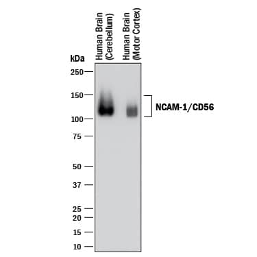
HAF018). A specific band was detected for NCAM-1/CD56 at approximately 100-150 kDa (as indicated). This experiment was conducted under reducing conditions and using
Immunoblot Buffer Group 1." alt="Western blot shows lysates of human brain (cerebellum) tissue and human brain (motor cortex) tissue. PVDF membrane was probed with 1 µg/mL of Mouse Anti-Human/Primate NCAM-1/CD56 Monoclonal Antibody (Catalog # MAB2408) followed by HRP-conjugated Anti-Mouse IgG Secondary Antibody (Catalog #
HAF018). A specific band was detected for NCAM-1/CD56 at approximately 100-150 kDa (as indicated). This experiment was conducted under reducing conditions and using
Immunoblot Buffer Group 1." class="big_thumb" />
MAB0041) followed by Anti-Mouse IgG APC-conjugated secondary antibody (
F0101B) and Mouse Anti-Human NKp46 PE-conjugated Monoclonal Antibody (
FAB1850P). Staining was performed using our Staining Membrane-associated Proteins protocol. " class="big_lightbox">
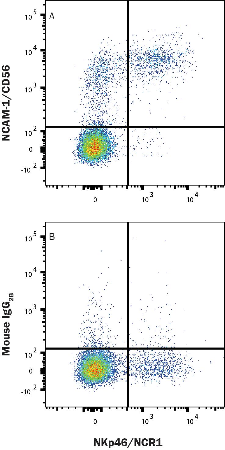
MAB0041) followed by Anti-Mouse IgG APC-conjugated secondary antibody (
F0101B) and Mouse Anti-Human NKp46 PE-conjugated Monoclonal Antibody (
FAB1850P). Staining was performed using our Staining Membrane-associated Proteins protocol. " class="big_thumb" />
CLD004) and were stained with either (A) Mouse Anti-Human NCAM-1/CD56 Monoclonal Antibody (Catalog # MAB24081) or (B) Mouse IgG2B Isotype Control (
MAB0041) followed by Anti-Mouse IgG APC-conjugated secondary antibody (
F0101B) and Mouse Anti-HumanCD3 PE-conjugated Monoclonal Antibody (
FAB100P). Staining was performed using our Staining Membrane-associated Proteins protocol. " class="big_lightbox">
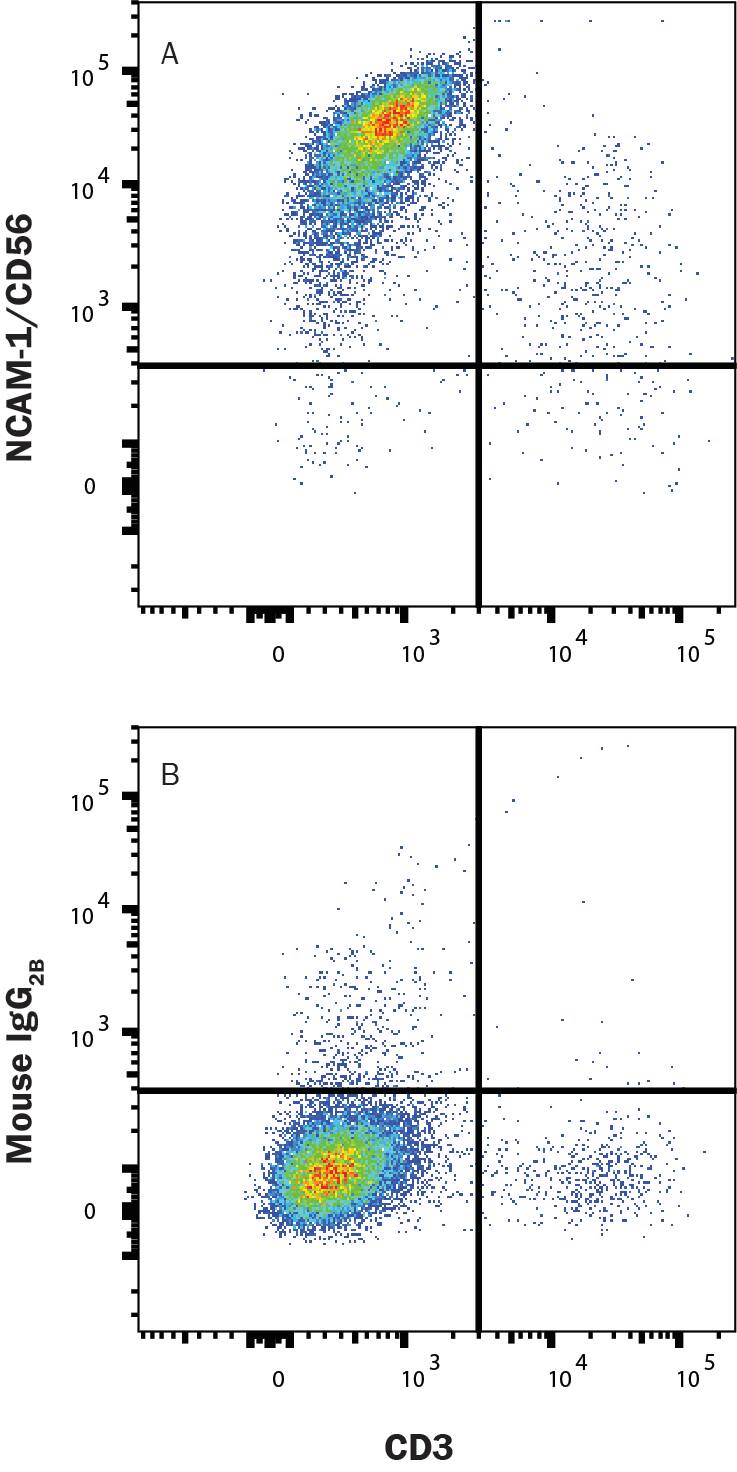
CLD004) and were stained with either (A) Mouse Anti-Human NCAM-1/CD56 Monoclonal Antibody (Catalog # MAB24081) or (B) Mouse IgG2B Isotype Control (
MAB0041) followed by Anti-Mouse IgG APC-conjugated secondary antibody (
F0101B) and Mouse Anti-HumanCD3 PE-conjugated Monoclonal Antibody (
FAB100P). Staining was performed using our Staining Membrane-associated Proteins protocol. " class="big_thumb" />
2408-NC) was serially diluted and captured by Mouse Anti-Human/Primate NCAM‑1/CD56 Monoclonal Antibody (Catalog # MAB2408) coated on a Clear Polystyrene Microplate (Catalog #
DY990). Goat Anti-Human/Mouse NCAM‑1/CD56 Antigen Affinity-purified Polyclonal Antibody (Catalog #
AF2408) was biotinylated and incubated with the protein captured on the plate. Detection of the standard curve was achieved by incubating Streptavidin-HRP (Catalog #
DY998)" class="big_lightbox">

2408-NC) was serially diluted and captured by Mouse Anti-Human/Primate NCAM‑1/CD56 Monoclonal Antibody (Catalog # MAB2408) coated on a Clear Polystyrene Microplate (Catalog #
DY990). Goat Anti-Human/Mouse NCAM‑1/CD56 Antigen Affinity-purified Polyclonal Antibody (Catalog #
AF2408) was biotinylated and incubated with the protein captured on the plate. Detection of the standard curve was achieved by incubating Streptavidin-HRP (Catalog #
DY998)" class="big_thumb" />
2408-NC) was serially diluted and captured by Mouse Anti-Human/Primate NCAM‑1/CD56 Monoclonal Antibody (Catalog # MAB2408) coated on a Clear Polystyrene Microplate (Catalog #
DY990). Goat Anti-Human/Mouse NCAM‑1/CD56 Antigen Affinity-purified Polyclonal Antibody (Catalog #
AF2408) was biotinylated and incubated with the protein captured on the plate. Detection of the standard curve was achieved by incubating Streptavidin-HRP (Catalog #
DY998)" class="big_lightbox">
2408-NC) was serially diluted and captured by Mouse Anti-Human/Primate NCAM‑1/CD56 Monoclonal Antibody (Catalog # MAB2408) coated on a Clear Polystyrene Microplate (Catalog #
DY990). Goat Anti-Human/Mouse NCAM‑1/CD56 Antigen Affinity-purified Polyclonal Antibody (Catalog #
AF2408) was biotinylated and incubated with the protein captured on the plate. Detection of the standard curve was achieved by incubating Streptavidin-HRP (Catalog #
DY998)" class="big_thumb" />
 HAF017). Specific bands were detected for NCAM-1/CD56 at approximately 100-150 kDa (as indicated). This experiment was conducted under reducing conditions and using Immunoblot Buffer Group 1." alt="Western blot shows lysates of human brain (cerebellum and motor cortex) tissue and mouse brain (cerebellum) tissue. PVDF membrane was probed with 0.5 µg/mL of Goat Anti-Human/Mouse NCAM-1/CD56 Antigen Affinity-purified Polyclonal Antibody (Catalog # AF2408) followed by HRP-conjugated Anti-Goat IgG Secondary Antibody (Catalog # HAF017). Specific bands were detected for NCAM-1/CD56 at approximately 100-150 kDa (as indicated). This experiment was conducted under reducing conditions and using Immunoblot Buffer Group 1." class="big_thumb" />
 DR647MS) and counterstained with DAPI (blue; Lunaphore Catalog # DR100). Specific staining was localized to the neuropil. Protocol available in COMET™ Panel Builder." alt="NCAM-1/CD56 was detected in immersion fixed paraffin-embedded sections of human brain Cortex using Mouse Anti-Human NCAM-1/CD56, Monoclonal Antibody (Catalog # MAB24081) at 25ug/mL at 37° Celsius for 8 minutes. Before incubation with the primary antibody, tissue underwent an all-in-one dewaxing and antigen retrieval preprocessing using PreTreatment Module (PT Module) and Dewax and HIER Buffer H (pH 9; Epredia Catalog # TA-999-DHBH). Tissue was stained using the Alexa Fluor™ 647 Goat anti-Mouse IgG Secondary Antibody at 1:200 at 37 ° Celsius for 2 minutes. (Yellow; Lunaphore Catalog # DR647MS) and counterstained with DAPI (blue; Lunaphore Catalog # DR100). Specific staining was localized to the neuropil. Protocol available in COMET™ Panel Builder." class="big_thumb" />
 HAF018). A specific band was detected for NCAM-1/CD56 at approximately 100-150 kDa (as indicated). This experiment was conducted under reducing conditions and using Immunoblot Buffer Group 1." alt="Western blot shows lysates of human brain (cerebellum) tissue and human brain (motor cortex) tissue. PVDF membrane was probed with 1 µg/mL of Mouse Anti-Human/Primate NCAM-1/CD56 Monoclonal Antibody (Catalog # MAB2408) followed by HRP-conjugated Anti-Mouse IgG Secondary Antibody (Catalog # HAF018). A specific band was detected for NCAM-1/CD56 at approximately 100-150 kDa (as indicated). This experiment was conducted under reducing conditions and using Immunoblot Buffer Group 1." class="big_thumb" />
 DR647RT) and counterstained with DAPI (blue; Lunaphore Catalog # DR100). Specific staining was localized to the neuropil. Protocol available in COMET™ Panel Builder." alt="CD56/NCAM1 was detected in immersion fixed paraffin-embedded sections of mouse Cerebellum using Rat Anti-Mouse CD56, Monoclonal Antibody (Catalog # MAB7820) at 15ug/mL at 37° Celsius for 4 minutes. Before incubation with the primary antibody, tissue underwent an all-in-one dewaxing and antigen retrieval preprocessing using PreTreatment Module (PT Module) and Dewax and HIER Buffer H (pH 9; Epredia Catalog # TA-999-DHBH).Tissue was stained using the Alexa Fluor™ 647 Goat anti-Rat IgG Secondary Antibody at 1:200 at 37 ° Celsius for 2 minutes. (Yellow; Lunaphore Catalog # DR647RT) and counterstained with DAPI (blue; Lunaphore Catalog # DR100). Specific staining was localized to the neuropil. Protocol available in COMET™ Panel Builder." class="big_thumb" />


 NL001). Actin filaments were stained with FITC-conjugated Phalloidin (green) and cell nuclei were counter-stained with DAPI (blue). NCAM-1/CD56 immuno-reactivity was localized to the plasma membrane. View our protocol for
  NL001) and counterstained with DAPI (blue). Specific staining was localized to cytoplasm. View our protocol for
  HAF109). This experiment was conducted under reducing conditions and using the 12-230 kDa separation system." class="big_thumb" />
 HAF017). A specific band was detected for NCAM‑1/CD56 at approximately 160 kDa (as indicated) in the parental U937 human histiocytic lymphoma cell line, but is not detectable in knockout U937 human histiocytic lymphoma cell line. GAPDH (
 2408-NC) was serially diluted and captured by Mouse Anti-Human/Primate NCAM‑1/CD56 Monoclonal Antibody (Catalog #
  2408-NC) was serially diluted and captured by Mouse Anti-Human/Primate NCAM‑1/CD56 Monoclonal Antibody (Catalog #
 ![Immunohistochemistry-Paraffin: NCAM-1/CD56 Antibody (123C3.D5) [NBP2-15184] - Analysis of NCAM-1/CD56 antibody (123C3.D5) on Human colon tissue. Incubation: 1 ug/ml for 30 minutes at room temperature. Image from verified customer review.](http://images.novusbio.com/fullsize/NCAM-1-CD56-Antibody-123C3-D5-Immunohistochemistry-Paraffin-NBP2-15184-img0019.jpg "Immunohistochemistry-Paraffin: NCAM-1/CD56 Antibody (123C3.D5) [NBP2-15184] - Analysis of NCAM-1/CD56 antibody (123C3.D5) on Human colon tissue. Incubation: 1 ug/ml for 30 minutes at room temperature. Image from verified customer review.")
![Flow Cytometry: NCAM-1/CD56 Antibody (123C3.D5) [NBP2-15184] - Flow cytometry of lymphocyte gated PBMCs unstained (gray) orstained with CF568-labeled NCAM-1/CD56 antibody (123C3.D5) (orange).](http://images.novusbio.com/fullsize/NCAM-1-CD56-Antibody-123C3-D5-Flow-Cytometry-NBP2-15184-img0020.jpg)
![Immunohistochemistry-Paraffin: NCAM-1/CD56 Antibody (123C3.D5) [NBP2-15184] - Formalin-fixed, paraffin-embedded human colon Ganglion stained with CD56 Monoclonal Antibody (123C3.D5)](http://images.novusbio.com/fullsize/NCAM-1-CD56-Antibody-123C3-D5-Immunohistochemistry-Paraffin-NBP2-15184-img0016.jpg)
![Flow Cytometry: NCAM-1/CD56 Antibody (123C3.D5) [NBP2-15184] - Analysis using the Azide and BSA Free version of NBP2-15184. Staining of AF700 conjugated CD56 in human PBMC using anti-CD56 antibody. The primary antibody was used at a dilution of 1:100, incubated for 25 min at 4C in 2% human serum, 0.5 mM EDTA in DPBS. Image from verified customer review.](http://images.novusbio.com/fullsize/NCAM-1-CD56-Antibody-123C3-D5-Flow-Cytometry-NBP2-15184-img0014.jpg)
![Simple Western: NCAM-1/CD56 Antibody (123C3.D5) [NBP2-15184] - Simple Western lane view shows a specific band for NCAM-1/CD56 in 0.2 mg/ml of IMR-32 lysate(s). This experiment was performed under reducing conditions using the 12-230 kDa separation system.](http://images.novusbio.com/fullsize/NCAM-1-CD56-Antibody-123C3-D5-Simple-Western-NBP2-15184-img0007.jpg)
![Simple Western: NCAM-1/CD56 Antibody (123C3.D5) [NBP2-15184] - Electropherogram image of the corresponding Simple Western lane. NCAM-1/CD56 antibody was used at 10 ug/ml dilution of IMR-32 lysates(s) respectively.](http://images.novusbio.com/fullsize/NCAM-1-CD56-Antibody-123C3-D5-Simple-Western-NBP2-15184-img0012.jpg)

 HAF018). A specific band was detected for NCAM‑1/CD56 at approximately 105-130 kDa (as indicated). This experiment was conducted under reducing conditions and using Western Blot Buffer Group 1." class="big_thumb" />
 MAB0041) followed by Anti-Mouse IgG APC-conjugated secondary antibody (
 CLD004) and were stained with either (A) Mouse Anti-Human NCAM-1/CD56 Monoclonal Antibody (Catalog # MAB24081) or (B) Mouse IgG2B Isotype Control (
 VC001) or the HRP-conjugated Anti-Mouse IgG Secondary Antibody (Catalog #
 ![Immunocytochemistry/Immunofluorescence: NCAM-1/CD56 Antibody (JF1021) [NBP2-66968] - Staining NCAM in N2A cells (red). The nuclear counter stain is DAPI (blue). Cells were fixed in paraformaldehyde, permeabilised with 0.25% Triton X100/PBS.](http://images.novusbio.com/fullsize/NCAM-1-CD56-Antibody-JF1021-Immunocytochemistry-Immunofluorescence-NBP2-66968-img0007.jpg "Immunocytochemistry/Immunofluorescence: NCAM-1/CD56 Antibody (JF1021) [NBP2-66968] - Staining NCAM in N2A cells (red). The nuclear counter stain is DAPI (blue). Cells were fixed in paraformaldehyde, permeabilised with 0.25% Triton X100/PBS.")
![Immunohistochemistry-Paraffin: NCAM-1/CD56 Antibody (JF1021) [NBP2-66968] - Analysis of paraffin-embedded human kidney tissue with Rabbit anti-NCAM-1/CD56 antibody washed with ddH2O and PBS, and then probed with the primary antibody at 1/200 dilution for 1 hour at room temperature. The detection was performed using an HRP conjugated compact polymer system. DAB was used as the chromogen. Tissues were counterstained with hematoxylin and mounted with DPX.](http://images.novusbio.com/fullsize/NCAM-1-CD56-Antibody-JF1021-Immunohistochemistry-Paraffin-NBP2-66968-img0011.jpg)
![Flow Cytometry: NCAM-1/CD56 Antibody (JF1021) [NBP2-66968] - Analysis of SH-SY-5Y cells with NCAM antibody at 1/50 dilution (red) compared with an unlabelled control (cells without incubation with primary antibody; black). Alexa Fluor 488-conjugated goat anti rabbit IgG was used as the secondary antibody.](http://images.novusbio.com/fullsize/NCAM-1-CD56-Antibody-JF1021-Flow-Cytometry-NBP2-66968-img0001.jpg)
![Immunocytochemistry/Immunofluorescence: NCAM-1/CD56 Antibody (JF1021) [NBP2-66968] - Staining NCAM in A549 cells (red). The nuclear counter stain is DAPI (blue). Cells were fixed in paraformaldehyde, permeabilised with 0.25% Triton X100/PBS.](http://images.novusbio.com/fullsize/NCAM-1-CD56-Antibody-JF1021-Immunocytochemistry-Immunofluorescence-NBP2-66968-img0002.jpg)
![Immunocytochemistry/Immunofluorescence: NCAM-1/CD56 Antibody (JF1021) [NBP2-66968] - Staining NCAM in SH-SY-5Y cells (red). The nuclear counter stain is DAPI (blue). Cells were fixed in paraformaldehyde, permeabilised with 0.25% Triton X100/PBS.](http://images.novusbio.com/fullsize/NCAM-1-CD56-Antibody-JF1021-Immunocytochemistry-Immunofluorescence-NBP2-66968-img0003.jpg)
![Immunohistochemistry-Paraffin: NCAM-1/CD56 Antibody (JF1021) [NBP2-66968] - Analysis of paraffin-embedded human tonsil tissue using anti-NCAM antibody. Counter stained with hematoxylin.](http://images.novusbio.com/fullsize/NCAM-1-CD56-Antibody-JF1021-Immunohistochemistry-Paraffin-NBP2-66968-img0004.jpg)
![Immunohistochemistry-Paraffin: NCAM-1/CD56 Antibody (JF1021) [NBP2-66968] - Analysis of paraffin-embedded zebrafish kidney tissue using anti-NCAM antibody. Counter stained with hematoxylin.](http://images.novusbio.com/fullsize/NCAM-1-CD56-Antibody-JF1021-Immunohistochemistry-Paraffin-NBP2-66968-img0005.jpg)
![<b>Biological Strategies Validation. </b>Immunohistochemistry-Paraffin: NCAM-1/CD56 Antibody (JF1021) [NBP2-66968] - Analysis of paraffin-embedded human tonsil tissue using anti-NCAM-1/CD56 antibody. The section was pre-treated using heat mediated antigen retrieval with Tris-EDTA buffer (pH 8.0-8.4) for 20 minutes.The tissues were blocked in 5% BSA for 30 minutes at room temperature, washed with ddH2O and PBS, and then probed with the primary antibody 1/50) for 30 minutes at room temperature. The detection was performed using an HRP conjugated compact polymer system. DAB was used as the chromogen. Tissues were counterstained with hematoxylin and mounted with DPX.](http://images.novusbio.com/fullsize/NCAM-1-CD56-Antibody-JF1021-Immunohistochemistry-Paraffin-NBP2-66968-img0010.jpg)
![Western Blot: NCAM-1/CD56 Antibody [NBP2-38452] - Analysis in human brain tissue.](http://images.novusbio.com/fullsize/NCAM-1-CD56-Antibody-Western-Blot-NBP2-38452-img0005.jpg "Western Blot: NCAM-1/CD56 Antibody [NBP2-38452] - Analysis in human brain tissue.")
![Immunocytochemistry/Immunofluorescence: NCAM-1/CD56 Antibody [NBP2-38452] - Staining of human cell line U-2 OS shows localization to plasma membrane & cytosol. Antibody staining is shown in green.](http://images.novusbio.com/fullsize/NCAM-1-CD56-Antibody-Immunocytochemistry-Immunofluorescence-NBP2-38452-img0004.jpg)


 MAB0041) followed by Anti-Mouse IgG APC-conjugated secondary antibody (
 CLD004) and were stained with either (A) Mouse Anti-Human NCAM-1/CD56 Monoclonal Antibody (Catalog # MAB24081) or (B) Mouse IgG2B Isotype Control (
 2408-NC) was serially diluted and captured by Mouse Anti-Human/Primate NCAM‑1/CD56 Monoclonal Antibody (Catalog # MAB2408) coated on a Clear Polystyrene Microplate (Catalog #
  2408-NC) was serially diluted and captured by Mouse Anti-Human/Primate NCAM‑1/CD56 Monoclonal Antibody (Catalog # MAB2408) coated on a Clear Polystyrene Microplate (Catalog #
  HAF005). A specific band was detected for NCAM-1/CD56 at approximately 120 kDa (as indicated). This experiment was conducted under reducing conditions and using
  MAB006, open histogram), followed by Allophycocyanin-conjugated Anti-Rat IgG Secondary Antibody (Catalog #
  VC005). Before incubation with the primary antibody, tissue was subjected to heat-induced epitope retrieval using VisUCyte Antigen Retrieval Reagent-Basic (Catalog #
  VC005). Before incubation with the primary antibody, tissue was subjected to heat-induced epitope retrieval using VisUCyte Antigen Retrieval Reagent-Basic (Catalog #
 ![Western Blot: NCAM-1/CD56 Antibody (NCAM1/795) [NBP2-44717] - Analysis of HCT116 Cell Lysate using CD56 Monoclonal Antibody (NCAM1/795)](http://images.novusbio.com/fullsize/NCAM-1-CD56-Antibody-NCAM1-795-Western-Blot-NBP2-44717-img0003.jpg "Western Blot: NCAM-1/CD56 Antibody (NCAM1/795) [NBP2-44717] - Analysis of HCT116 Cell Lysate using CD56 Monoclonal Antibody (NCAM1/795)")
![Immunohistochemistry-Paraffin: NCAM-1/CD56 Antibody (NCAM1/795) [NBP2-44717] - Formalin-fixed, paraffin-embedded human Pancreas stained with NCAM-1/CD56 Antibody (NCAM1/795).](http://images.novusbio.com/fullsize/NCAM-1-CD56-Antibody-NCAM1-795-Immunohistochemistry-Paraffin-NBP2-44717-img0004.jpg)
![Immunohistochemistry-Paraffin: NCAM-1/CD56 Antibody (NCAM1/795) [NBP2-44717] - Human Lung Carcinomastained with CD56 Monoclonal Antibody (NCAM1/795)](http://images.novusbio.com/fullsize/NCAM-1-CD56-Antibody-NCAM1-795-Immunohistochemistry-Paraffin-NBP2-44717-img0002.jpg)
![N/A NCAM-1/CD56 [Biotin]](http://images.novusbio.com/fullsize/elisa/DATA_NCAM1_DY2408_ELISA_1823.jpg "N/A NCAM-1/CD56 [Biotin]")

![Western Blot: NCAM-1/CD56 Antibody (1B9) [NBP2-46107] - Analysis of HEK293T cells were transfected with the pCMV6-ENTRY control (Left lane) or pCMV6-ENTRY NCAM1.](http://images.novusbio.com/fullsize/NCAM-1-CD56-Antibody-1B9-Western-Blot-NBP2-46107-img0006.jpg "Western Blot: NCAM-1/CD56 Antibody (1B9) [NBP2-46107] - Analysis of HEK293T cells were transfected with the pCMV6-ENTRY control (Left lane) or pCMV6-ENTRY NCAM1.")
![Immunohistochemistry: NCAM-1/CD56 Antibody (1B9) [NBP2-46107] - Analysis of Human embryonic cerebellum tissue. (Heat-induced epitope retrieval by 1 mM EDTA in 10mM Tris, pH8.5, 120C for 3min)](http://images.novusbio.com/fullsize/NCAM-1-CD56-Antibody-1B9-Immunohistochemistry-NBP2-46107-img0005.jpg)
![Immunohistochemistry: NCAM-1/CD56 Antibody (1B9) [NBP2-46107] - Analysis of Human adult heart tissue. (Heat-induced epitope retrieval by 1 mM EDTA in 10mM Tris, pH8.5, 120C for 3min)](http://images.novusbio.com/fullsize/NCAM-1-CD56-Antibody-1B9-Immunohistochemistry-NBP2-46107-img0001.jpg)
![Immunohistochemistry: NCAM-1/CD56 Antibody (1B9) [NBP2-46107] - Analysis of Human adult brain tissue. (Heat-induced epitope retrieval by 1 mM EDTA in 10mM Tris, pH8.5, 120C for 3min)](http://images.novusbio.com/fullsize/NCAM-1-CD56-Antibody-1B9-Immunohistochemistry-NBP2-46107-img0002.jpg)
![Immunohistochemistry: NCAM-1/CD56 Antibody (1B9) [NBP2-46107] - Analysis of Human muscle tissue. (Heat-induced epitope retrieval by 1 mM EDTA in 10mM Tris, pH8.5, 120C for 3min)](http://images.novusbio.com/fullsize/NCAM-1-CD56-Antibody-1B9-Immunohistochemistry-NBP2-46107-img0003.jpg)
![Immunohistochemistry: NCAM-1/CD56 Antibody (1B9) [NBP2-46107] - Analysis of Human embryonic brain cortex tissue. (Heat-induced epitope retrieval by 1 mM EDTA in 10mM Tris, pH8.5, 120C for 3min)](http://images.novusbio.com/fullsize/NCAM-1-CD56-Antibody-1B9-Immunohistochemistry-NBP2-46107-img0004.jpg)


